
Separations platform based upon electroosmosis-driven planar chromatography

Inactive Publication Date: 2005-12-08
INCHROMATICS
View PDF33 Cites 37 Cited by
  • Summary
  • Abstract
  • Description
  • Claims
  • Application Information

AI Technical Summary

Benefits of technology

[0012] One aspect of the present invention provides a high resolution protein, peptide and glycan separation system that employs a solid phase support and simple combinations of organic and aqueous mobile phases to facilitate the fractionation of biological species by a combination of electrophoretic and / or chromatographic mechanisms. The separation system includes mechanical stability of the separating medium, accessibility of the analytes to post-separation characterization techniques (immunodetection, mass spectrometry), ability to fractionate hydrophobic analytes and large molecular complexes, and minimizes sample consumption, number of manual manipulations and timelines for performing the actual fractionation.
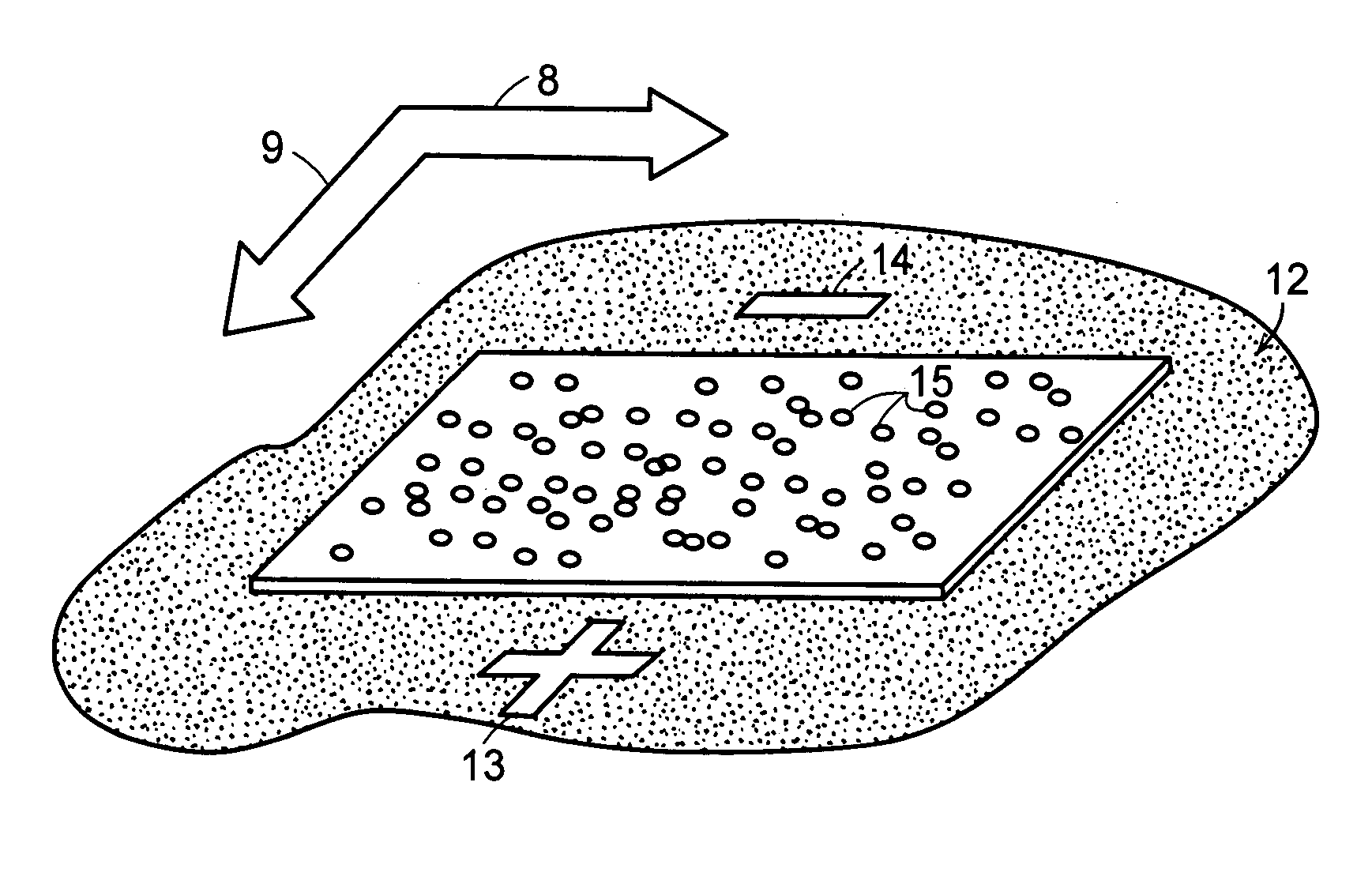
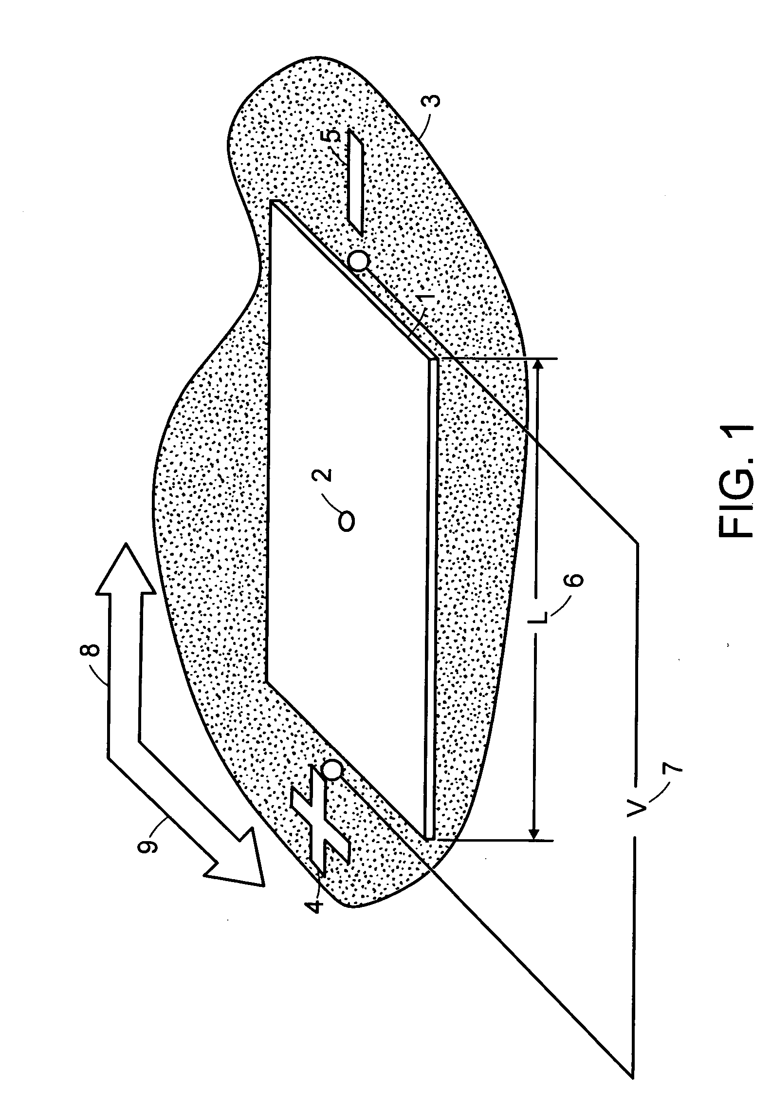
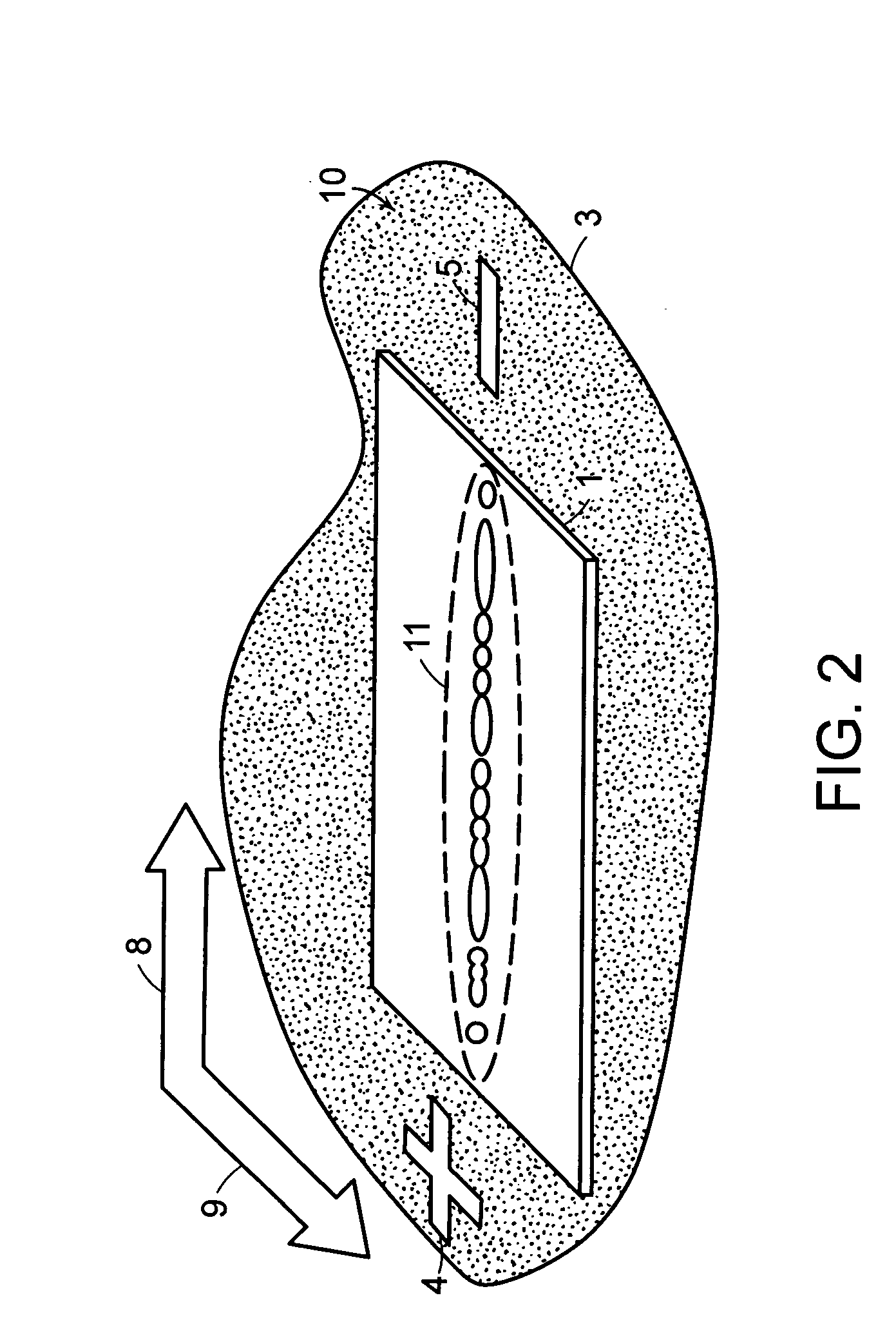
[0013] In one aspect of the invention, a method of separating biomolecules is provided. The method includes the steps of providing a sample comprising one or more biomolecules, loading the sample on a planar stationary phase, wherein the stationary phase is amphiphilic; contacting the stationary phase with a first liquid mobile phase, providing a first and a second electrode in electronic contact with opposing edges of the stationary phase; and creating an electrical field between the first electrode and the second electrode so as to cause the first liquid mobile phase to be advanced across the length of the stationary phase, whereby one or more biomolecules are separated.

Problems solved by technology

The level of complexity, coupled with the relative abundances of different proteins, presents unique challenges in terms of separations technologies.
Polyacrylamide gels are mechanically fragile, susceptible to stretching and breaking during handling.
Other limitations include difficulty in automating the separation process, low throughput of samples, and difficulty in detecting low abundance, extremely basic, very hydrophobic, very high molecular weight or very low molecular weight proteins.
While detection of proteins directly in gels with labeled antibodies or lectins has been accomplished, the approach is not generally applicable to every antigen and is relatively insensitive.
The polyacrylamide gel also poses difficulties in the identification of proteins by microchemical characterization techniques, such as mass spectrometry, since the gels must be macerated and rinsed, the proteins must be incubated with proteolytic enzymes, and peptides must be selectively retrieved and concentrated using a reverse-phase column prior to identification.
The 2DGE technique is poorly suited for the fractionation of hydrophobic proteins, particularly proteins containing two or more alpha-helical transmembrane domains, because the technique is based upon aqueous buffers and hydrophilic polymers.
The remaining 95-98% of the peptides are discarded, thus prohibiting a comprehensive analysis of the sample.
Additionally, such procedures are unable to distinguish among the various protein isoforms exhibited in a proteome that arise from differential mRNA splicing and post-translational modification due to a combination of poor sequence coverage and the sequence scrambling arising from the fragmentation process itself.
Another technique applied to the analysis of peptides and proteins is capillary electrochromatography (CEC), but its use has been limited to 1-D capillary separations of model analytes.
Thus, the proteins interact minimally with filter paper in aqueous medium, and once the applied current is removed the separation pattern will degrade rapidly due to diffusion.
The cellulose acetate membranes are considered extremely fragile for diagnostic applications in clinical settings and the generated profiles of very hydrophilic proteins, such as urinary and serum proteins, are poor compared to those generated with polyacrylamide gels.

Method used


Image

  • Separations platform based upon electroosmosis-driven planar chromatography
  • Separations platform based upon electroosmosis-driven planar chromatography
  • Separations platform based upon electroosmosis-driven planar chromatography

Examples


Embodiment Construction

[0048] System and methods for separation of biomolecules, e.g., proteins, peptides, amino acids, oligosaccharides, glycans and even small drug molecules, using electroosmosis-driven planar chromatography are described. In electroosmosis-driven planar chromatography an amphiphilic polymeric membrane, amphiphilic thin-layer chromatography plate or similar planar substrate provides the stationary phase for the separation platform. The planar substrate surface is characterized by a combination of charge carrying groups (ion exchangers), non-covalent groups (counterions), and nonionic groups that facilitate chemical interactions with the analyte, e.g., proteins or peptides. In a method for the separation of biomolecules using a planar electrochromatographic system, electroosmotic flow is generated by application of a voltage across the planar support in the presence of a miscible organic solvent-aqueous buffer mobile phase. Charged ions accumulate at the electrical double layer of the so...


PUM

PropertyMeasurementUnit
Diameteraaaaaaaaaa
Diameteraaaaaaaaaa
Diameteraaaaaaaaaa

Abstract

The present invention describes a system and method for separation of proteins, peptides and glycans by one-dimensional or two-dimensional electroosmosis-driven planar chromatography. Separation is performed using amphiphilic polymeric membranes, amphiphilic thin-layer chromatography plates or other planar amphiphilic surfaces as the stationary phase with a combination of organic and / or aqueous buffers as the mobile phase. Systematic selection of stationary phase supports, mobile phase buffers and operating conditions allow for the adaptation of the invention to a broad range of applications in proteomics, mass spectrometry, drug discovery and the pharmaceutical sciences.

Description

CROSS-REFERENCE TO RELATED APPLICATIONS [0001] The application claims benefit of U.S. provisional patent application No. 60 / 521,250, filed Mar. 19, 2004.FIELD OF THE INVENTION [0002] The present invention generally relates to the separation of proteins, peptides and glycans using electroosmosis-driven planar chromatography. The present invention also relates to systems and methods for separating biomolecules using planar electrochromatography. BACKGROUND OF INVENTION [0003] The human proteome is known to contain approximately 30,000 different genes. But, due to post-translational modifications and differential mRNA splicing, the total number of distinct proteins is most likely to be close to one million. The level of complexity, coupled with the relative abundances of different proteins, presents unique challenges in terms of separations technologies. Analytical methods for the simultaneous quantitative analysis of the abundances, locations, modifications, temporal changes and inter...

Claims


Application Information

Patent Timeline
IPC IPC(8): B01D15/08C07K1/26G01N27/447
CPCC07K1/26C07K1/285G01N27/44747G01N2030/945G01N30/92G01N2030/8813G01N30/58
Inventor PATTON, WAYNE F.SCHERMER, MACK J.
Owner INCHROMATICS
Learn More