
Method and kit for donor specific complement-fixing antibodies crossmatch

a technology of complement-fixing antibodies and kits, which is applied in the field of complement-fixing antibodies (cfabs) measurement, can solve the problems of neither fixing complements nor inducing cdc effects, organ transplantation failure, and low sensitivity

Inactive Publication Date: 2005-12-15
CHEN GE
View PDF1 Cites 19 Cited by
  • Summary
  • Abstract
  • Description
  • Claims
  • Application Information

AI Technical Summary

Benefits of technology

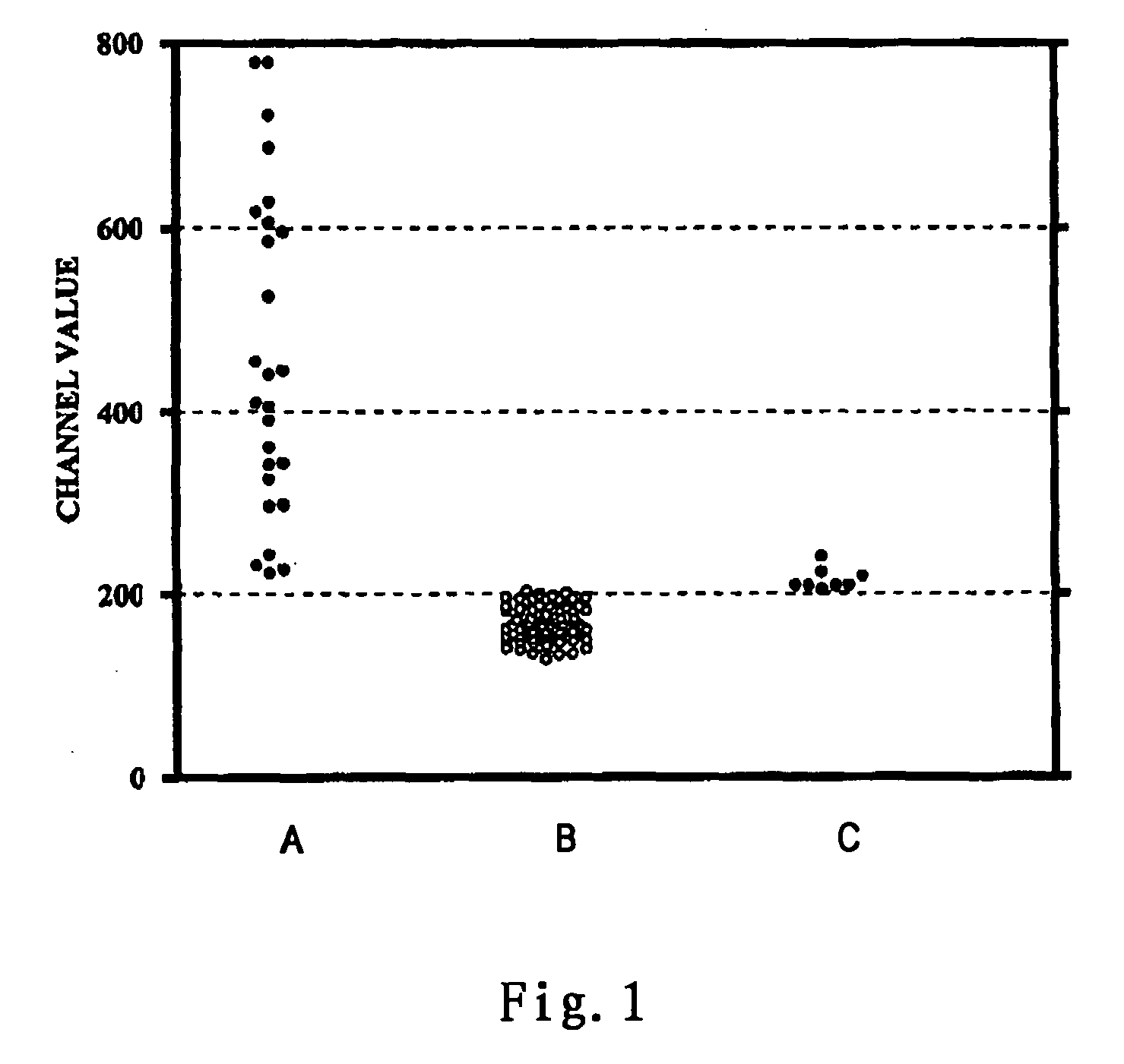
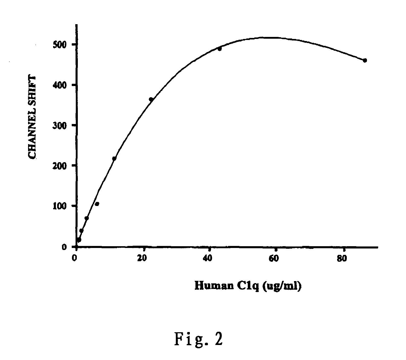
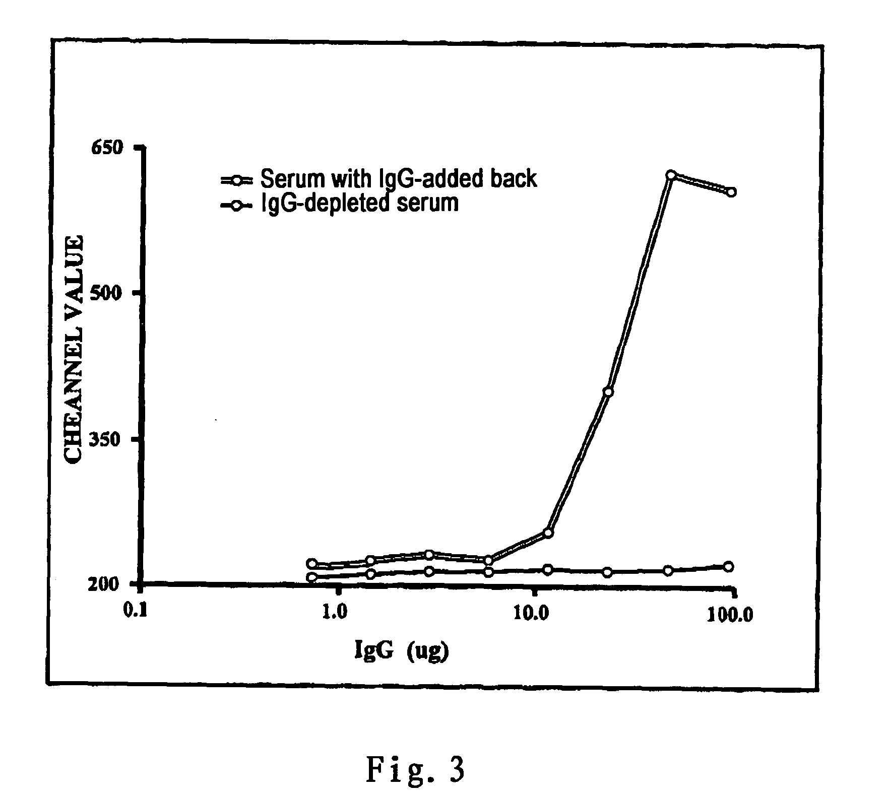
[0015] According to the basic principles of CDC effects, the present invention establishes a method based upon immunological methodology for evaluating CDC effects by detecting the quantity of the complements fixed by CFAbs. During the initial stage of CDC effect, CFAbs bind with corresponding HLA antigens on cell surface and fix complements, the quantity of complements fixed by CFAbs has directly correlated to the strength of CDC effect (cells death). Therefore, CDC effects can be directly evaluated by determining the quantity of complements fixed by CFAbs. On the other hand, labeled antibodies against the characteristic antigens on cell surface can be introduced into the same reaction system of the method to simultaneousely identify CDC effects on different particular cell populations. Consequently, the method can selectively determine CDC effects on different cell populations in a same tube.
[0050] Because binding activity of the CFAbs and complements in serum can be kept at low temperature for a long time (several years), the method of the invention has stability and good reproducibility in methodology.

Problems solved by technology

This recognition sequentially activates both the humoral and cellular immune systems of the recipient to attack and destroy the graft and result in organ transplantation failure.
Although they are able to specifically bind to HLA molecules, they neither fix complements nor induce CDC effects.
Besides, it has other disadvantages like time-consuming, low sensitivity, technical complexity, allogeneic complements use, and many other uncontrollable test conditions.
The major disadvantage of this category of methods is that the existence of natural HLA antibodies (Non-CFAbs) have been neglected.
Therefore, the above-mentioned methods have obvious disadvantages in terms of principles of methodology, and can't differentiate the above two categories of antibodies that have totally different biological functions.
In clinical organ transplantation, the results obtained by these methods are often showing false positives, which mislead the selection of donors and result in exacerbation or death of certain patients in urgent need of graft due to losing the chance of transplantation.
The above studies show that non-CFAbs are also detected in flow cytometry crossmatching which leads to false positive results, and that non-CFAbs are not closely correlated to clinical outcome or even contrary to the same.
However, these two types of antibodies can't be detected by routine flow cytometry cross-match.
However, IVIG binding is detectable by flow cytometry IgG crossmatch and gives a false positive crossmatch.
In this case, a positive crossmatch is often contrary to clinical outcome in the patient who was under the IVIG treatment, and may mislead physicians in connection with the real imunological state of the patients and therefore immunosuppressive treatment.
Another significant disadvantage of the existing methods for determining HLA antibodies is time-consuming.
All of the crossmatch methods that are currently used in transplantation practice are not perfectly meet the demands mentioned above.

Method used


Image

  • Method and kit for donor specific complement-fixing antibodies crossmatch
  • Method and kit for donor specific complement-fixing antibodies crossmatch
  • Method and kit for donor specific complement-fixing antibodies crossmatch

Examples


example 1

T Cell Crossmatch Method

[0066] (1) To a 1.5 ml Eppendorf centrifuge vial, 0.1˜0.25×10⁶ mononuclear cells of the donor prepared by Ficoll-Hypaque density-gradient centrifugation method was added.

[0067] (2) The cells were centrifuged at 800 g for 5 minutes and the supernatant was aspirated.

[0068] (3) 25 ul test serum of the recipient was added to the vial and mixed well genteelly. Then the mixture was incubated for 10 minutes at room temperature (˜25° C.).

[0069] (4) 30 ul FITC-anti human C1q (BIODESIGN; 1:30 diluted in 5% FCS / HBSS) and 10 ul PerCP CD3 (BD Biosciences) were added and mixed well. The mixture was incubated for an additional 20 minutes at 4° C.

[0070] (5) 0.5 ml wash buffer (5% FCS / HBSS) was added to the reaction mixture. The mixture was centrifuged and the supernatant was aspirated. The washing was repeated once again.

[0071] (6) After the second wash, the cell pellet was resuspended in 0.5 ml 1% paraformaldehyde. The samples were analyzed on a flow cytometer (FACSca...

example 2

Direct Cross-Match Method

[0072] (1) The serum of the recipient was incubated at 56° C. for 30 minutes to inactivate autologous complements.

[0073] (2) 25 ul of the above serum and 10 ul of FITC-C1q were added to a test tube containing 0.1˜0.25×10⁶ mononuclear cells from the donor, and the mixture was incubated at room temperature for 10 minutes.

[0074] (3) 10 ul PerCP CD3 (BD Biosciences) was added to the tube and mixed well. The mixture was incubated for an additional 20 minutes at 4° C.

[0075] (4) The samples were washed and analyzed according to the same process as in the steps (5) and (6) in Example 1

example 3

B Lymphocyte Cross-Match

[0076] The substantially same procedures as in Example 1 were carried out except that PerCP CD3 was replaced with PE labeled CD19.


PUM


Abstract

Provided is a flow cytometry method and kit composition for semi-quantitatively determining complement fixing antibodies (CFAbs). The said antigen specific CFAbs in the sample will react with the antigens on the solid carriers or the surfaces of target cells as well as the labeled complement or labeled anti-complement antibody at the same time; and the labeled target cell specific antibody will bind to the target cell surfaces. Then the sample is analyzed to determine the amount of complements fixed by CFAbs, especially the amount of complements fixed by CFAbs on the surfaces of particular target cells or donor specific HLA antigens pre-captured by corresponding antibodies fixed on solid microparticles for evaluating whether there is CFAbs and their relative concentration.

Description

[0001] This specification is related to provisional application No. 60 / 578,354 filed on Jun. 10, 2004.TECHNICAL FIELD [0002] The invention relates to a method for complement-fixing antibodies (CFAbs) measurement. Complement-fixing antibodies are directly or indirectly measured by detecting the complements fixed by CFAbs in an antigen-antibody reaction system. The method of the invention is particularly suitable for cross-match in organ transplantation. TECHNICAL BACKGROUND [0003] Rejection in organ transplantation is the immunological responses to the allogeneic graft (non-self) by the recipient's immune system. When the proteins on the surface of graft (non-self antigens) contact the immune system of the recipient, the non-self antigens are immunologically recognized. This recognition sequentially activates both the humoral and cellular immune systems of the recipient to attack and destroy the graft and result in organ transplantation failure. [0004] In the graft rejection of organ...

Claims


Application Information

Patent Timeline
IPC IPC(8): G01N33/53G01N33/555G01N33/564G01N33/567
CPCG01N2333/4716G01N33/564
Inventor CHEN, GE
Owner CHEN GE
Learn More