Count adaptive noise reduction method of x-ray images

a noise reduction and x-ray imaging technology, applied in image enhancement, image analysis, instruments, etc., can solve the problems of reducing the contrast of useful information, reducing the accuracy of x-ray image processing, and exhibiting such noise in a readily visible manner, so as to reduce noise, reduce noise, and smooth high attenuated x-ray image regions

Inactive Publication Date: 2006-01-12
GE MEDICAL SYST GLOBAL TECH CO LLC
View PDF14 Cites 24 Cited by
  • Summary
  • Abstract
  • Description
  • Claims
  • Application Information

AI Technical Summary

Benefits of technology

[0010] The embodiments of the present invention provide several advantages. One such advantage is the provision of generating a noise reduced image that is structure dependent and count dependent. This dependency prevents the removal or filtering of at least somewhat visible structures with relatively low counts or pixel intensities.
[0011] Another advantage that is provided by an embodiment of the present invention is the provision of smoothing highly attenuated x-ray image regions more than relatively low attenuated regions. Noise reduction is performed while minimizing contrast degradation in high signal-to-noise regions of the x-ray images.
[0012] Yet another advantage provided by an embodiment of the present invention is the provision of performing limiting noise reduction in low signal-to-noise regions where significant structures are present, thus maintaining clinical detail.
[0013] A further advantage provided by an embodiment of the present invention is the provision of modulating smoothing in response to visually relevant structures and graininess pertaining to noise of the x-ray images. This aids in inadvertently filtering desired structure data.
[0014] Moreover, the embodiments of the present invention provide an adaptive filtering technique that is easily implemented, computationally efficient, and offers options for image enhancement and time optimization.

Problems solved by technology

Modalities such as x-ray imaging and optical imaging, where image data is directly acquired, exhibit such noise in a readily visible manner.
However, imaging methods requiring image reconstruction, such as MRI, CT, and ultrasound, convert point or spike noise into splotches or small streaks that are usually hidden with the patterned noise.
Unfortunately, current methods designed to mitigate patterned noise do not adequately mitigate point noise without blurring or decreasing the contrast of the useful information in the processed image.
During the display processing of the x-ray images, the image noise at highly attenuated regions in the images becomes more noticeable, and can therefore decrease the perceived quality of the final images.
This perceived image degradation is mainly due to the contrast enhancement that occurs in the highly attenuated regions of the image.
However, this noise reduction decreases the contrast at lowly attenuated regions of the images, such as in lung parenchyma regions.
However, certain structures that are desired for evaluation and that have relatively low counts, such that they are partially visible, when heavily filtered are also undesirably removed from an image.

Method used


Image

  • Count adaptive noise reduction method of x-ray images
  • Count adaptive noise reduction method of x-ray images
  • Count adaptive noise reduction method of x-ray images

Examples


Embodiment Construction

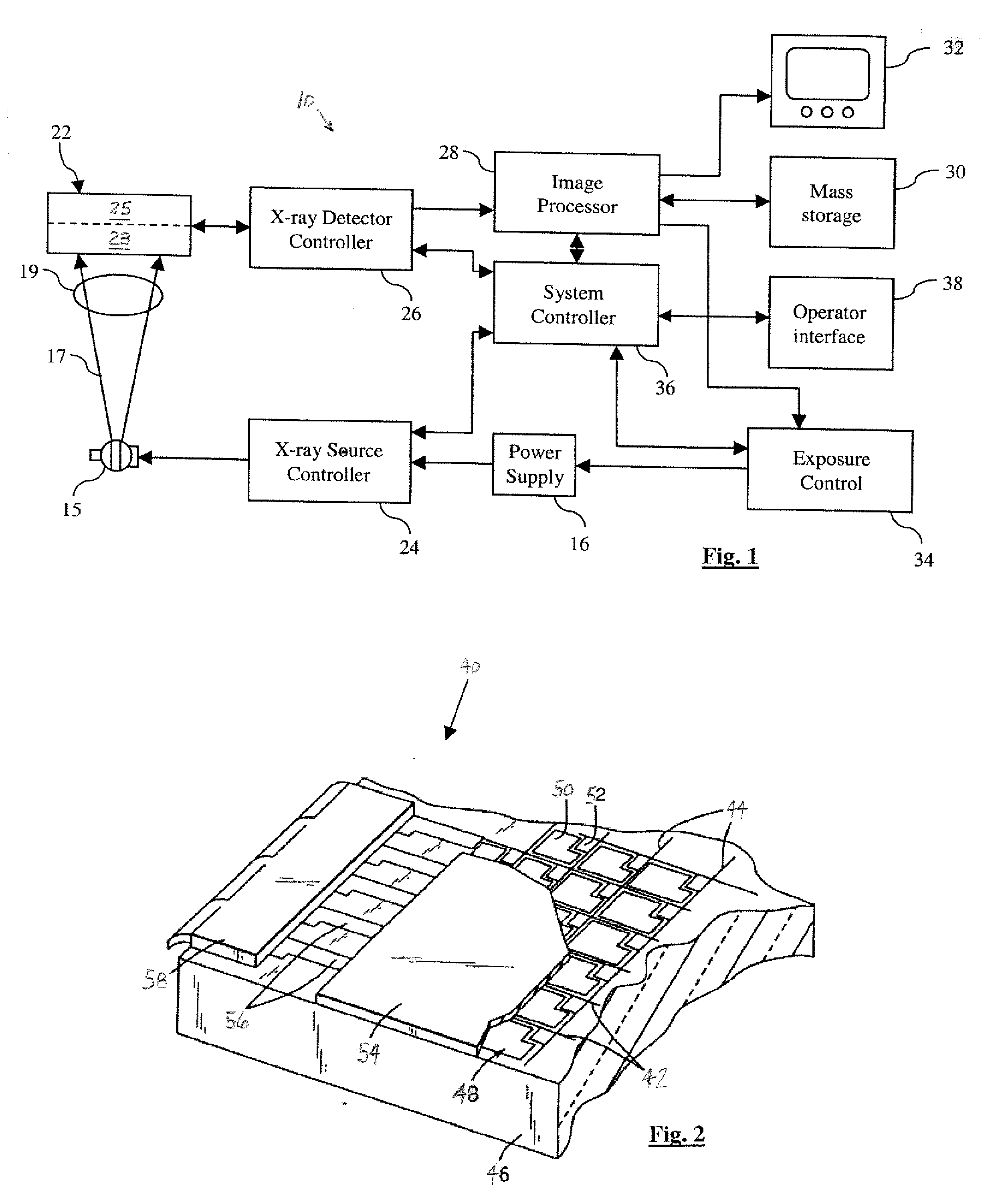
[0029] In the following figures, the same reference numerals will be used to refer to the same components. While the present invention is described with respect to the reduction of noise within x-ray images of an x-ray system, the present invention is capable of being adapted for various purposes and is not limited to the following applications: computed tomography systems, radiotherapy or radiographic systems, x-ray imaging systems, ultrasound imaging systems, magnetic resonance imaging systems, positron emission tomography systems, electron beam imaging systems, tomosynthesis systems, and other applications known in the art.

[0030] In the following description, various operating parameters and components are described for one constructed embodiment. These specific parameters and components are included as examples and are not meant to be limiting.

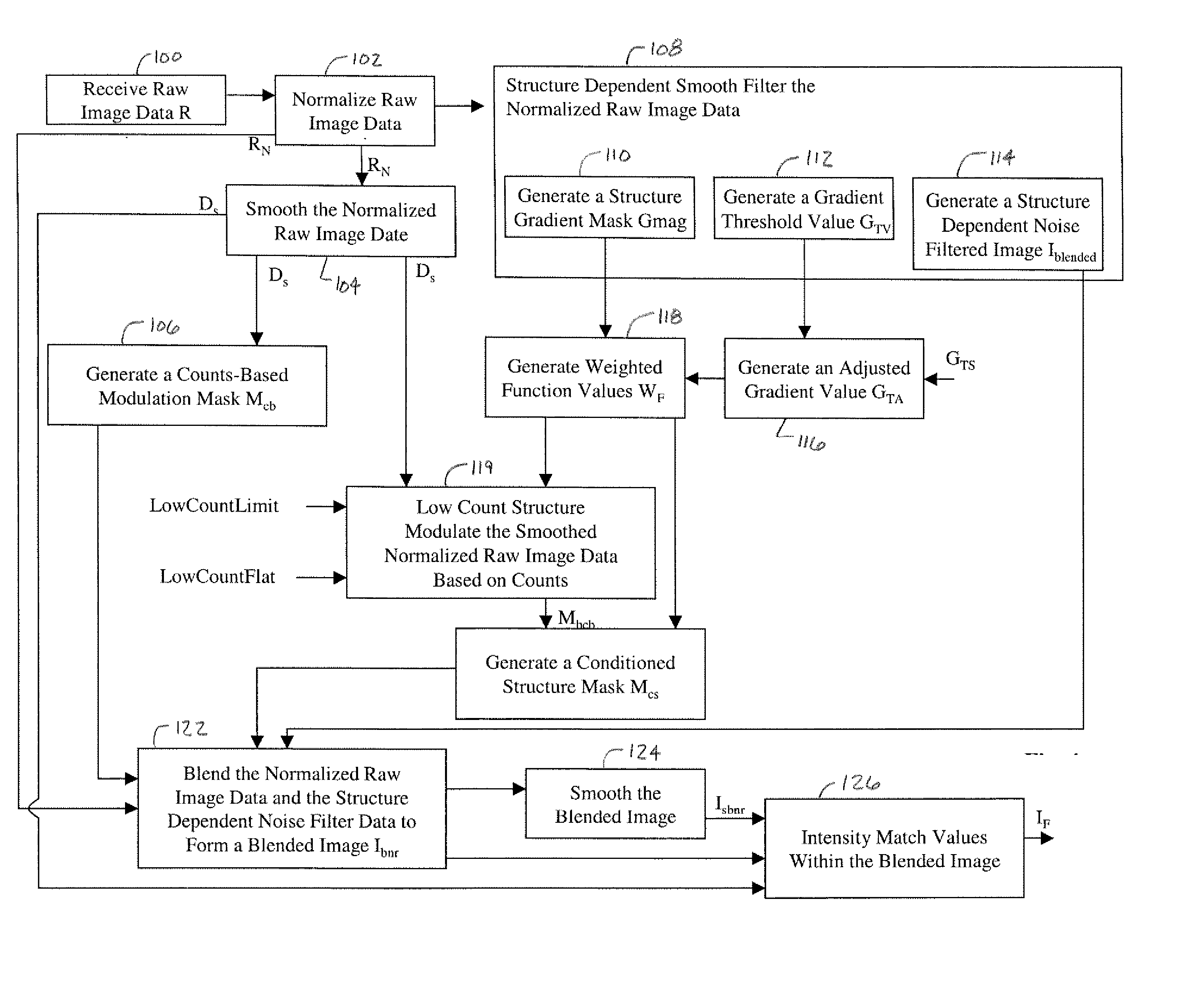
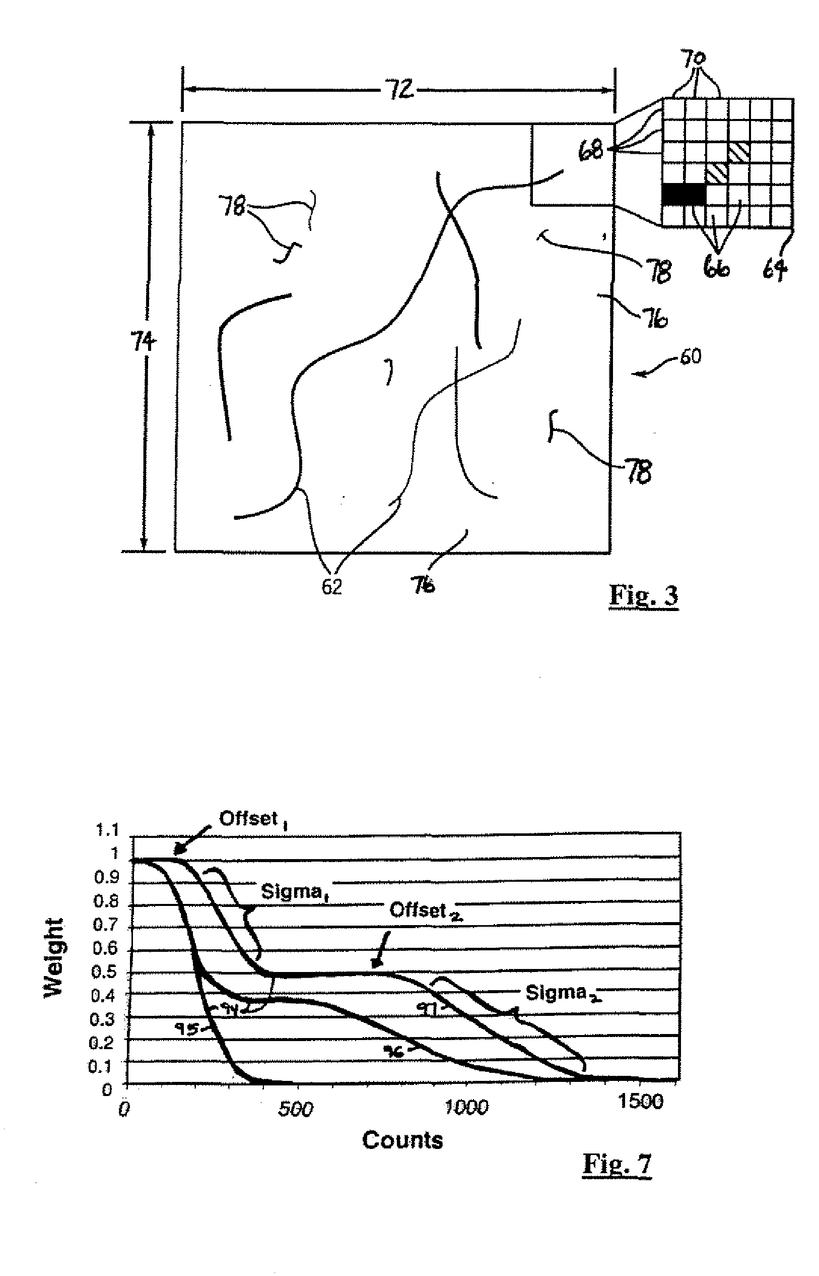
[0031] Also, in the following description the term “mask” may refer to a “look-up” table, a conversion scale, a function, or the like. ...


PUM


Abstract

A method of adaptively reducing noise within an x-ray image includes receiving raw data (R) representing a detected x-ray signal from an object. A counts-based modulation mask (Mcb) is generated in response to the raw data (R). In one embodiment, a structure dependent noise filtered image (Iblended) is generated in response to the raw data. A noise-reduced image (IF) is generated in response to the counts-based modulation mask (Mcb) and the structure dependent noise filtered image (Iblended). In another embodiment, a structure gradient mask (Mcs) is generated in response to the raw data (R). The noise-reduced image (IF) is generated in response to the counts-based modulation mask (Mcb) and the structure gradient mask (Mcs).

Description

BACKGROUND OF INVENTION [0001] The present invention relates generally to x-ray imaging systems. More particularly, the present invention relates to the reduction of noise within x-ray images at highly attenuated regions without affecting image contrast at relatively low attenuated regions. [0002] Many techniques are known and are presently in use for the generation of digital image data. Such techniques range from the use of simple charge coupled device apparatuses, such as digital cameras, to more complex imaging systems, such as those used for part inspection and medical diagnostics purposes. The stated systems, in general, form a matrix of discrete picture elements or pixels that have individual values over a range of intensities. Raw image data acquired by the stated systems may be processed to clarify an image, to enhance image features, or to improve the image quality from various points of view. In general, the goal of image enhancement and quality improvement is to provide ...

Claims


Application Information

Patent Timeline
Patent Type & Authority Applications(United States)
IPC IPC(8): G06K9/40
CPCG06T5/20G06T5/002G06T2207/30004G06T2207/20012G06T2207/10116
Inventor AVINASH, GOPAL B.MARINELLI, NICHOLAS L.JABRI, KADRI N.
Owner GE MEDICAL SYST GLOBAL TECH CO LLC
Why Eureka
Try Eureka