

Method and composition for skin grafts
a skin graft and composition technology, applied in the field of skin graft composition, can solve the problems of skin integrity loss, mortality or morbidity, sepsis or systemic infection,
- Summary
- Abstract
- Description
- Claims
- Application Information
AI Technical Summary
Problems solved by technology
Method used
Image

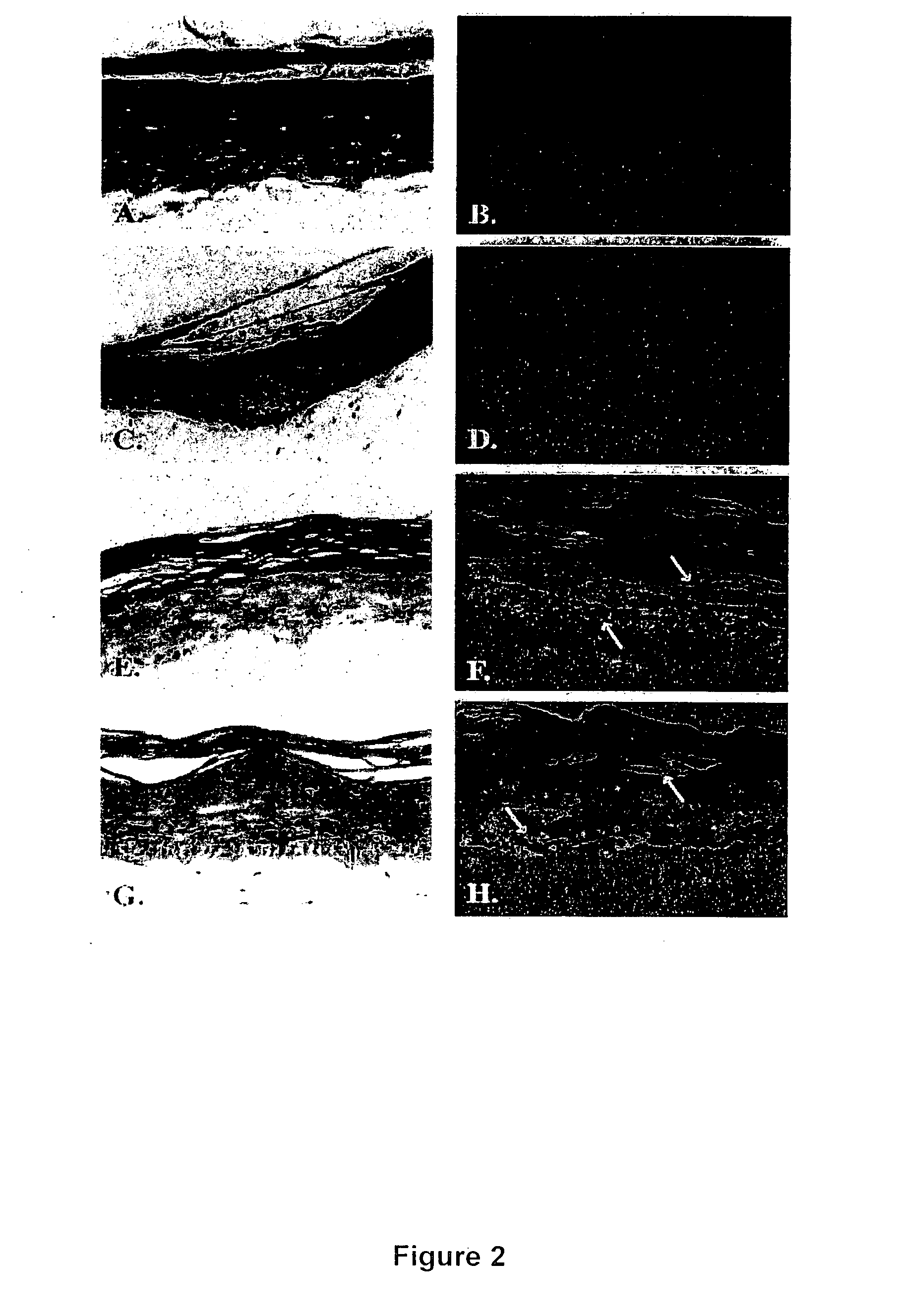
Examples
examples
Methods
Cell Culture Methods:
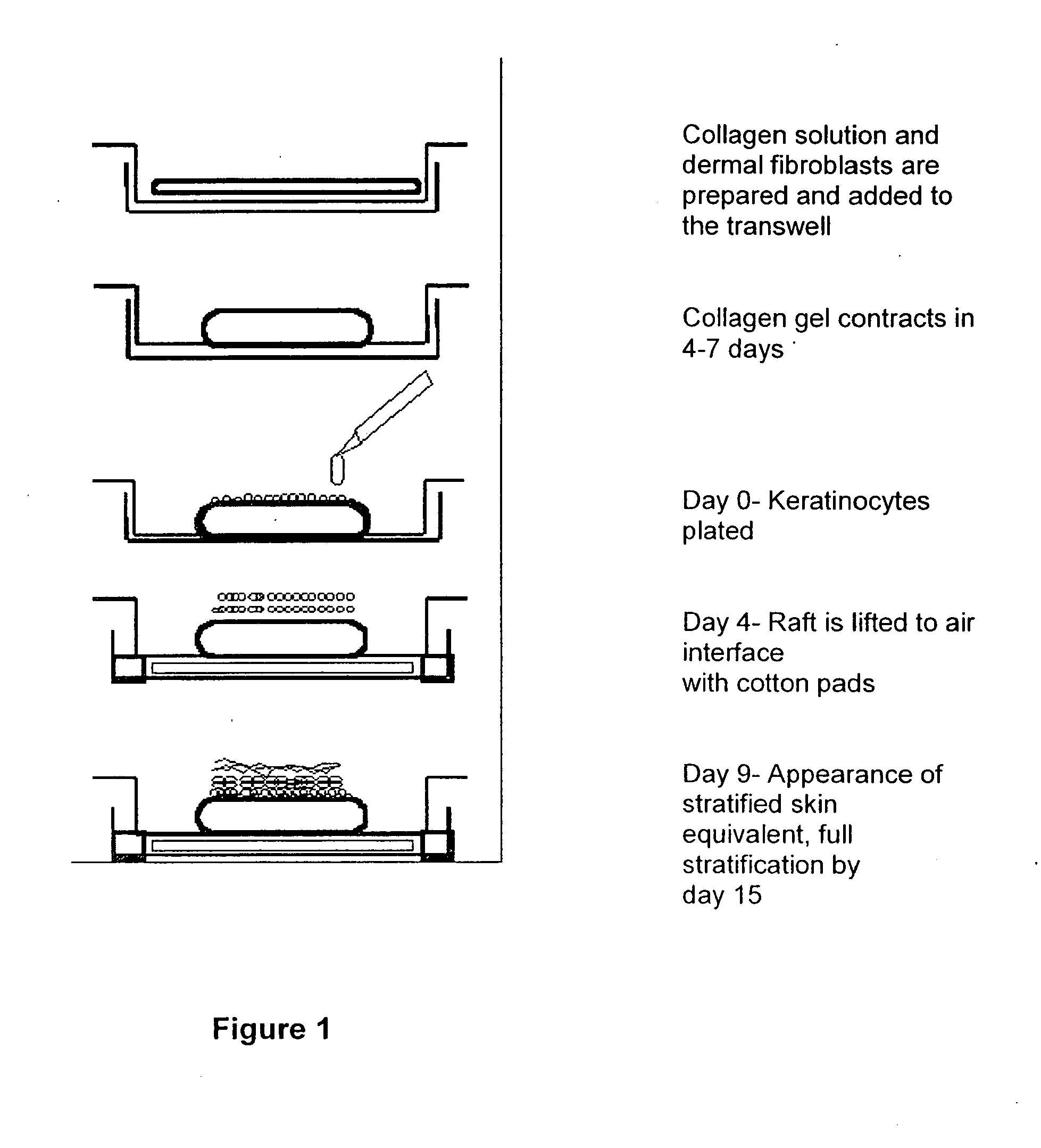
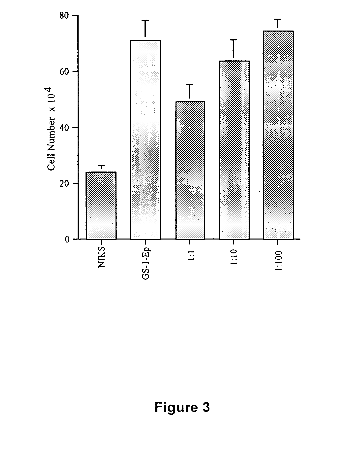
[0067] Donor keratinocytes (GS-1-EP, LAW-1-EP) were isolated from newborn human foreskins. Samples were obtained after circumcision under the approval of the hospital's Human Subjects Committee and Institutional Biosafety Committee. GS-1-EP, LAW-1-EP and NIKS keratinocyte cultures were established by plating aliquots of a single cell suspension in the presence of mitomycin C-treated Swiss mouse 3T3 fibroblasts (mito-3T3) as previously described (6). The standard keratinocyte culture medium was composed of a mixture of Ham's F-12 medium:Dulbecco's modified Eagle's medium (DME), (3:1, final calcium concentration 0.66 mM) supplemented with 2.5% fetal calf serum (FCS), 0.4 μg / ml hydrocortisone (HC), 8.4 ng / ml cholera toxin (CT), 5 μg / ml insulin (Ins), 24 μg / ml adenine (Ade), 10 ng / ml epidermal growth factor (EGF), 100 units penicillin and 100 μg / ml streptomycin (P / S). Cells were sub-cultured at weekly intervals at 3×10⁵ cells per 100-mm tissue culture dis...
PUM
| Property | Measurement | Unit |
|---|---|---|
| time | aaaaa | aaaaa |
| molecular weight | aaaaa | aaaaa |
| concentration | aaaaa | aaaaa |
Abstract
Description
Claims
Application Information