Method for detecting acetyltransferase and deacetylase activities and method for screening inhibitors or enhancers of these enzymes

a technology which is applied in the field of acetyltransferase and deacetylase activities detection and screening methods for inhibitors or enhancers of these enzymes, can solve the problems of difficult assaying many samples under numerous conditions, cumbersome methods known for measuring acetyltransferase and deacetylase activities, etc., and achieves convenient screening and convenient detection of a

Inactive Publication Date: 2006-02-16
MEDICAL & BIOLOGICAL LAB CO LTD
View PDF10 Cites 2 Cited by
  • Summary
  • Abstract
  • Description
  • Claims
  • Application Information

AI Technical Summary

Benefits of technology

[0008] An objective of the present invention is to provide a method for conveniently detecting acetyltransferase and deacetylase activities, a method for conveniently screening inhibitors or enhancers for acetyltransferase and deacetylase, and a kit for detecting and screening these.
[0009] As a result of strenuous research to solve the above problems, the present inventors came upon the idea of using an antibody specifically binding to an acetylated peptide (an anti-acetylated peptide antibody) for the detection of the acetyltransferase and deacetylase activities. Thus, an anti-acetylated peptide antibody was prepared, and the acetylation reaction of a peptide substrate by acetyltransferase, and the deacetylation reaction of an acetylated peptide substrate by deacetylase were executed. After the completion of the reactions, the acetyl group bound to the peptide substrate was detected. As a result, the present inventors found that the acetyltransferase and deacetylase activities of proteins can be conveniently detected by using an anti-acetylated peptide antibody.
[0010] Moreover, the present inventors discovered that inhibitors and enhancers for acetyltransferase and deacetylase could be conveniently screened by using the system for detecting the acetyltransferase and deacetylase activities utilizing the anti-acetylated peptide antibody.

Problems solved by technology

The methods known for measuring the acetyltransferase and deacetylase activities are, however, very cumbersome.
These measurement systems are so cumbersome that assaying many samples under numerous conditions is difficult.

Method used


Image

  • Method for detecting acetyltransferase and deacetylase activities and method for screening inhibitors or enhancers of these enzymes
  • Method for detecting acetyltransferase and deacetylase activities and method for screening inhibitors or enhancers of these enzymes
  • Method for detecting acetyltransferase and deacetylase activities and method for screening inhibitors or enhancers of these enzymes

Examples


example 1

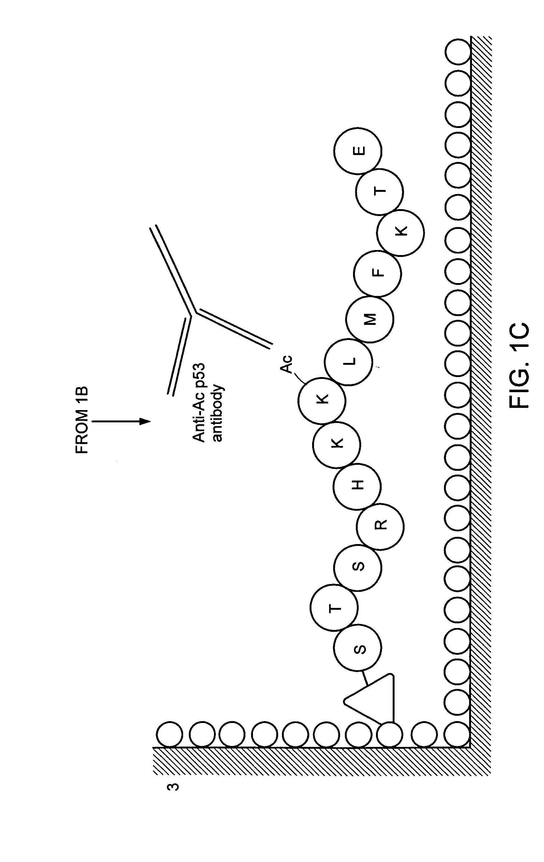
Preparation of an Anti-Acetylated Peptide Antibody

1. Preparation of an Immunogen

(1) Preparation of a Peptide

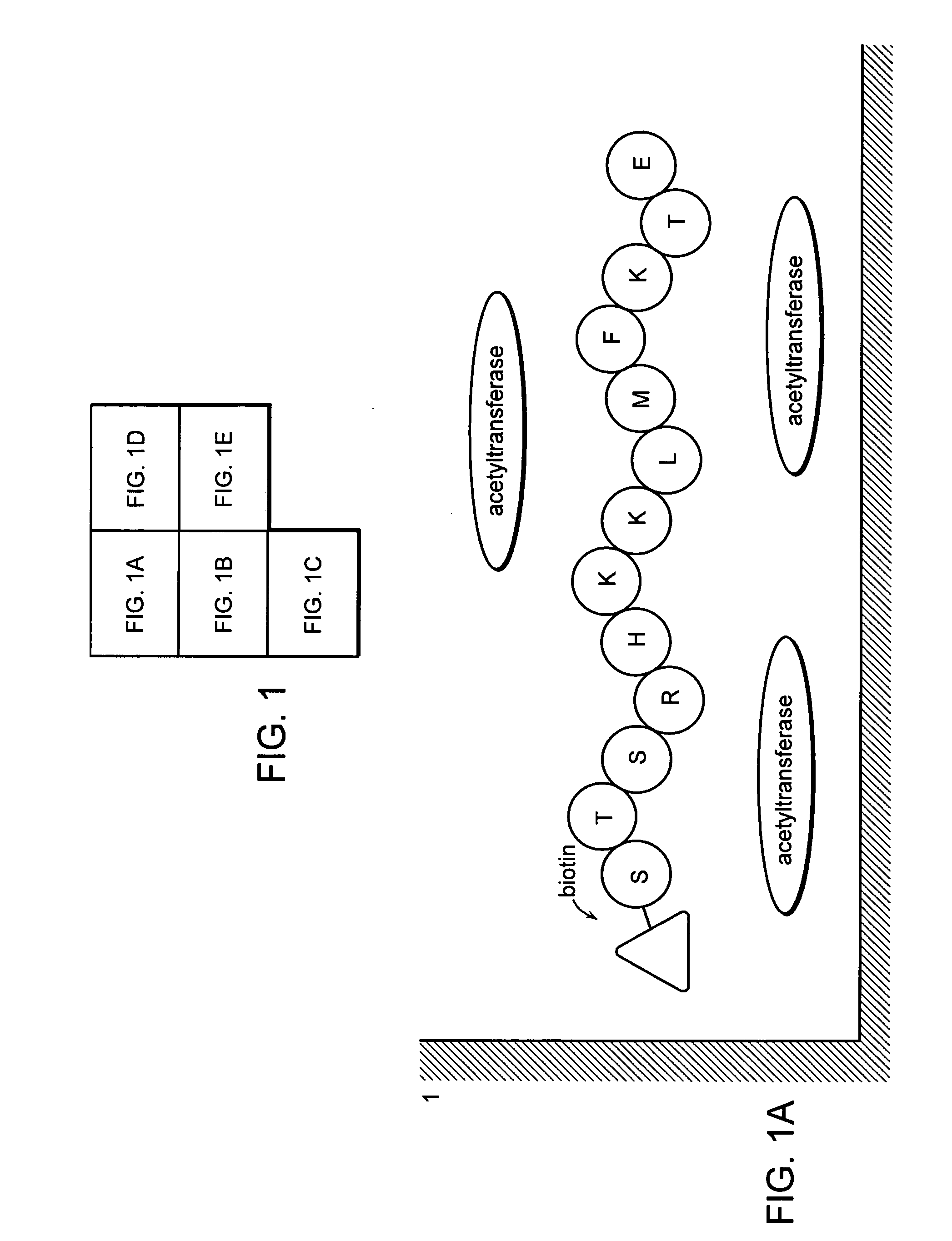
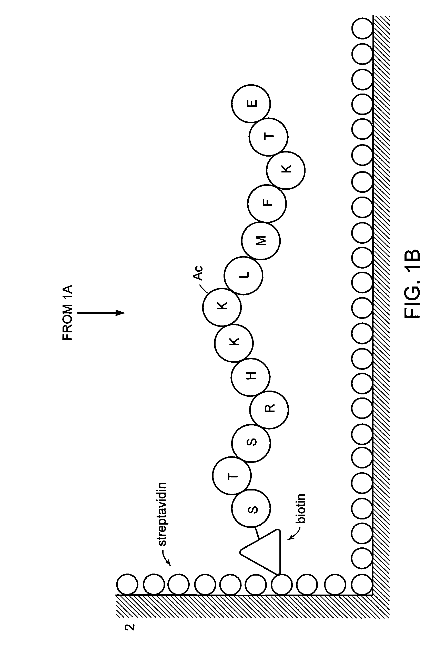
[0103] Four peptides comprising the 373rd and 382nd lysine residues of human p53, reported as acetylation sites were prepared by a peptide synthesizer. These peptides are “Ac p53-1” (SEQ ID NO: 1 / STSRHKK (Ac) LMFKTEC), “p53-1” (SEQ ID NO: 2 / STSRHKKLMFKTEC), “Ac p53-2” (SEQ ID NO: 3 / SHLKSKK (Ac) GQSTSRC) and “p53-2” (SEQ ID NO: 4 / SHLKSKKGQSTSRC). Amino acids are given in one-letter codes, and K(Ac) indicates an acetylated lysine residue. HPLC confirmed that the purity of synthesized peptides was 90% or more. “Ac p53-1” and “p53-1” are composed of amino acids residues 367 to 388 of human p53, “Ac p53-2” and “p53-2” are composed of amino acid residues 367 to 379 of human p53. The cysteine residue at the carboxyl terminus in all peptides was inserted for binding a peptide into a carrier protein.

(2) Binding a Peptide into a Carrier Protein

[0104] Each acetylated peptide (“Ac...

example 2

Preparation of Acetyltransferase and Deacetylase by Genetic Engineering

1. Isolation of Genes by PCR

(1) Acetyltransferase

[0112] Up to now, as acetyltransferases of p53 and mammalian histone, five genes: P300 / CBP, Gcn5, TAFII250, P / CAF, and Tip 60 have been reported. Among them, P300 and CBP are different genes, however, their nucleotide and amino acid sequences are highly homologous. Among these acetyltransferases, P300 / CBP and Gcn5 were amplified and isolated by PCR method. The following PCR primers were prepared. For amplifying P300 / CBP, a forward primer “CBPF” [SEQ ID NO: 5 / 5′-GCGGGATCCCAGAATAGGTATCATTTCTGTGAG-3′ [the three nucleotides (GCG) at the 5′ end were added for enhancing the treatment with restriction enzymes, and forth to ninth nucleotides (GGATCC) from the 5′ end is the restriction enzyme BamHI site] and a reverse primer “CBPR” [SEQ ID NO: 6 / 5′-AGACTCGAGCTTGCACTCGTTGCAGGTGTAGAC-3′ (the three nucleotides (AGA) at the 5′ end were added for enhancing the treatment wit...

example 3

System for Measuring the Acetyltransferase Activity

1. Construction of an ELISA System for Measuring the Acetyltransferase Activity

(1) Construction of a Peptide Substrate and Recombinant p53-C ter Protein

[0122] Two peptide substrates “Sub p53-1” (SEQ ID NO: 13 / Bio-STSRHKKLMFKTE) and “Sub p53-2” (SEQ ID NO: 14 / Bio-SHLKSKKGQSTSR) comprising the 373rd and 382nd lysine residues of the amino acid sequence of human p53, respectively, reported as an acetylation sites of acetyltransferase, were prepared by a peptide synthesizer. Amino acids in the peptide are shown in one-letter codes, and “Bio” at the amino ends means biotin. Ninety percent or higher purity was confirmed by HPLC. “Sub p53-1” and “Sub-p53-2” were composed of amino acids 376 to 388 and amino acids 367 to 379, respectively. The DNA comprising the genetic information of 110 amino acids (residues 284 to 393) at the carboxyl end in p53 was amplified using the primer set “p53cF” [SEQ ID NO: 15 / 5′-TATGGATCCACAGAGGAAGAGAATCTCCG...


PUM

PropertyMeasurementUnit
pHaaaaaaaaaa
pHaaaaaaaaaa
concentrationaaaaaaaaaa

Abstract

A method for simply and conveniently detecting acetyltransferase and deacetylase activities of proteins by executing an acetylation reaction of a peptide substrate with an acetyltransferase, or a deacetylation reaction of an acetylated peptide substrate with a deacetylase, and after the completion of these reactions, detecting the acetyl group bound to the peptide substrate by using an anti-acetylated peptide antibody. This system for detecting acetyltransferase and deacetylase activities using the anti-acetylated peptide antibody enables screening inhibitors or enhancers of acetyltransferase and deacetylase. A system for screening deacetylase inhibitors or acetyltransferase enhancers using cultured cells is also provided.

Description

TECHNICAL FIELD [0001] The present invention relates to a method for detecting acetyltransferase and deacetylase activities by using an anti-acetylated peptide antibody, a method for screening inhibitors or enhancers of acetyltransferase and deacetylase, and a kit for detecting and screening these enzymes. BACKGROUND ART [0002] A protein is synthesized (transcription and translation) based on the nucleotide information of DNA, the entity of a gene. It is known that activities and functions in most proteins are regulated by further modification after translation. Phosphorylation is one of the most investigated posttranslational modifications of proteins. Many of the oncogene family proteins, such as c-Src and c-Raf, which manage intracellular signal transduction are regulated by phosphorylation and dephosphorylation, and these intracellular signal transductions themselves are conduced by a sequence of phosphorylation and dephosphorylation (Morrison, D. K., Kaplan, D. R. et al. (1989)...

Claims


Application Information

Patent Timeline
Patent Type & Authority Applications(United States)
IPC IPC(8): G01N33/537A61K31/665G01N33/53G01N33/543C07K16/18C12Q1/48
CPCC07K16/18C12Q1/48G01N2500/02Y10S435/81Y10S435/975Y10S435/969Y10S436/80Y10S436/815Y10S436/805Y10S436/823Y10S435/814
Inventor TAYA, YOICHITAMAI, KATSUYUKIMIYAZAKI, TOSHIAKI
Owner MEDICAL & BIOLOGICAL LAB CO LTD
Why Eureka
Try Eureka