Analysis of volume elements for tissue characterization

a volume element and tissue technology, applied in the field of volume element analysis for tissue characterization, can solve the problems of inability to characterize tissue based on representative samples, inability to perform tissue biopsy and laboratory analysis, and inability to perform tissue biopsies

Inactive Publication Date: 2006-03-09
MEDISPECTRA
View PDF2 Cites 59 Cited by
  • Summary
  • Abstract
  • Description
  • Claims
  • Application Information

AI Technical Summary

Benefits of technology

[0030] The sequential illumination of a plurality of volume elements can be carried out with a variety of devices. In some embodiments of the invention, an array of optical shutters is interposed between the light source and the sample, each shutter serving as either a field stop or an aperture stop for a specific optical assembly. In some embodiments, a single array of optical shutters is provided, while in other embodiments two arrays of optical shutters are provided. In yet another embodiment of the invention, an array of micromirrors is used to control the sequential illumination and response collection of the various volume elements in the sample. In yet another embodiment of the invention, an arrayed bundle of optical fibers is used to sequentially illuminate an array of volume elements in the sample and to collect sequentially responses from the volume elements. Appropriate movement of the optics so as to probe various depths of the sample is provided.
[0031] The optical responses from the selected volume elements bear important information about the volume elements, such as chemistry, morphology, and in general the physiological nature of the volume elements. When the sample is spectrally simple, these optical responses are analyzed by classical spectral techniques of peak matching, deconvolution or intensity determination at selected wavelengths. One such system could be the determination of the degree of homogeneity of a mixture or a solution of a plurality of compounds. However, when the samples are complex biological specimens, as mentioned above, the spectral complexity is often too great to obtain meaningful diagnosis. When such biological specimens are analyzed for subtle characteristics, we surprisingly found that the application of correlation transforms to spatially filtered optical responses obtained from an array of discrete volume elements, or the use of such transforms in conjunction with data obtained through non imaging microscopy, yields diagnostically meaningful results.
[0032] Specifically, we first select a training sample of a specific target pathology. Such a sample will preferably have at least 10 specimens. Optical responses are first collected from well

Problems solved by technology

These prior art techniques, however, contain serious drawback as documented in copending application Ser. No. 08/510,041 filed Aug. 1, 1995 and Ser. No. 08/510,043 filed Aug. 1, 1995, which are incorporated herein by reference.
For example, performing a tissue biopsy and analyzing the extracted tissue in the laboratory requires a great deal of time.
In addition, tissue biopsies can only characterize the tissue based upon representative samples taken from the tissue.
In addition, tissue biopsies are subject to sampling and interpretation errors.
Magnetic resonance imaging is a successful tool, but is expensive and has serious limitations in detecting pathologies that are very thin or in their early stages of development.
Alfano, however, fails to teach a method capable of distinguishing between normal, malignant, benign, tumorous, dysplastic, hyperplastic, inflamed, or infected tissue.
Failure to define these subtle distinctions in diagnosis makes appropriate treatment choices nearly impossible.
While the simple ratio, difference and comparison analysis of Alfano and others have proven to be useful tools in cancer research and provocative indicators of tissue status, these have not, to date, enabled a method nor provided means which are sufficiently accurate and robust to be clinically acceptable for cancer diagnosis.
It is quite evident from the above that the actual spectra obtained from biological tissues are extremely complex and thus difficult to resolve by standard peak matching programs, spectral deconvolution or comparative spectral analysis.
Furthermore, spectral shifting further complicates such attempts at spectral analysis.
Last, laser fluorescence and other optical responses from tissues typically fail to achieve depth resol

Method used


Image

  • Analysis of volume elements for tissue characterization
  • Analysis of volume elements for tissue characterization
  • Analysis of volume elements for tissue characterization

Examples


Embodiment Construction

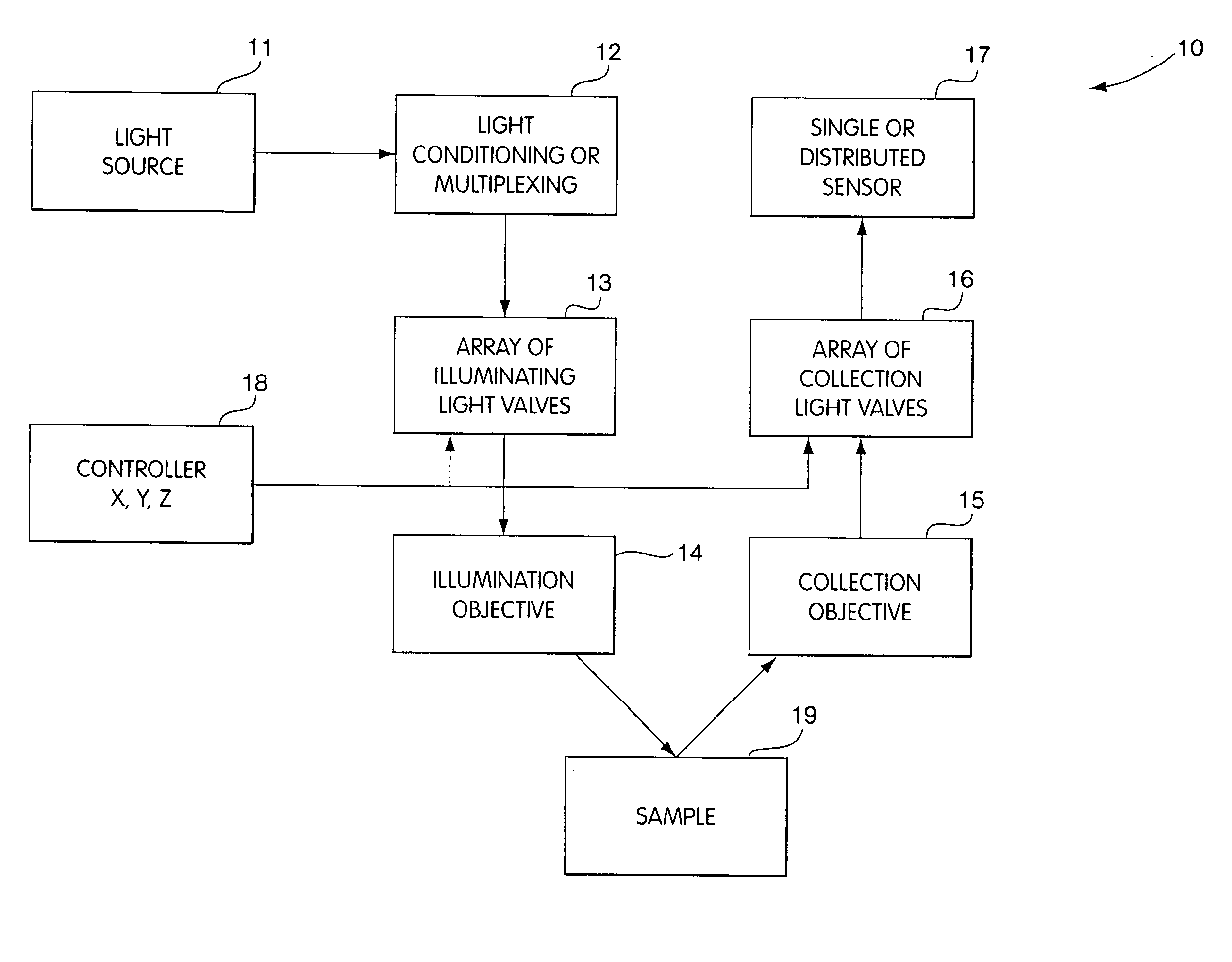
[0052] In FIG. 1 we show a generalized schematic volume probe array, 10, whose function is to collect data from a plurality of points in a target sample. The system generally includes an appropriate light source 11, whose light output is conditioned and may be multiplexed in block 12 to create a plurality of light sources to be relayed to an array 13 of light valves. These light valves can act as illuminating field stops or aperture field stops, and only one valve is open at a given time, thus providing for sequential illumination of volume elements in sample 19. The light emanating from each light valve is then directed to a targeted volume element in the sample 19 with an appropriate illumination objective 14. In some embodiments, a single objective lens is used, while in other embodiments, we incorporate an array of objective microlens having the same periodicity as that of the light valve array.

[0053] Responses from each targeted volume element in the form of light emanating fr...


PUM


Abstract

Methods and apparatus are provided for determining a characteristic of a sample of a material by the interaction of electromagnetic radiation with the sample. The apparatus includes a source of electromagnetic radiation, an optical assembly and a detector. The optical assembly sequentially illuminates a plurality of volume elements in the sample with an intensity distribution in the sample that drops off substantially monotonically from a first region in a first optical path and collects electromagnetic radiation emanating from each of the volume elements. The optical assembly collects the electromagnetic radiation emanating from each of the volume elements with a collected distribution that drops off substantially monotonically from a second region in a second optical path. The first and second regions at least partially overlap in each of the volume elements. The detector detects the collected electromagnetic radiation emanating from each of the sequentially illuminated volume elements to produce responses representative of the characteristic in each of the volume elements.

Description

PRIOR APPLICATIONS [0001] This application claims the benefit of U.S. Provisional Patent Application 60 / 115,373; Jan. 11, 1999, and is a continuation-in-part of U.S. patent application Ser. No. 08 / 782,936, filed Jan. 13, 1997.FIELD OF THE INVENTION [0002] The present invention provides apparatus and methods to derive spatially differentiated analytical information from an exposed surface by analyzing the results of the interaction of electromagnetic radiation with discrete volume elements of the sample. This is achieved by spatially limiting the probing beam to a small volume element and limiting the accepted response detected from the same volume element only, scanning the sample at various depths along the axis of the optical assembly formed by the beam to determine the interaction from volume elements at the different depths and collecting such data from a plurality of points in a plane generally perpendicular to the probing beam. BACKGROUND OF THE INVENTION [0003] An important r...

Claims


Application Information

Patent Timeline
IPC IPC(8): A61B6/00A61B1/00A61B5/00G01N21/47
CPCA61B5/0066A61B5/0075G01N21/4795A61B2562/08A61B5/0084
Inventor DEBARYSHE, GREGORYMODELL, MARKHED, A. ZE'EV
Owner MEDISPECTRA
Why Eureka
Try Eureka