

Interstitial fluid analyzer
a technology of interstitial fluid and analyzer, which is applied in the field of medical devices, can solve the problems of limited monitoring methods of blood glucose levels and devices of the prior art that are not designed or mean
- Summary
- Abstract
- Description
- Claims
- Application Information
AI Technical Summary
Benefits of technology
Problems solved by technology
Method used
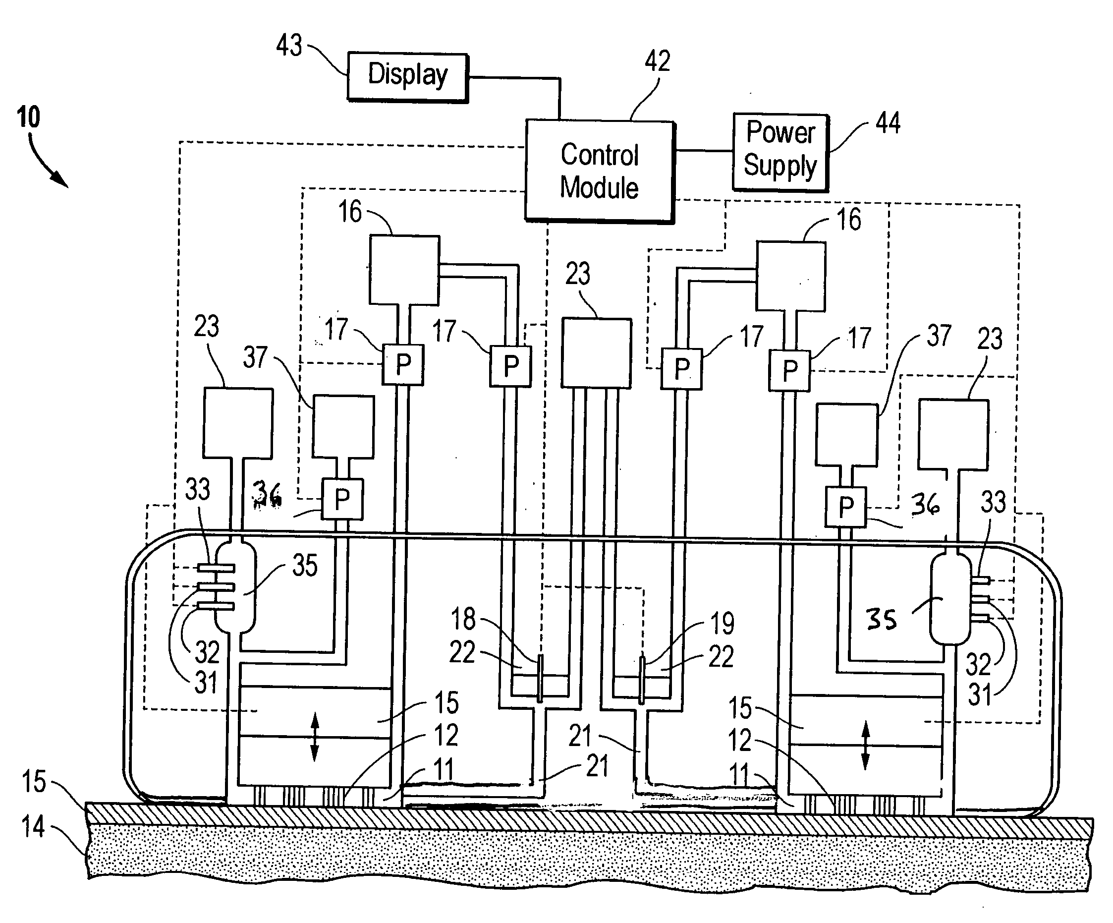
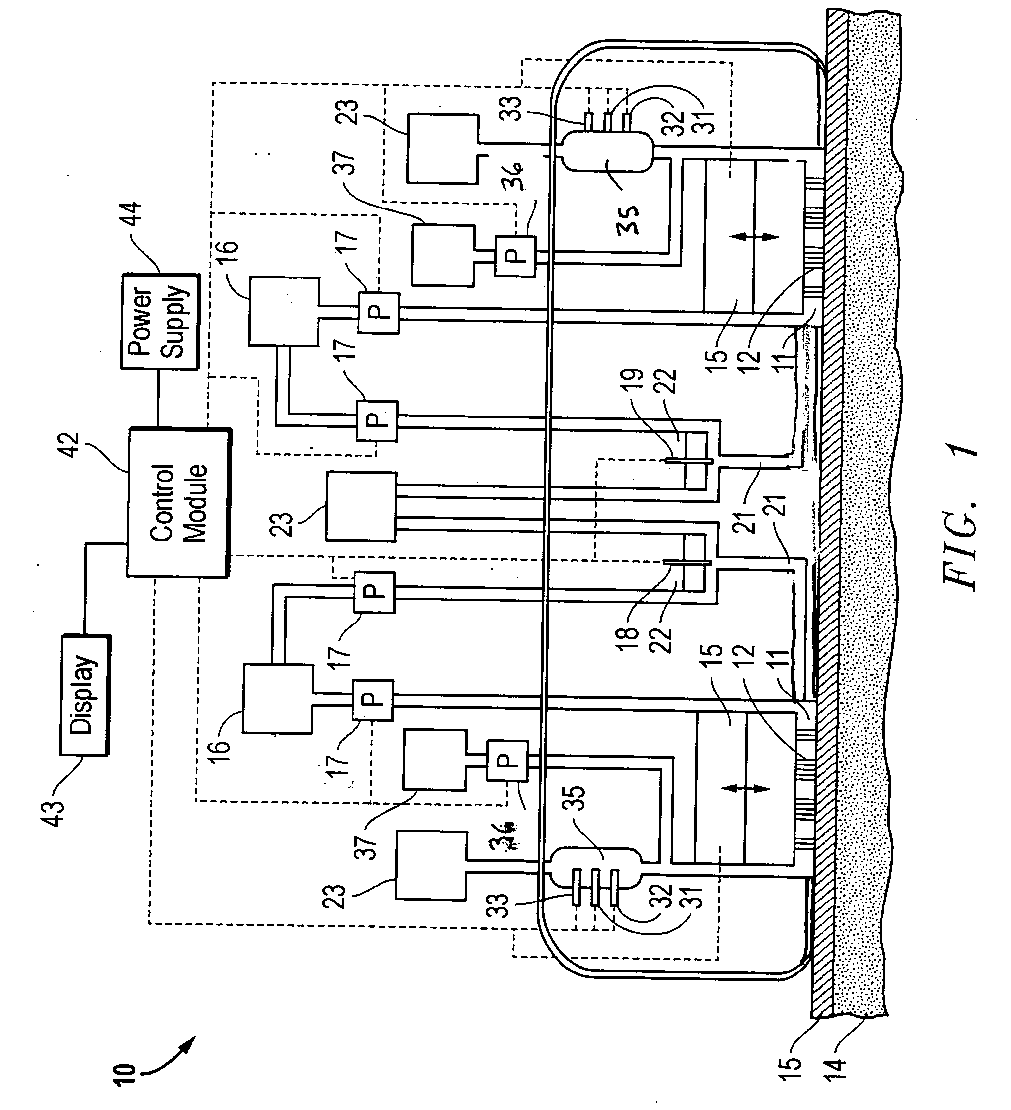
Image

Examples
example 1
[0077] Microprojections were prepared using the following method. A small glass vial, having a diameter of about 2.5 cm, was filled with a solution used for electroetching wire into the microprojections. The vial was then closed with a plastic cap containing a septum. The solution used for electroetching depends upon the type of wire used for making the microprojections. For tungsten microprojections, the solution used was 0.1 M NaOH. For platinum microprojections, the solution used was saturated NaNO2 solution. For gold microprojections, the solution used contained 10 g KCN and 5 g KOH per 40 mL of water.
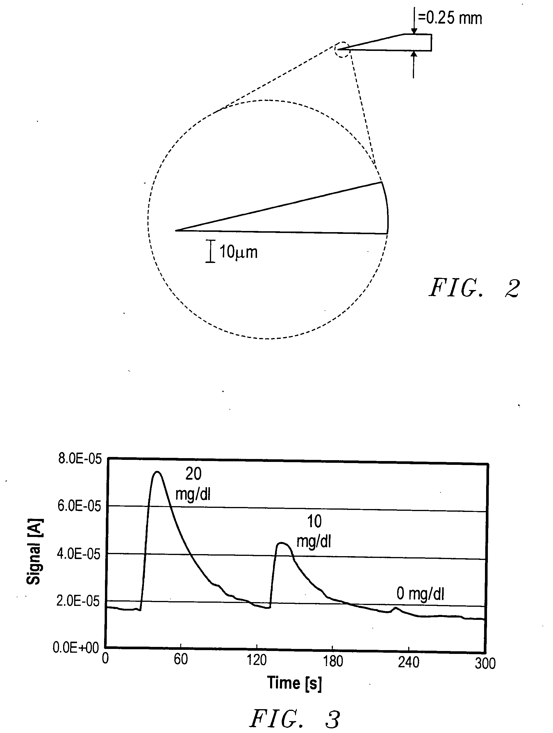
[0078] Three stainless steel needles were inserted through the septum so that the ends were about 5 mm above the solution level in the vial. A length of wire about 5 cm in length and having a thickness of about 0.25 mm, was inserted into the solution through the first needle to a depth of about 2 mm. A second wire was inserted through the second needle and immersed in the solution...
example 2
[0080] A microfluidic flow cell was assembled. The assembly consisted of a dual glassy carbon electrode with a thin Teflon sheet, which provided the microfluidic channel structure. The channels were cut into the Teflon sheet and provided the flow from the inlet over the electrodes to the outlet. The Teflon sheet was sandwiched between two polycarbonate plates. A Ag / AgCl reference electrode was placed in the channel inlet and positioned near the working electrode.
[0081] A solution containing 0.86 mM ferrocene and 0.8 mg / mL glucose oxidase in 0.1 M phosphate buffer, pH=7, was pumped through the microfluidic flow cell using a syringe pump. Glucose solutions were prepared in 0.1 M phosphate buffer. He flow cell was tested for glucose detection using the following solutions: 0.05 mL of 20 mg / mL glucose, 0.05 mL of 10 mg / mL glucose, and 0.05 mL of phosphate buffer. The amperometric method was used for glucose detection.
[0082] The results are shown in FIG. 3. As may be seen from the grap...
example 3
[0084] Four samples of pigskin were cut from one piece to minimize sample error. Each sample of pigskin was placed in the testing device. For each experiment, the lower compartment of the testing device was filled with 0.15 M glucose solution prepared in 0.05 M phosphate buffer, pH=7. For each data point, the cathode and the anode compartments were washed with DI water, wiped with a Kimwipe, the anode compartment was filled up with 0.1 mL of 0.8 M phosphate buffer and the cathode compartment was filled with 0.1 mL of 40 mg / dL glucose in 0.8 M phosphate buffer and charged with 0.02 mL of glacial ascetic acid. The pH in both compartments was confirmed to be neutral.
[0085] The three samples were contacted with 0.5 mm tungsten microneedle arrays for 0.5, 10, and 30 minutes, respectively during 30 minutes of electroosmosis at a current of 2 mA controlled by a Solartron Potentiostat / Galvanostat, Model 173. The fourth sample of pigskin was subjected to identical experimental conditions bu...
PUM
 Abstract
Description
Claims
Application Information