Systems and methods of identifying biomarkers for subsequent screening and monitoring of diseases
- Summary
- Abstract
- Description
- Claims
- Application Information
AI Technical Summary
Benefits of technology
Problems solved by technology
Method used
Image

Examples
examples
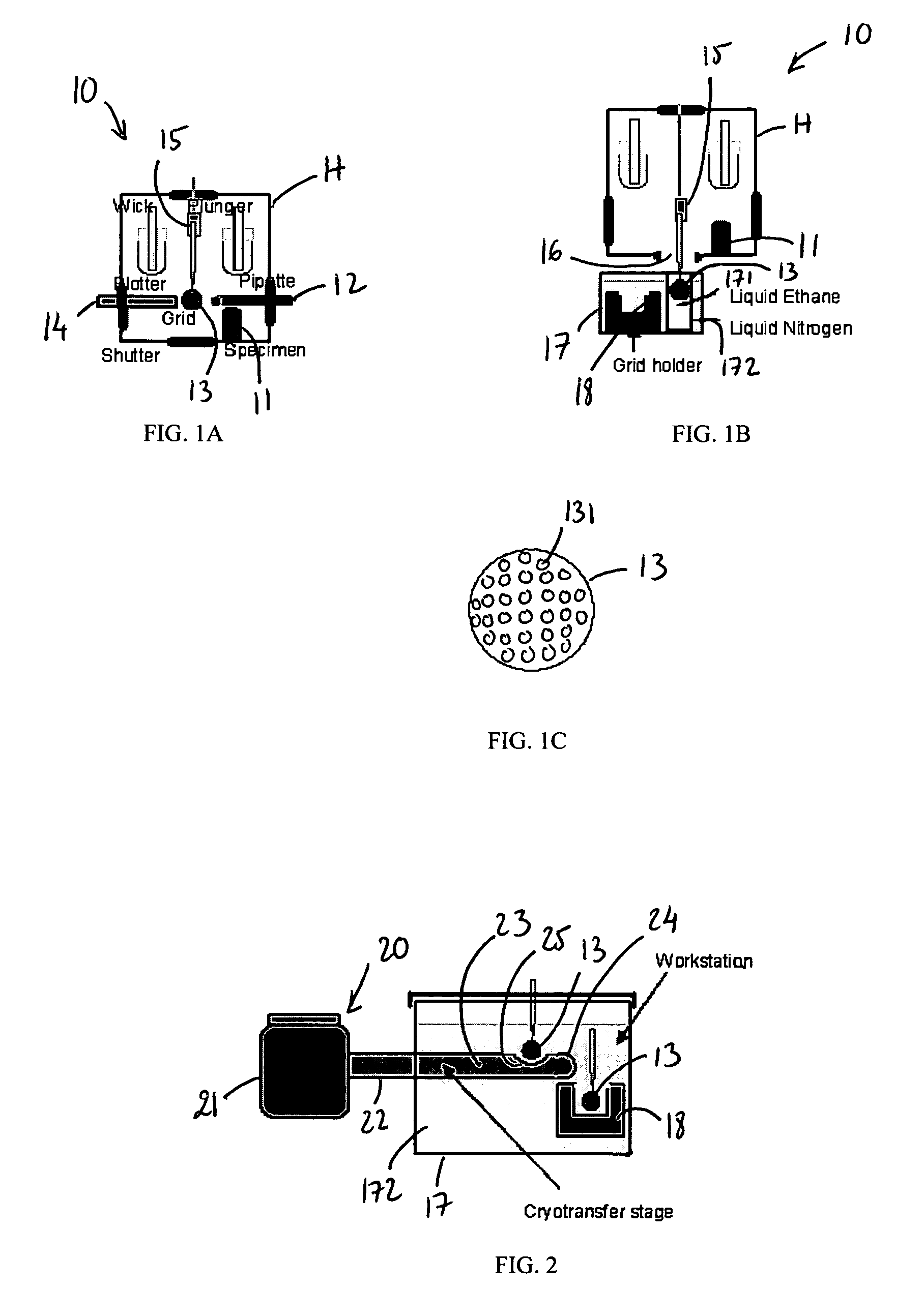
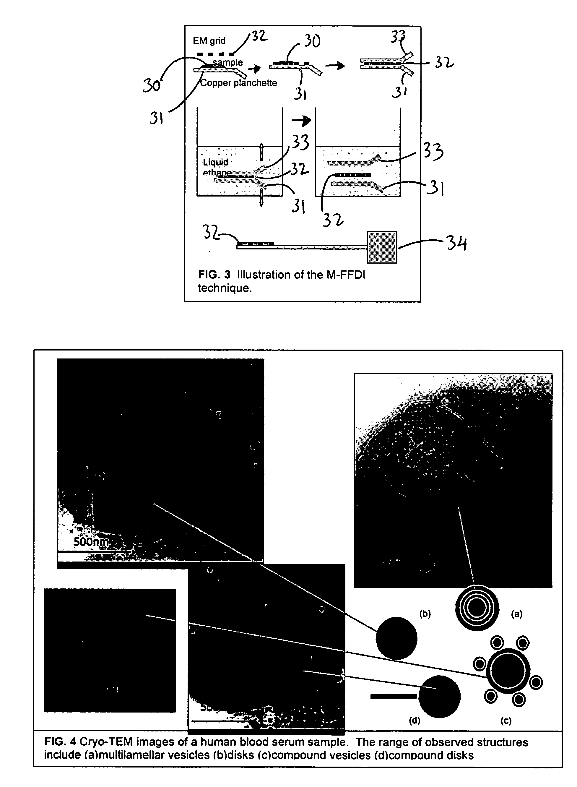
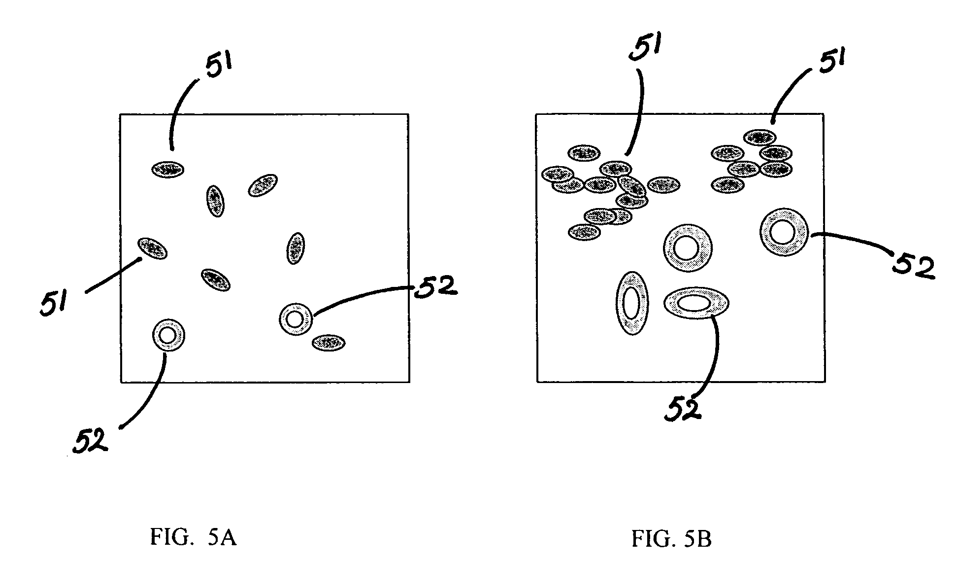
[0033] In an experiment, a serum sample from a test subject was prepared using cryogenic vitrification in the manner set forth above and subsequently imaged through the use of cryo-TEM. The images of the serum sample are illustrated in FIG. 4. In particular, the images are taken from different regions of the same holey carbon grid (i.e. different relatively thin regions of the thin film on the grid). As can be seen, the images show a relatively rich range of structures, including (a) multilamellar vesicles, (b) single and (d) compound discs, and (c) compound vesicles, all at nanoscale resolution of approximately 500 nm or less.
[0034] It should be noted that serum typically contains macromolecules, such as metabolites, lipids, hormones, peptides, and proteins. Certain of these biological macromolecules can also organize into 3-D complexes, which can be biochemically homogeneous or heterogenous in nature. For examples, serum can contain many glycoproteins, glycopeptides, lipoproteins...
PUM
| Property | Measurement | Unit |
|---|---|---|
| Temperature | aaaaa | aaaaa |
| Temperature | aaaaa | aaaaa |
| Thickness | aaaaa | aaaaa |
Abstract
Description
Claims
Application Information
 - R&D
- Intellectual Property
- Life Sciences
- Materials
- Tech Scout
- Unparalleled Data Quality
- Higher Quality Content
- 60% Fewer Hallucinations
Browse by: Latest US Patents, China's latest patents, Technical Efficacy Thesaurus, Application Domain, Technology Topic, Popular Technical Reports.
© 2025 PatSnap. All rights reserved.Legal|Privacy policy|Modern Slavery Act Transparency Statement|Sitemap|About US| Contact US: help@patsnap.com