Detection and analysis of ophthalmically-relevant fluorescent molecules

a fluorescent and fluorescent technology, applied in the field of ophthalmic conditions, can solve the problems of severe damage to the central vision, gradual loss of vision, and vision loss, and achieve the effect of lowering the level of serum retinol

Inactive Publication Date: 2006-05-11
REVISION THERAPEUTICS INC
View PDF18 Cites 16 Cited by
  • Summary
  • Abstract
  • Description
  • Claims
  • Application Information

AI Technical Summary

Benefits of technology

[0090] In further embodiments, the administration of a compound of Formula (I) is used to treat ophthalmic conditions by (a) lowering the levels of serum retinol in the body of a patient.

Problems solved by technology

Vision loss can then occur when drusen interfere with the function of photoreceptors in the macula.
The dry form of ARMD results in the gradual loss of vision over many years.
The wet form of ARMD can progress rapidly and cause severe damage to central vision.

Method used


Image

  • Detection and analysis of ophthalmically-relevant fluorescent molecules
  • Detection and analysis of ophthalmically-relevant fluorescent molecules
  • Detection and analysis of ophthalmically-relevant fluorescent molecules

Examples


example 1

Fluorescence Analysis of A2E and A2PE-H2 in Tissue Extracts

[0209] A. Preparing the Tissue Extract. Eyes are enucleated from euthanized mice and hemisected to reveal retina and retinal pigment epithelium (RPE). Retina is removed cleanly from underlying RPE with dissecting forceps. RPE is brushed from the underlying scleral tissue into 100-200 μl of PBS, pH 7.2 using a #2 camel hair brush. RPE cells are aspirated from the eyecup using a micro-pipette. Human post-mortem tissue is processed in a similar fashion. Tissues are homogenized by hand using a Duall glass-glass homogenizer following the addition of 500 μl chloroform / methanol (2:1, v / v). Samples are transferred to a borosilicate tube and lipids are extracted into 1 ml of chloroform. The organic extract is washed with 1 ml PBS, pH 7.2 and the samples are centrifuged at 3,000×g, 10 min. The chloroform phase is decanted and the aqueous phase is re-extracted with another 1 ml of chloroform. Following centrifugation, the chloroform p...

example 2

Fluorescence Analysis of A2PE-H2 in Whole Retina Explant

[0211] A. Preparing the Whole Retina and Retinal Epithelium Explants. Eyes are enucleated from euthanized mice and hemisected to reveal retina and retinal pigment epithelium (RPE). Retina is removed cleanly from underlying RPE with dissecting forceps. The remaining RPE / sclera are saved and stored separately. Samples of post-mortem human retina tissue are obtained as described above. The retina and RPE / sclera samples are moistened with PBS, pH 7.2 and placed separately into a solid phase sample mount so that the sample is oriented perpendicular to the incoming light (FIG. 8A). Emission spectra are obtained from the samples as described (FIG. 10A).

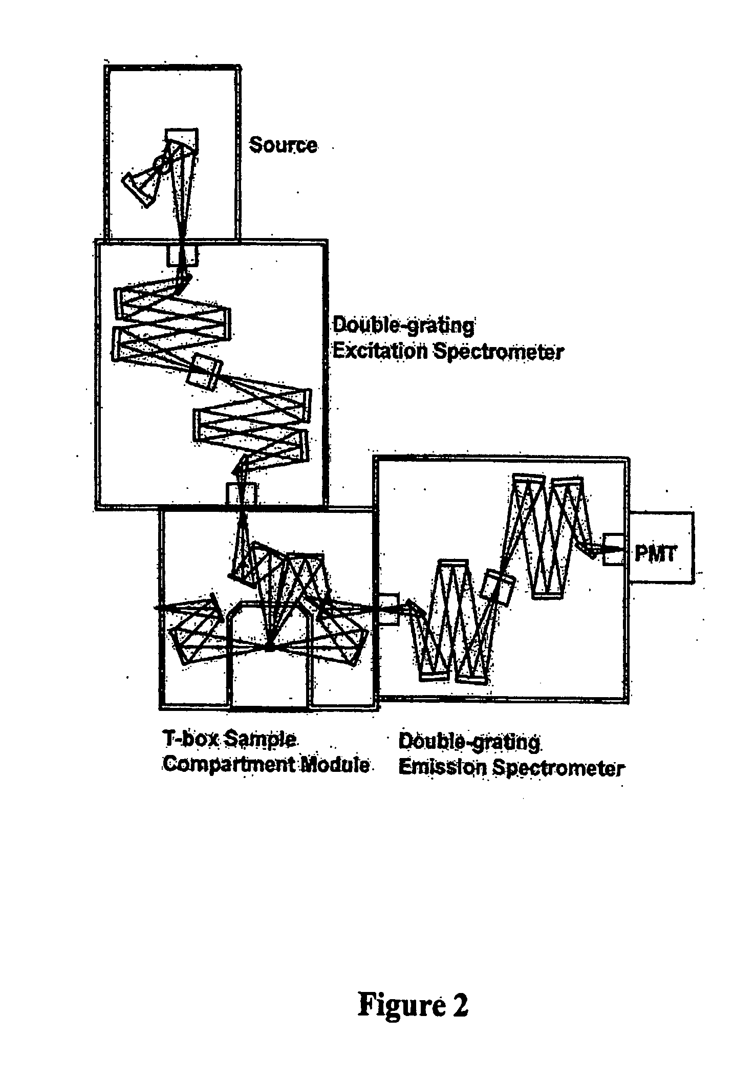
[0212] B. Fluorescence Analysis of A2PE-H2. Front face fluorescence emission from retina samples are acquired at 22.5° relative to the incoming light. Excitation light is set to 480 nm and emission spectra are acquired from 500 nm to 650 nm using a Jobin-Yvon Fluorolog 3 spectrofluoro...

example 3

Fluorescence Analysis of A2E and A2PE-H2 in the Intact Eye of a Live Animal

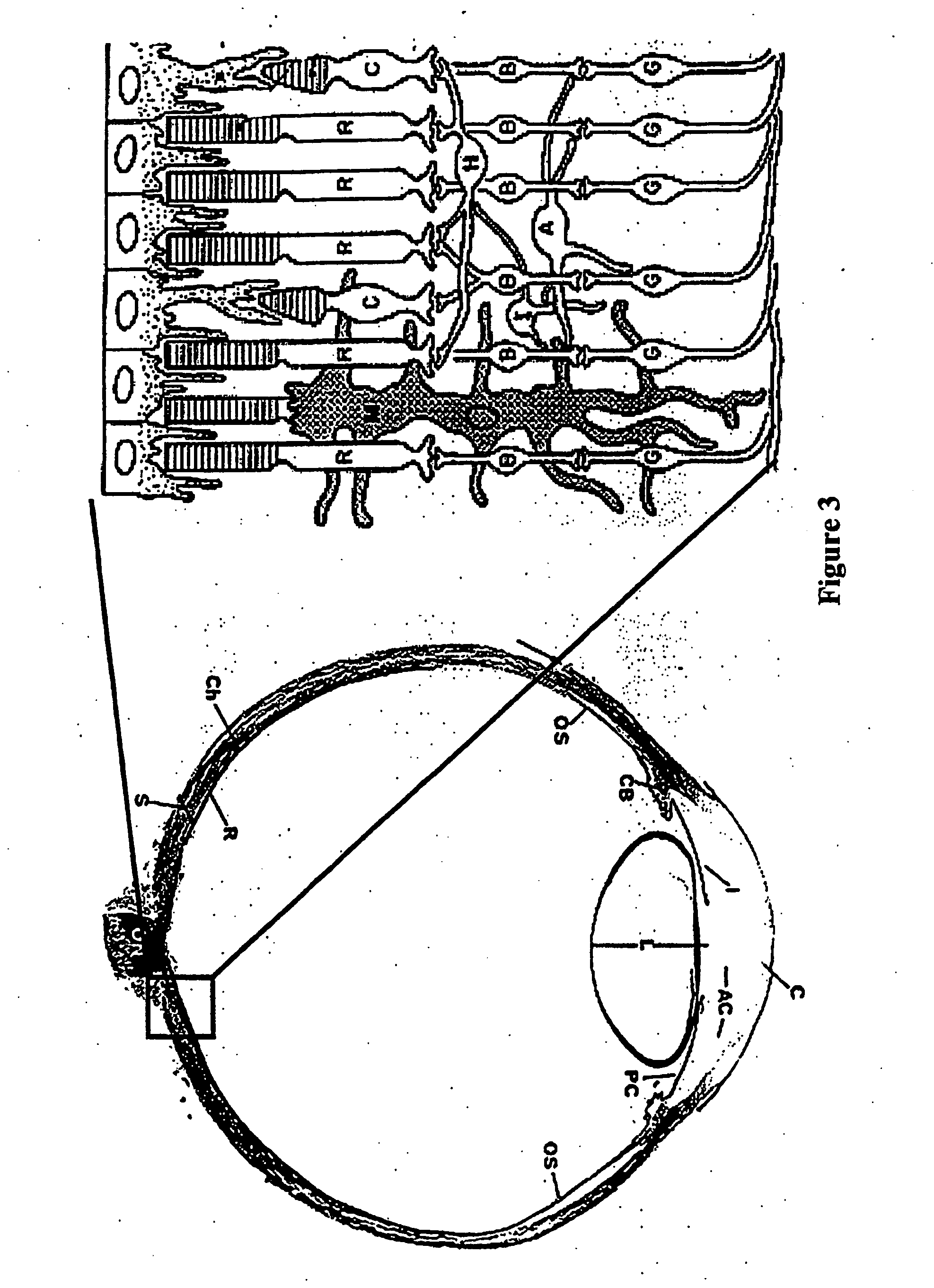
[0213] A. Preparing the Intact Eye of a Live Animal. Live mice are treated with a mydriatic (e.g., atropine) in order to dilate the pupil. The mice are anesthetized and placed onto a modified sample cell carriage such that the right or left eye is oriented toward the incoming light. See FIG. 8b.

[0214] B. Fluorescence Analysis of A2E and A2PE-H2 in the Intact Eye of a Live Animal. Front face fluorescence emission from intact eyes are acquired at 22.5° relative to the incoming light. Three analyses are performed: 1) Excitation light is set to 450 nm and emission spectra are acquired from 460 nm to 650 nm; 2) Excitation light is set to 480 nm and emission spectra are acquired from 490 nm to 650 nm; 3) Excitation light is set to 500 nm and emission spectra are acquired from 510 nm to 700 nm;

[0215] All data are acquired using a Jobin-Yvon Fluorolog 3 spectrofluorometer. Bandpass filters (slit widths) are adjust...


PUM


Abstract

Disclosed herein are methods and devices for detecting fluorescent molecules that are relevant to the health of the eye and related tissues. The presence of such molecules in the eye and related tissues can be used to diagnose whether the patient has certain diseases, including the macular degenerations and macular dystrophies. The amount of such molecules in the eye and related tissues can be used to determine the extent and stage of these diseases, to monitor the progress of these diseases, to design treatment strategies, to monitor the effectiveness of such treatments and to develop new therapies.

Description

CROSS-REFERENCE TO RELATED APPLICATIONS [0001] This Application claims the benefit of U.S. Provisional Applications Ser. Nos. 60 / 622,213 filed Oct. 25, 2004, 60 / 629,695, filed on Nov. 19, 2004, 60 / 660,904, filed on Mar. 11, 2005, and 60 / 672,405, filed on Apr. 18, 2005, the disclosures of all of which are hereby incorporated by reference in their entirety.FIELD OF THE INVENTION [0002] The methods and compositions described herein are directed to the treatment of ophthalmic conditions. BACKGROUND OF THE INVENTION [0003] The early diagnosis of macular degenerations and / or macular dystrophies is important in order to initiate prompt therapeutic interventions. Macular degenerations include age-related macular degenerations (ARMD), which include wet and dry forms of ARMD. The dry form of ARMD, which accounts for about 90 percent of all cases, is also known as atrophic, nonexudative, or drusenoid macular degeneration. With the dry form of ARMD, drusen typically accumulate in the retinal pi...

Claims


Application Information

Patent Timeline
Patent Type & Authority Applications(United States)
IPC IPC(8): G01N33/50
CPCA61K31/355G01N21/6456G01N21/6486Y10T436/17G01N2800/16G01N2800/164G01N33/50
Inventor MATA, NATHAN L.WIDDER, KENNETHLICHTER, JAY
Owner REVISION THERAPEUTICS INC
Why Eureka
Try Eureka