Treatments for a patient with congestive heart failure

a technology for congestive heart failure and treatment, applied in the field of therapeutic procedures for patients heart, can solve problems such as the population of patients who are otherwise unsuitable for conventional surgical treatmen

Inactive Publication Date: 2006-05-11
TRANSCARDIAC THERAPEUTICS
View PDF35 Cites 22 Cited by
  • Summary
  • Abstract
  • Description
  • Claims
  • Application Information

AI Technical Summary

Benefits of technology

[0009] To the extent required, the patient's deflated lung is moved out of the way, and then the pericardium on the patient's heart wall is removed to expose a region of the epicardium. The patient's heart wall is pierced at the exposed epicardial location to provide a passageway through the heart wall to a heart cavity such as the left ventricle, defined in part by the pierced heart wall. Preferably, the passageway is formed through a region of the heart wall at or near the apex of the patierit's heart. Suitable piercing elements include a 14 gauge needle. A guide wire is advanced through the inner lumen of the needle into the heart chamber and further advanced through the valve to be treated, e.g. the mitral valve, into an adjacent heart chamber. The penetrating needle may then be removed leaving the guide wire in place. A valve for the ventricular wall is advanced over the guide wire and installed in the ventricular wall passageway formed by the needle. The valve is configured to enable passage of instruments for the procedure through the heart wall into the heart chamber while preventing loss of blood through the passageway. The valve may be disposed permanently or temporarily within the heart wall passageway.
[0013] An elongated grasping device with at least a pair of grasping members such as jaws on the distal end thereof for grasping tissue structure is advanced through the ventricular wall valve until the distal end of the device extends into the heart chamber. The grasping members or jaws of the grasping device are operated from the proximal end of the grasping device which extends out of the patient's chest. The jaws of the grasping device are opened to receive the stabilized valve leaflets in the grasping location and then closed to grip the leaflets so that the free edges of the valve leaflets are placed into an operative position for the “Bow-Tie” repair. The free edges of the grasped valve leaflets may be joined or otherwise secured together by suitable connecting elements. Once the free edges of the valve leaflets are secured together, the instruments for the procedure may be withdrawn through the ventricular wall valve and then the opening in the patient's chest. The ventricular wall valve will close upon instrument removal to eliminate or at least minimize blood leakage through the valve. The ventricular wall valve may be left in place or the valve may be removed and the passageway sutured or otherwise suitably closed.
[0015] In that instance, it has been found that providing an artificial cordae tendenae such as a strand with one end secured to the secured valve leaflets and another end secured to the heart wall, particularly in the same orientation as the natural cordae tendenae, will support the connected valve leaflets in more or less a normal manner to minimize ventricular deformation (e.g. dilated cardiomyopathy) which leads to decreased output. One end of the strand is secured to the connecting element securing the free edges of the valve leaflets or to the connected free edges themselves and the other end of the strand is secured to a location on the heart wall, preferably on the exterior of the heart wall passing through the ventricular wall valve or if the valve is removed through the ventricular wall passageway. The strand should be relatively inelastic or non-compliant to ensure an effective closed position of the leaflets. A suitable strand material is polytetrafluoroethylene (PTFE). In this case it is preferred that the passageway through the ventricular wall pass through the apex region of the heart between the two papillary muscles in the left ventricle, so that the pull on the valve leaflets by the strand secured to the leaflets is in approximately the same angle or orientation as the natural pull by the competent cordae tendenae. This provides for a better seal of the leaflets and thereby minimizes leakage through the valve. The “Bow-Tie” procedure in conjunction with the use of an artificial cordae tendenae extending between the secured valve leaflets and the heart wall may be used with mini-thoracotomy procedures and open chest procedures, both on and off pump, in addition to the minimally invasive procedures described herein.
[0019] The blood flow output from the CHF patient's heart due to the pacing and valve repair in accordance with the present invention is greatly increased, and leads to significant improvement in the physical well being, the life extension and the quality of life of the CHF patient.

Problems solved by technology

Moreover, due to the minimally invasive nature of these procedures, many of the CHF patient population, who are otherwise unsuitable for conventional surgical treatments, may be treated with the present procedures.

Method used


Image

  • Treatments for a patient with congestive heart failure
  • Treatments for a patient with congestive heart failure
  • Treatments for a patient with congestive heart failure

Examples


example

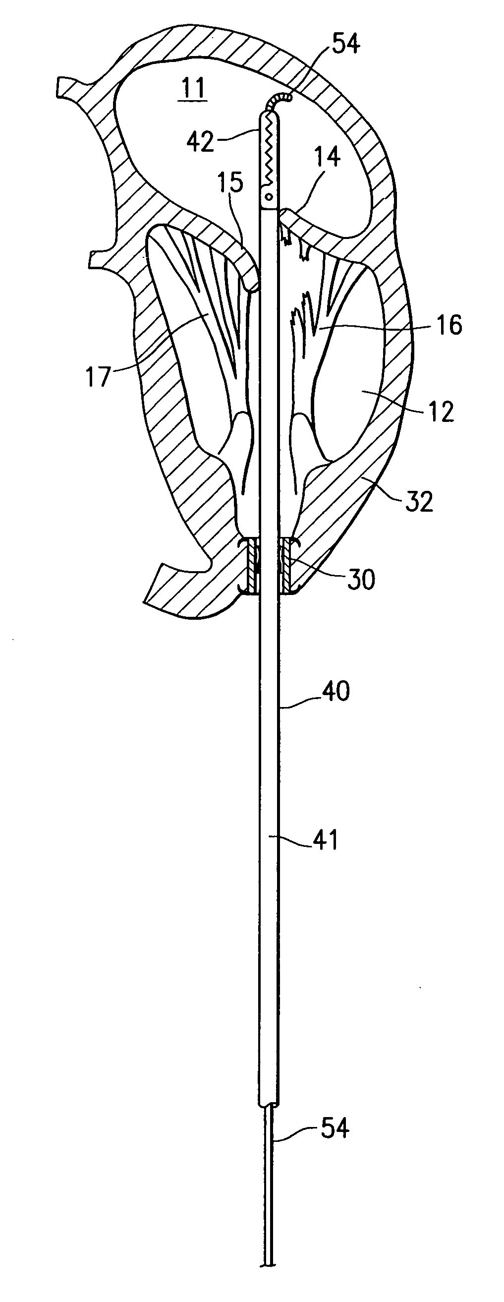
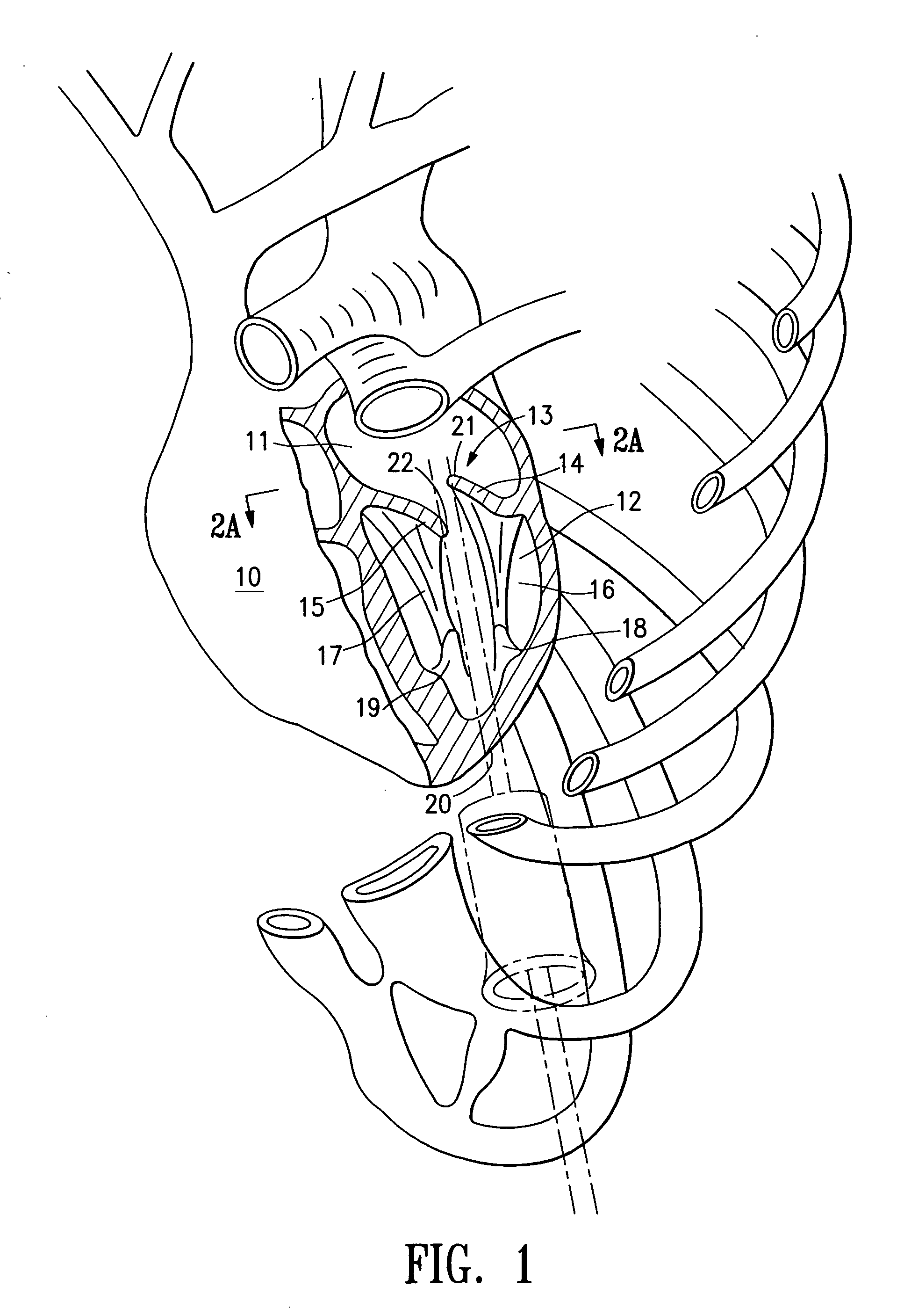
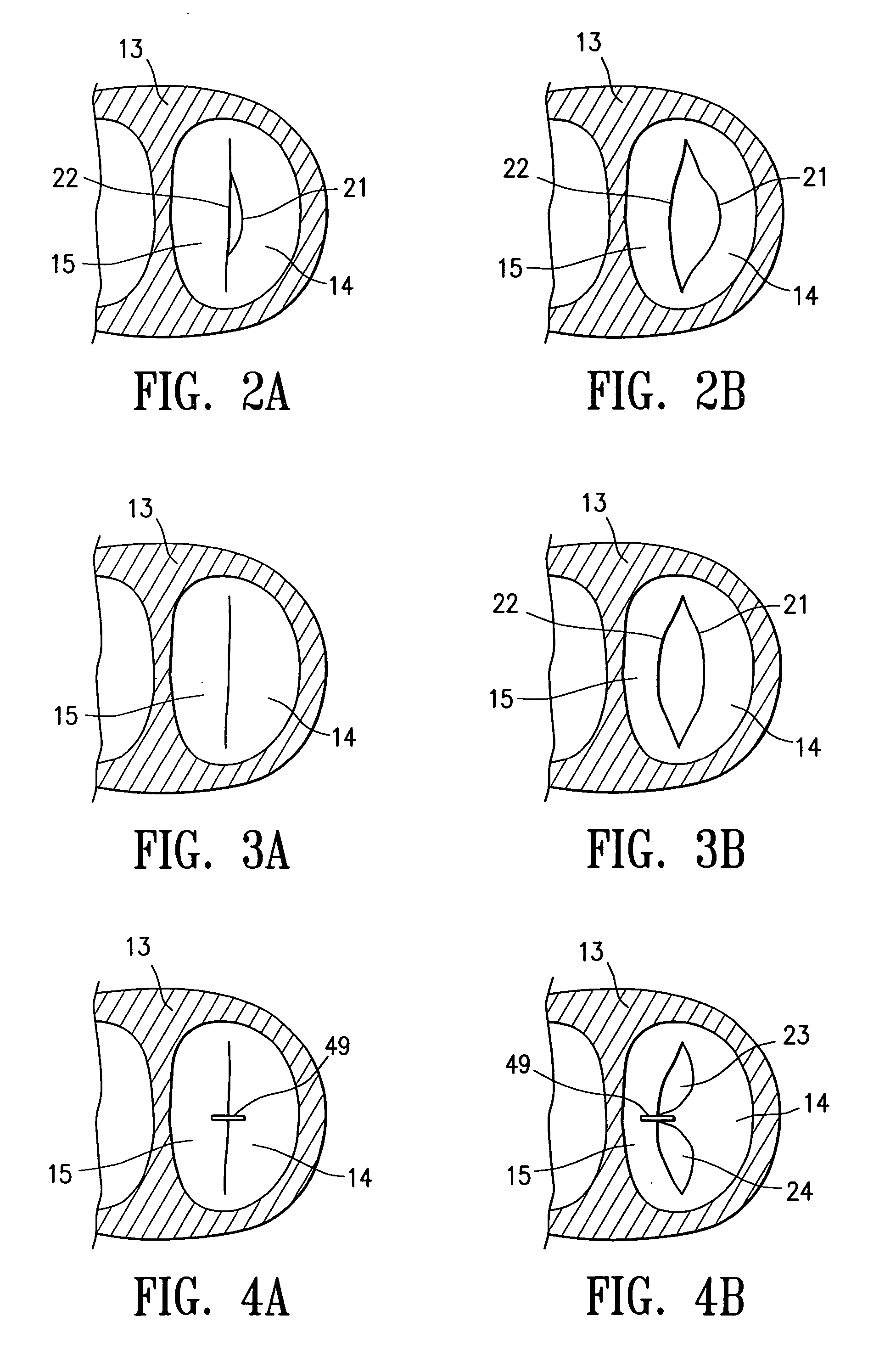
[0077] Twenty patients were selected (12 men, 8 women) for thorocoscopically direct left ventricular lead placement. The patients had New York Heart Association Class III or IV congestive heart failure with a mean ejection fraction of 20%±8%. All of the patients had previously undergone transvenous right-ventricular lead placement and subcutaneous implantation of a dual or triple chamber pacemaker but had failed transvenous left-ventricular lead placement due to suboptimal coronary vein anatomy. Surgical entry into the left chest was carried out through a 2 cm incision in the mid acillary line at the sixth intercostal space, following collapse of the left lung. A 15 mm thoracoport (trocar 87 in FIG. 32) from U.S. Surgical was inserted with the tip of the trocar pointing to the left minimize contact with the heart. A 5 mm rigid port (trocar 88 in FIG. 32) was inserted inferolateral to the left nipple of the patient in the sixth intercostal space to allow insertion of a grasper such a...


PUM


Abstract

The invention is directed to two minimally invasive therapeutic procedures, particularly for patients with congestive heart failure, and devices and systems for such procedures. One procedure involves providing a valved passageway through the patient's left ventricular wall at the apex of the patient's heart and advancing instruments through the valved passageway to connect the valve leaflets of the patient's heart valve, e.g. the mitral valve, in a “Bow-Tie” configuration to prevent or minimize regurgitation through the valve. The second procedure involves advancing a pacing lead and a pacing lead implanting device through a trocar in the patient's chest and implanting the pacing lead on an exposed epicardial region of the patient's heart wall. The pacing lead has a penetrating electrode which is secured within the heart wall. One or both procedures may be performed on a patient with CHF. Improved devices for these procedures include a minimally invasive grasping device having an inner lumen for advancing connecting members and other instruments through the device to the distal end thereof. Other improved devices include a pacing lead implant instrument which is releasably secured by its distal end to the exposed heart wall to facilitate penetration of the pacing lead electrode into the heart wall. Other improved instruments include a leaflet connector with an artificial cordae tendenae strand secured to an end of the leaflet connector.

Description

CROSS-REFERENCE TO RELATED APPLICATIONS [0001] This application is a continuation-in-part of application Ser. No. 10 / 295,390, filed on Nov. 15, 2002 which is related to and claims the priority of Provisional Application No. 60 / 340,062, filed Dec. 8, 2001, Provisional Application Ser. No. 60 / 365,918, filed Mar. 20, 2002, and Provisional Application Ser. No. 60 / 369,988, filed Apr. 4, 2002. The entire contents of these applications are incorporated herein by reference.BACKGROUND OF THE INVENTION [0002] This invention is directed to therapeutic procedures for a patient's heart and to instruments and systems for such procedures. The invention is particularly suitable for treating a patient suffering from the symptoms of congestive heart failure (CHF), and particularly to those CHF patients exhibiting mitral valve regurgitation (MVR) and / or those exhibiting intraventricular conduction delay with resulting disturbance of the synchronous right and / or left ventricular contractility. [0003] T...

Claims


Application Information

Patent Timeline
Patent Type & Authority Applications(United States)
IPC IPC(8): A61F2/24A61B17/00A61B17/064A61B17/068A61B17/08A61B17/122A61B17/128A61B17/30A61B17/34A61F2/02A61F2/06A61N1/05
CPCA61B17/00234A61B17/0401A61B17/064A61B17/0644A61B17/068A61B17/0682A61B17/08A61B17/10A61B17/122A61B17/1285A61B17/29A61B17/3421A61B17/3423A61B2017/00243A61B2017/00247A61B2017/00398A61B2017/00539A61B2017/00557A61B2017/0647A61B2017/0649A61B2017/22068A61B2017/22069A61B2017/306A61B2017/320064A61B2017/3425A61B2017/3488A61B2018/00392A61F2/06A61F2/24A61N1/056A61N1/0573A61N1/0587A61N2001/0578A61N2001/058Y10S623/904
Inventor LATTOUF, OMAR M.
Owner TRANSCARDIAC THERAPEUTICS
Why Eureka
Try Eureka