Folic acid-chitosan-DNA nanoparticles

Inactive Publication Date: 2006-05-18
VALORISATION RECH LLP +1
View PDF0 Cites 34 Cited by
  • Summary
  • Abstract
  • Description
  • Claims
  • Application Information

AI Technical Summary

Benefits of technology

[0008] The present invention provides a nanoparticle made of a folic acid-chitosan conjugate. The nanoparticle comprises one or more therapeut

Problems solved by technology

Although these systems demonstrate high transfection efficiency when compared to non-viral vectors, they are accompanied by a number of drawbacks that severely hinder their use in vivo (Luo, D. et al., 2000, Nat. Biotechnol. 18: 33-37).
Such limitations include their rapid clearance from the circulation, the reduced capacity to carry a large amount of genetic information and the associated risks of toxicity and immunogenic ity, which limits the possibility of subsequent administra

Method used


Image

  • Folic acid-chitosan-DNA nanoparticles
  • Folic acid-chitosan-DNA nanoparticles
  • Folic acid-chitosan-DNA nanoparticles

Examples


example 1

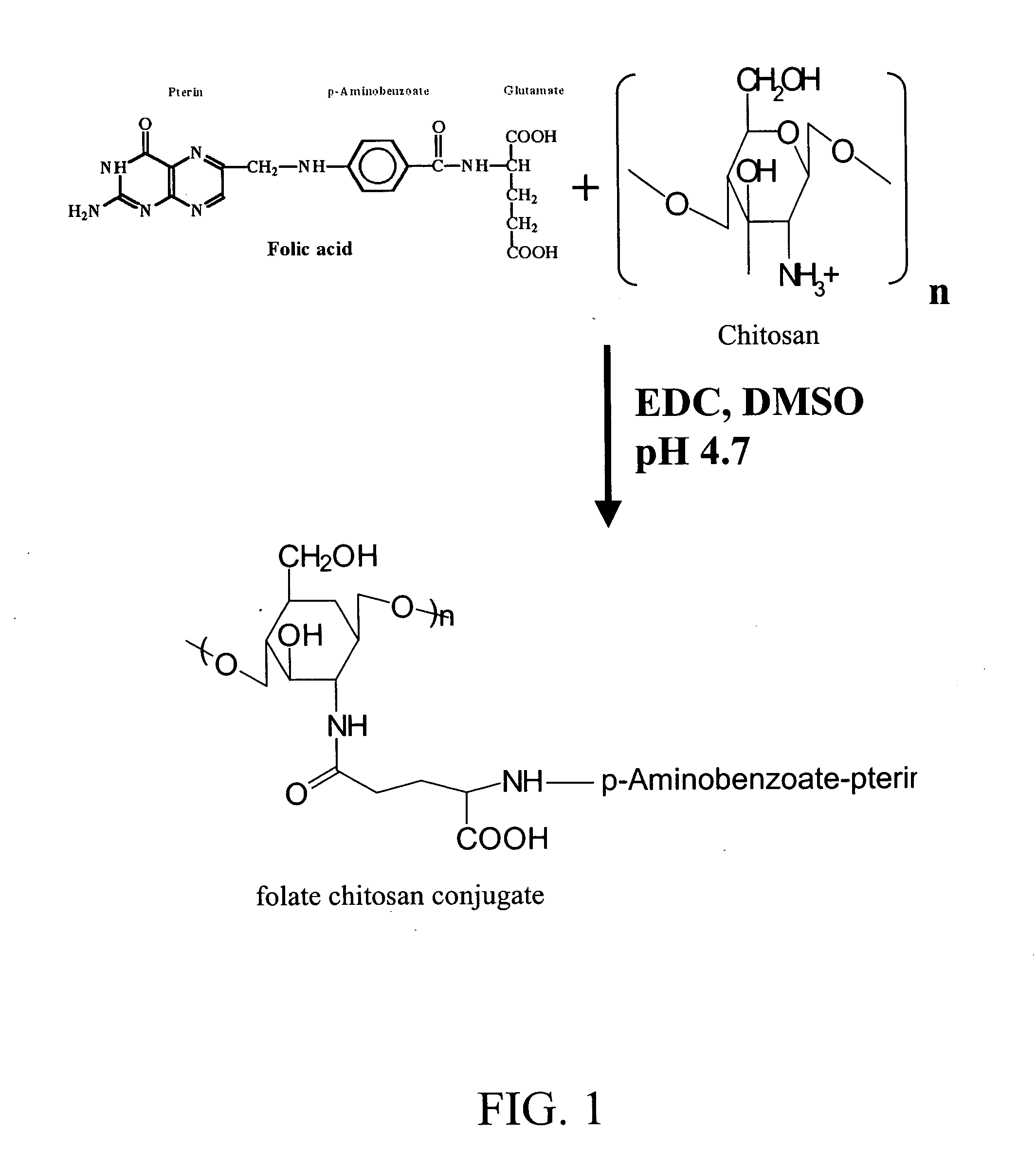
Synthesis of Folic Acid-Chitosan Conjugate.

[0024] A solution of 500 mg of 1-(3-dimethylaminopropyl)-3-ethylcarbodiimide hydrochloride (EDC) and 500 mg of folic acid (Sigma-Aldrich, St. Louis, Mo., USA) in 12 ml of anhydrous dimethylsulfoxide (DMSO) (Sigma) is prepared and stirred for 1 hour at room temperature until the folic acid is dissolved. The folic acid preparation is added to a solution of 0.1% (w / v) chitosan (MW: 150 kDa, 85% degree of deacetylation obtained from Fluka Biokemica, Buchs, Switzerland) in acetate buffer (pH 4.7) and stirred, in the dark, for 16 hours at room temperature. The pH of the solution is brought to 9.0 by dropwise addition of diluted aqueous NaOH. The resulting mixture is dialyzed for a period of 3 days against phosphate buffer at pH 7.4 and 3 days against water. The resulting folic acid-chitosan polymer is then isolated by lyophilization. The reaction scheme and resulting polymer are illustrated in FIG. 1.

[0025] A preferred folic acid-chitosan conj...

example 2

1. Preparation of DNA Plasmid.

[0027] The VR1412 DNA plasmid (VICAL Inc., San Diego, Calif., USA) was purified using the Qiagen QIAfilter plasmid Giga kit (Mississauga, ON, Canada) according to the manufacturer's instructions and resuspended in water. The integrity of DNA plasmid was analyzed on a 0.8% agarose gel and DNA concentration was measured by UV absorbance at 260 nm (Corsi, K. et al., 2003, Biomaterials 24: 1255-1264).

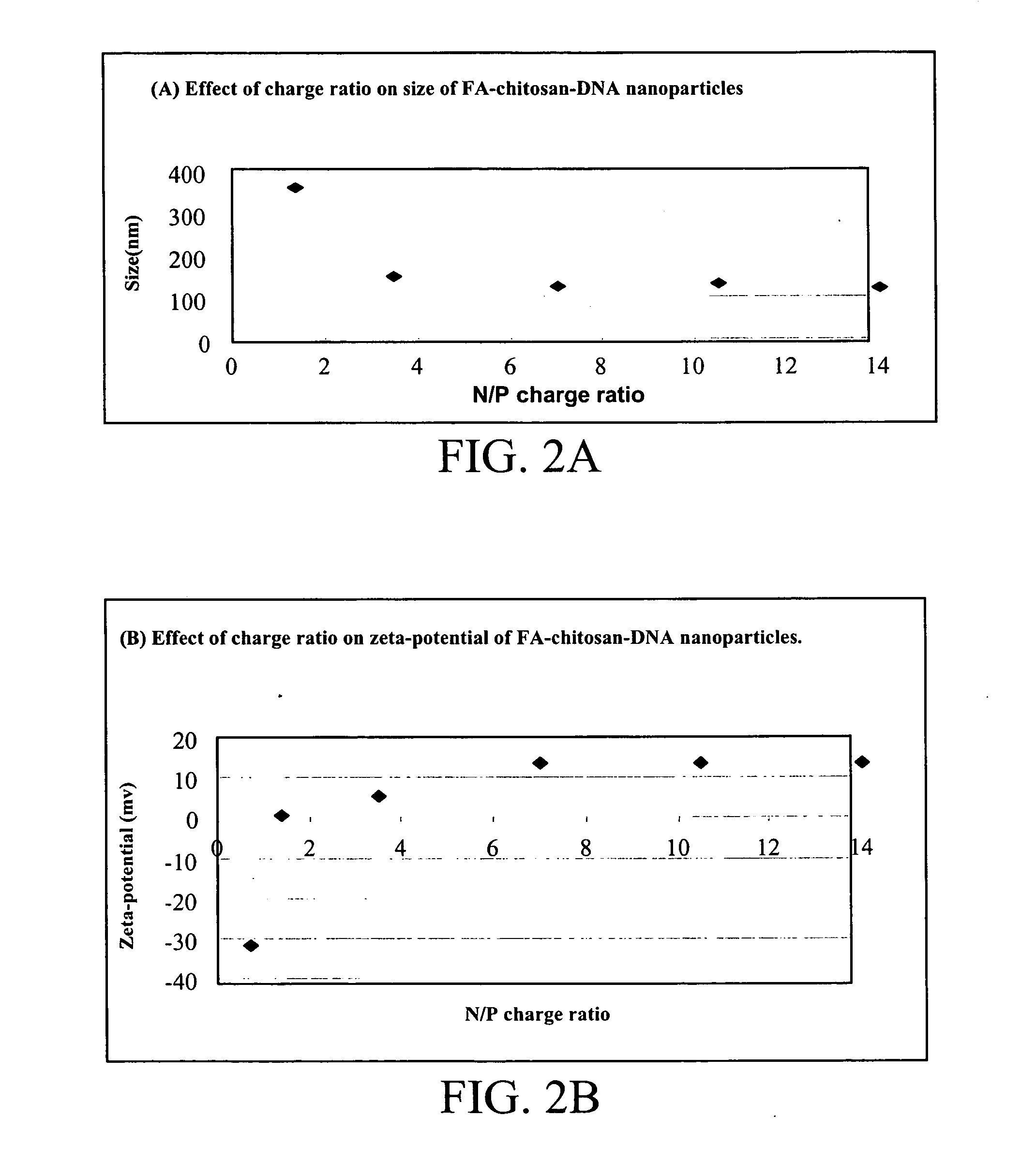
2. Synthesis of Folic Acid-Chitosan-DNA Nanoparticles.

[0028] a) The folic acid-chitosan conjugate of Example 1 was dissolved in 20 mM acetic acid at pH 5.5 under low heating (inferior to 45° C.). The solution was then adjusted to a final concentration of 0.01% chitosan in 5 mM acetic acetate and sterile filtered through a 0.22 μm filter. The DNA plasmid solution was diluted in a 4.3 mM sodium sulfate solution to a concentration of 200 mg / ml. The folic acid-chitosan-DNA complex formation was achieved by a coacervation technique as described by Mao et al. (2...


PUM

PropertyMeasurementUnit
Sizeaaaaaaaaaa
Sizeaaaaaaaaaa
Ratioaaaaaaaaaa

Abstract

The present invention relates to a non-viral novel drug delivery system. Nanoparticles comprising folic acid and chitosan are used to deliver a therapeutic agent of interest to the cell for various therapeutic applications.

Description

BACKGROUND OF THE INVENTION [0001] Gene therapy involves the introduction of exogenous genes into target cells for the purpose of achieving a therapeutic effect for the treatment of inherited and acquired diseases. Gene therapy relies on carriers such as viral or non-viral vectors for delivery. Viral gene delivery systems have been well characterized and include retroviruses, adenoviruses, adeno-associated viruses, herpes simplex virus and lentivirus (Oligino, T. J. et al., 2000, Clin. Orthop. 379 Suppl.: S17-30). Although these systems demonstrate high transfection efficiency when compared to non-viral vectors, they are accompanied by a number of drawbacks that severely hinder their use in vivo (Luo, D. et al., 2000, Nat. Biotechnol. 18: 33-37). Such limitations include their rapid clearance from the circulation, the reduced capacity to carry a large amount of genetic information and the associated risks of toxicity and immunogenic ity, which limits the possibility of subsequent ad...

Claims


Application Information

Patent Timeline
IPC IPC(8): A61K48/00A61K9/14C08B37/08
CPCA61K9/5161A61K31/519A61K31/722A61K47/48107A61K47/48923A61K48/00C08B37/003C08L5/08A61K47/551A61K47/6939
Inventor FERNANDES, JULIOWINNIK, FRANCOISEMANSOURI, SANIAYAN, CUIE
Owner VALORISATION RECH LLP
Why Eureka
Try Eureka