Minimally invasive medical device with helical pattern for indicating distance of movement

a technology of helical pattern and medical device, which is applied in the field of minimally invasive medical device, can solve the problems of inability to quantify the amount of movement or make measurements within the body, the inability to direct measurement, and the tendency to become displaced

Inactive Publication Date: 2006-07-06
WILSONCOOK MEDICAL
View PDF19 Cites 25 Cited by
  • Summary
  • Abstract
  • Description
  • Claims
  • Application Information

AI Technical Summary

Benefits of technology

[0007] The present invention provides a minimally invasive medical device, such as a wire guide, catheter, or sphincterotome, having an easy-to-use visual indicia system for monitoring distance of advancement. The visual indicia system includes a multi-colored or multi-patterned helix, or a series of colored / patterned dots, on the minimally invasive medical device and having a regular interval in the color / pattern so that the distance between the stripes on the helix (or between the different dots) indicates the distance of advancement. One advantage of the visual indicia system of the present invention is that minimally invasive medical devices may use a system of different colors and / or patterns to indicate total length and / or to indicate the widths of the helical stripes or the sizes of (and increment between) the dots useful in determining incremental advancement.
[0009] When the indicia pattern of the present invention is used with an exchange wire guide, it permits the measurement of anatomical structures when used with an endoscope having an accessory channel for introducing ancillary devices or instrumentation. This obviates the need for separate measuring devices, and makes it especially useful for Endoscopic Retrograde Cholangiopancreatography (ERCP) procedures, which often requires the step of measuring the length of biliary strictures. Biliary strictures typically occur in the common bile duct. The common bile duct provides a path from the gall bladder to the duodenum. More specifically, the common bile duct proceeds from the junction of the common hepatic duct with cystic duct, which is open to the gall bladder, and merges with the pancreatic duct, forming the ampulla of Vater, which itself opens into the duodenum at the papilla of Vater. The sphincter of Oddi is a muscular ring that controls passage of fluid from the ampulla of Vater into the duodenum.
[0012] In one example of how the present invention can be utilized, the clinician determines the position of the wire guide in a biliary duct using an endoscope, then withdraws the wire guide, counting the number of bands of one color that are exposed on the distal portion of the wire guide, until the radio-opaque tip marks the proximal boundary of the stricture. Alternatively, the number of helical bands may be counted at the proximal end of the endoscope where the wire guide protrudes from and is manipulated through an access port. The length associated with the helical bands on the wire guide permits the length of the stricture readily to be ascertained. This information can be important in subsequent treatment or procedures, such as determining the correct sizing of a biliary stent for placement across the stricture. Once the stricture has been measured, the wire guide can be maintained in place to serve as an exchange wire for introducing other instruments. Other applications of the present invention can be made in endovascular and other minimally invasive procedures.

Problems solved by technology

One example is the use of an exchange wire guide, which has a tendency to become displaced as catheters or other instruments are advanced or withdrawn over the wire guide.
However, such systems typically do not have a standardized helical color pattern width and therefore do not permit quantifying the amount of movement or making measurements within the body such as measuring the length of a stricture or lesion within a duct or vessel.
Direct visualization of indicia on the exchange wire guide via an endoscope offers some advantages; however, during procedures that access certain treatment sites such as the Papilla of Vater, mucous and other material can obscure the view, making direct measurement impossible.
In addition, a prior art exchange wire guide that is useful for measuring strictures for quantifying distance can be inadequate for discerning movement when the visible area of the wire guide under endoscopy corresponds to gaps between markings.

Method used


Image

  • Minimally invasive medical device with helical pattern for indicating distance of movement
  • Minimally invasive medical device with helical pattern for indicating distance of movement
  • Minimally invasive medical device with helical pattern for indicating distance of movement

Examples


Embodiment Construction

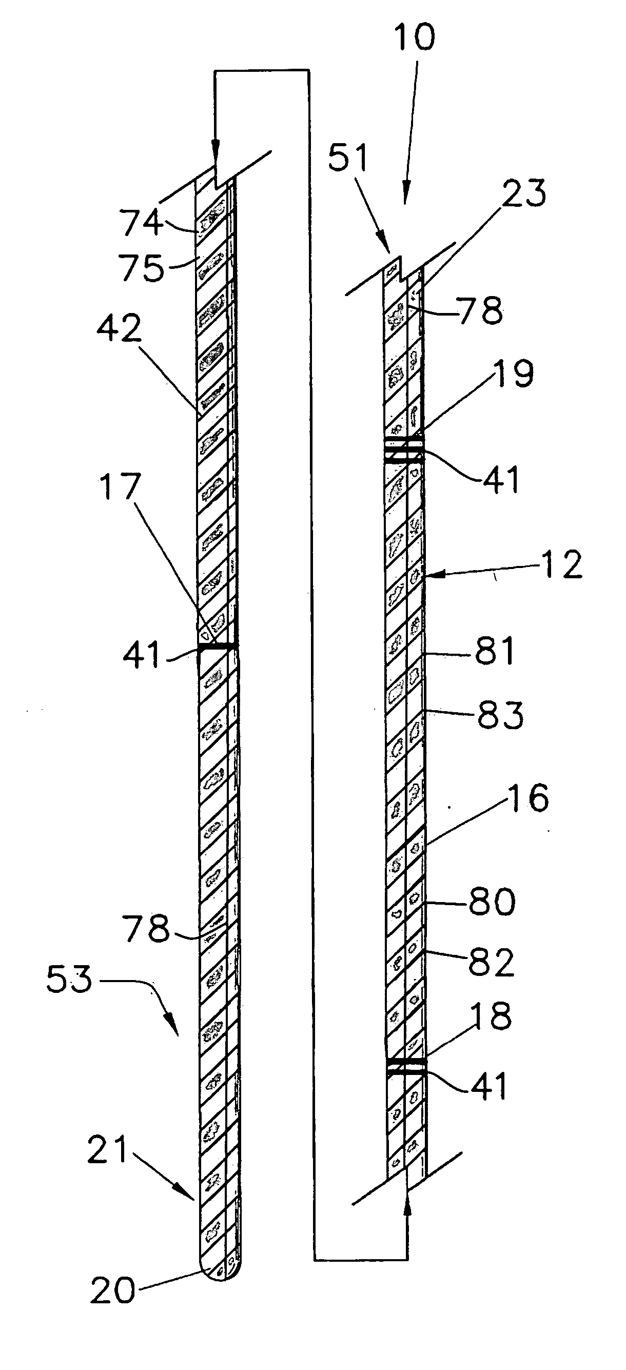
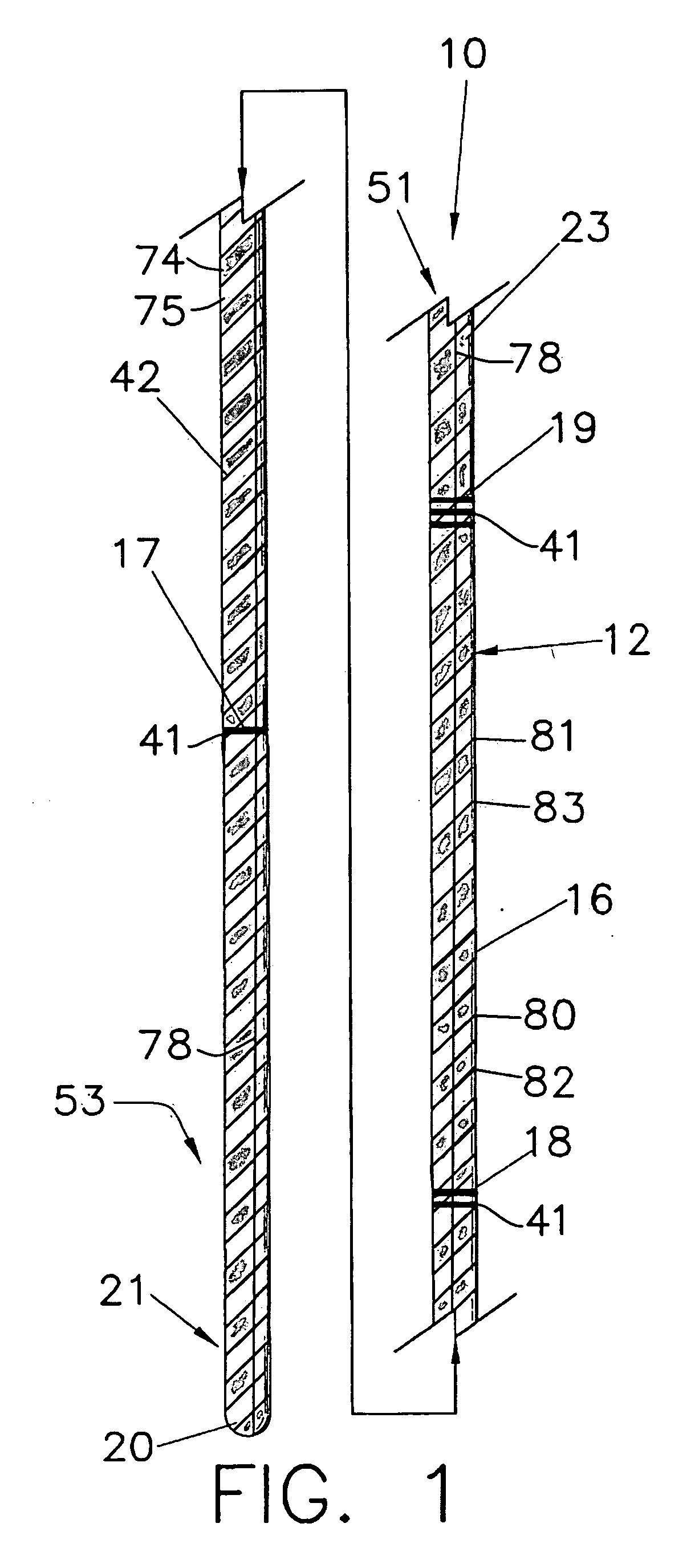
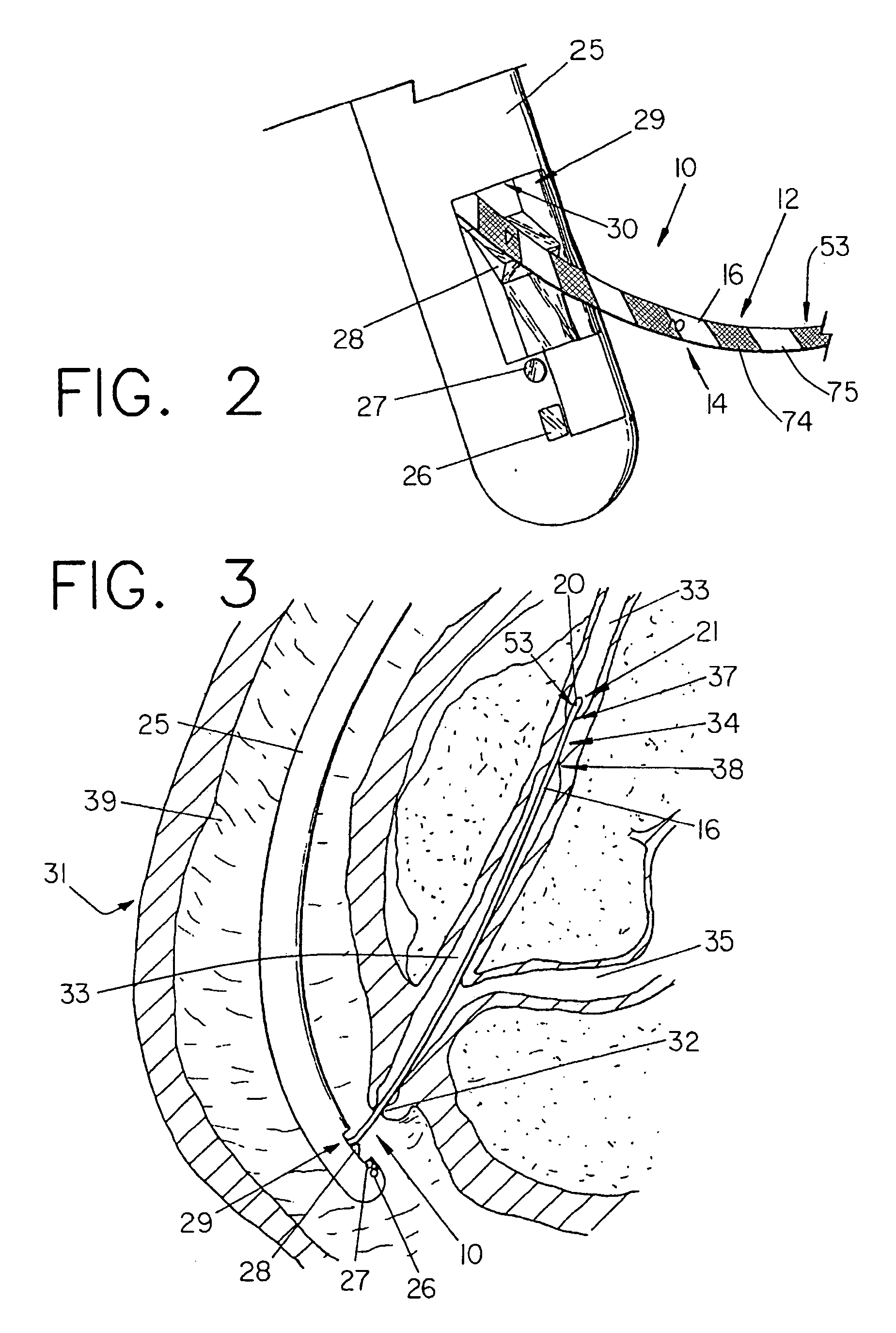
[0020]FIGS. 1-4 depict a minimally invasive medical device 10 comprising a wire guide 16 or similar elongate member having an indicia pattern comprising a multi-colored system of helical indicia 12 that allows the wire guide 16 to be used with an endoscope 25 to measure anatomical structures within a patient. The illustrative device 10 preferably comprises a standard exchange wire guide 16, e.g., 480 cm or 260 cm in length, with a solid core wire 50, such as nitinol, and an outer surface coating 42, such as PET or PTFE, that is shrink-wrapped over the wire (as is best shown in FIG. 4). To aid in fluoroscopic positioning of the wire guide 16, a distal portion of the device includes a marker material 21, either as a single marker, a plurality of markers, or an extended radio-opaque region that is several centimeters long (e.g., the distal 5 cm). Typical methods of providing radio-opacity include the addition of a distal platinum coil, adding gold or other radio-opaque material markers...


PUM


Abstract

A minimally invasive medical device, including an elongate shaft that has a helical color pattern of at least an alternating first color band having a first width and second color band having a second width. Along a surface parallel to a central longitudinal axis of the shaft, a length interval between each of a plurality of occurrences of the first color band is sufficiently uniform to allow use for measurement.

Description

CROSS-REFERENCE TO RELATED APPLICATIONS [0001] This application claims priority to U.S. Provisional Application Ser. No. 60 / 636,187, filed Dec. 15, 2004.TECHNICAL FIELD [0002] This invention relates to minimally invasive medical devices and particularly to wire guides and related devices. BACKGROUND [0003] It has been recognized that the placement of a series of markings on a surgical instrument such as a wire guide, catheter, needle, etc., can aid a physician in proper placement of the device in the body of a patient during a medical procedure. These markings can include bands, dots, lettering, numbering, colors, or other types of indicia to indicate position or movement of the device within the body. Visually distinguishable marks are often located at regular predetermined intervals, e.g., placement of one dot or circumferential band at the 5 cm mark, two dots or circumferential bands at 10 cm, etc. Such a system of indicia can be made to be visible under fluoroscopy by the use of...

Claims


Application Information

Patent Timeline
Patent Type & Authority Applications(United States)
IPC IPC(8): A61M25/00
CPCA61B1/018A61B5/1076A61B2019/462A61M25/00A61M25/09A61M2025/0008A61B2090/062
Inventor KENNEDY, KENNETH C. IIHARDIN, DAVID M.
Owner WILSONCOOK MEDICAL
Why Eureka
Try Eureka