Cartilage replacement implant and method for producing a cartilage replacement implant

a cartilage replacement and cartilage technology, applied in the field of cartilage replacement implants, can solve the problems of biomaterials used developing the tendency to contract, affecting the suction effect of implants, and affecting the suction effect, so as to facilitate the suction effect of implants

Active Publication Date: 2006-10-26
TETEC TISSUE ENG TECH
View PDF7 Cites 51 Cited by
  • Summary
  • Abstract
  • Description
  • Claims
  • Application Information

AI Technical Summary

Benefits of technology

[0064] To ensure that the implant retains its position at the defective cartilage area, it is advantageous for the cartilage replacement implant to be anchored to the cartilage defect with resorbable suture material.
[0065] Alternatively or add

Problems solved by technology

However, with biomaterials for a cell-free implantation with or without growth factors or in the carrier-coupled transplantation of cells, for example, chondrocytes obtained and proliferated from the body's own cells or mesenchymal stem cells, into a tissue defect, for example, a damaged cartilage area, there is the problem that in the course of their resorption, the biomaterials used develop the tendency to contract.
An undesired consequence of this is that upon commencement of resorption of the biomaterials,

Method used


Image

  • Cartilage replacement implant and method for producing a cartilage replacement implant
  • Cartilage replacement implant and method for producing a cartilage replacement implant
  • Cartilage replacement implant and method for producing a cartilage replacement implant

Examples


Embodiment Construction

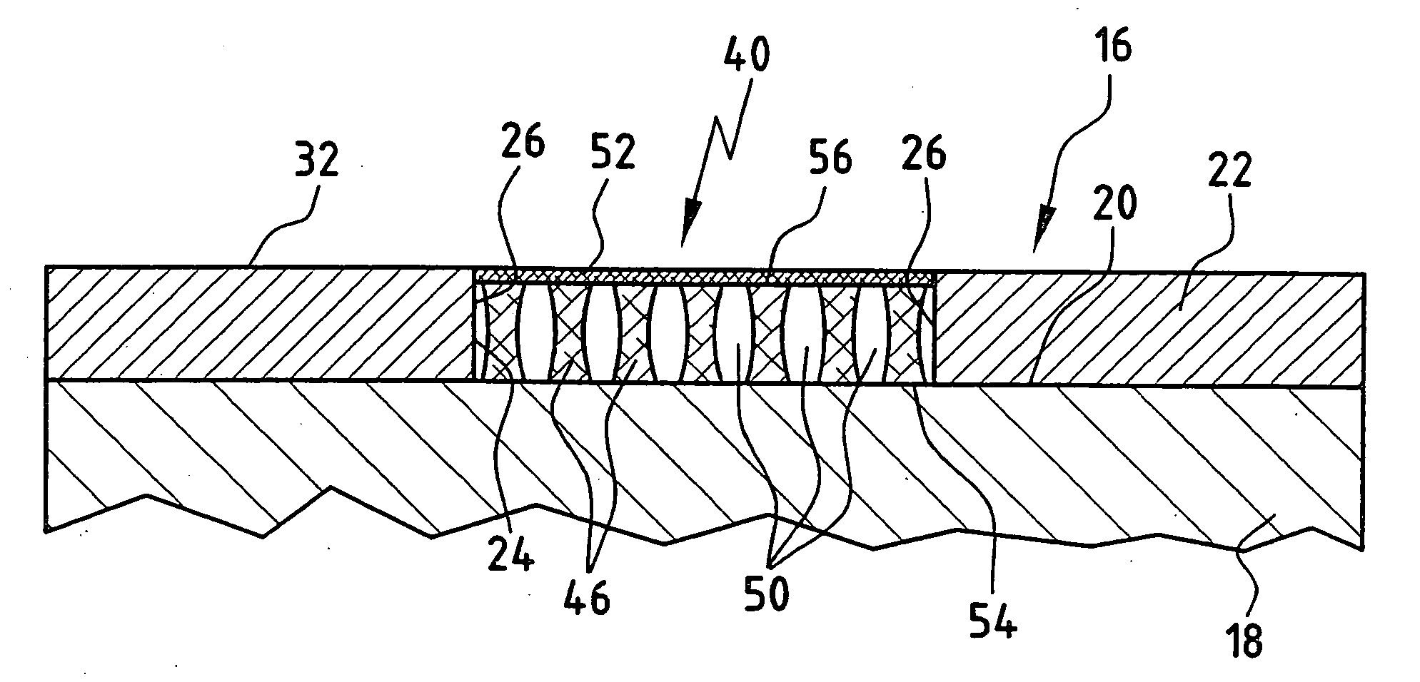
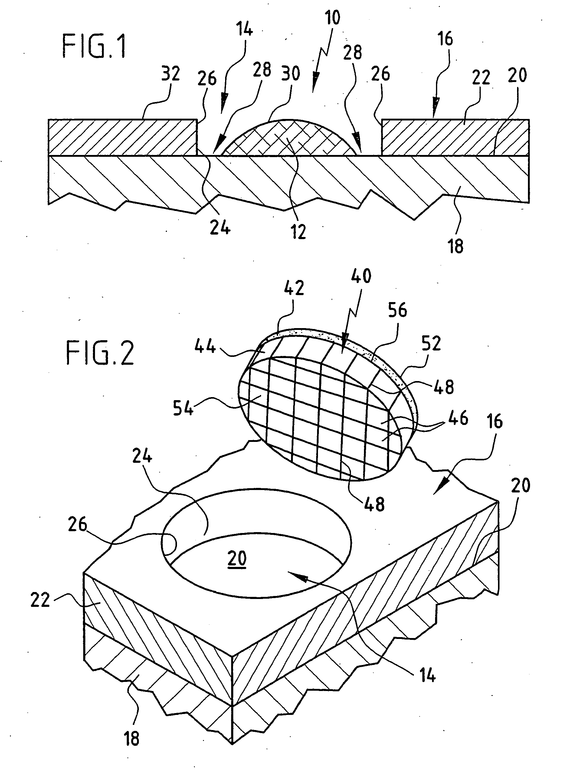
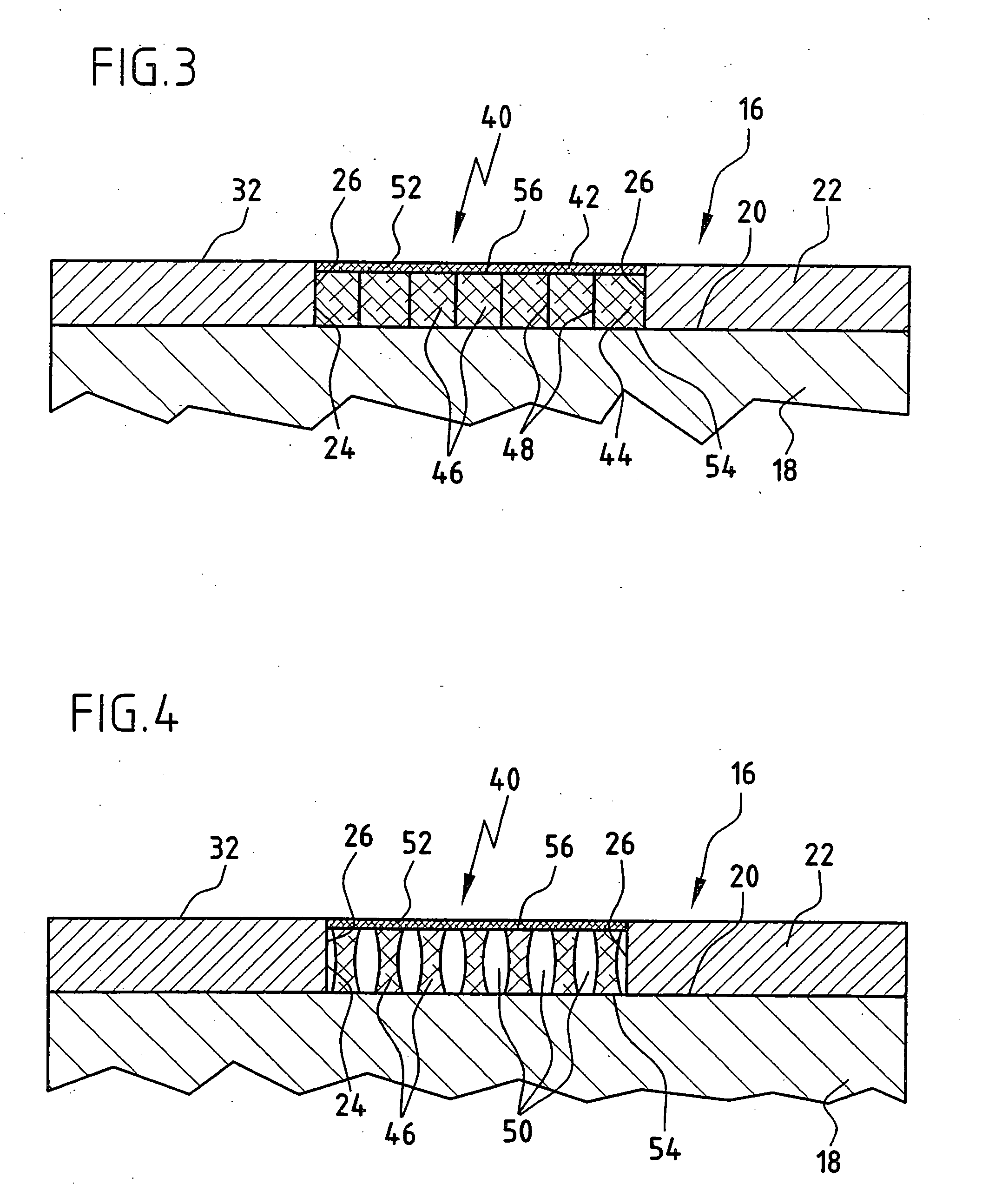
[0076]FIG. 1 shows a cartilage replacement implant 10, as it is known from the prior art. It consists of only one cell carrier 12, which may be inoculated with cells or not prior to implantation. In FIG. 1, the cartilage replacement implant 10 is inserted into a cartilage defect 14 of an otherwise intact cartilage area 16 of a joint in the human body. The cartilage area 16 is formed by a bone 18 and articular cartilage 22 covering the surface 20 thereof. For the sake of simplicity, the structure of the bone 18 and the articular cartilage 22 covering the surface 20 thereof is represented as a two-layer model. In nature, the transition from bone to cartilage usually occurs via a gradient of several millimeters in length. The cartilage defect 14 may have been caused by, for example, traumatic or inflammatory, degenerative processes. As shown schematically in FIG. 1, the cartilage defect 14 forms a gap in the cartilage area 16, which is delimited at the sides by cartilage edges 26 of th...


PUM

PropertyMeasurementUnit
Thicknessaaaaaaaaaa
Thicknessaaaaaaaaaa
Thicknessaaaaaaaaaa

Abstract

To improve a cartilage replacement implant for the biological regeneration of a damaged cartilage area of articular cartilage in the human body, comprising a cell carrier which has a defect-contacting surface for placement on the damaged cartilage area and is formed and designed for colonization with human cells, so that after implantation of the cartilage replacement implant, formation of a gap between adjacent contact surfaces of the implant and surrounding recipient tissue is minimized, it is proposed that the cell carrier rest with surface-to-surface contact on a carrier and be joined to the carrier at a cell carrier surface that faces away from the defect-contacting surface. A method for producing a cartilage replacement implant is also proposed.

Description

[0001] This application is a continuation of international application number PCT / EP2004 / 011240 filed on Oct. 8, 2004. [0002] The present disclosure relates to the subject matter disclosed in international application number PCT / EP2004 / 011240 of Oct. 8, 2004 and German application number 103 48 219.9 of Oct. 13, 2003, which are incorporated herein by reference in their entirety and for all purposes.BACKGROUND OF THE INVENTION [0003] The invention relates to a cartilage replacement implant for the biological regeneration of a damaged cartilage area of articular cartilage in the human body, comprising a cell carrier which has a defect-contacting surface for placement on the damaged cartilage area and is formed and designed for colonization with human cells. [0004] The invention further relates to a method for producing a cartilage replacement implant for the biological regeneration of damaged articular cartilage in the human body, wherein a cell carrier is used, which has a defect-con...

Claims


Application Information

Patent Timeline
IPC IPC(8): A61F2/28A61B17/00A61B17/06A61B17/68A61F2/00A61F2/02A61F2/30A61K35/12A61L27/38A61L27/50C12N5/077
CPCA61B17/06166C12N5/0655A61B2017/00004A61F2/30756A61F2/30767A61F2002/30006A61F2002/30011A61F2002/30032A61F2002/30062A61F2002/30225A61F2002/30324A61F2002/30448A61F2002/30785A61F2002/30971A61F2210/0004A61F2220/005A61F2230/0069A61F2250/0015A61F2250/0018A61F2250/0023A61F2250/003A61F2250/0036A61F2310/00365A61F2310/00383A61K35/12A61L27/3817A61L27/3852A61L27/50A61L2430/06A61B17/68
Inventor FRITZ, JUERGENGAISSMAIER, CHRISTOPHAICHER, WILHELM KARL
Owner TETEC TISSUE ENG TECH
Why Eureka
Try Eureka