

Xenograft tissue control for histology
- Summary
- Abstract
- Description
- Claims
- Application Information
AI Technical Summary
Benefits of technology
Problems solved by technology
Method used
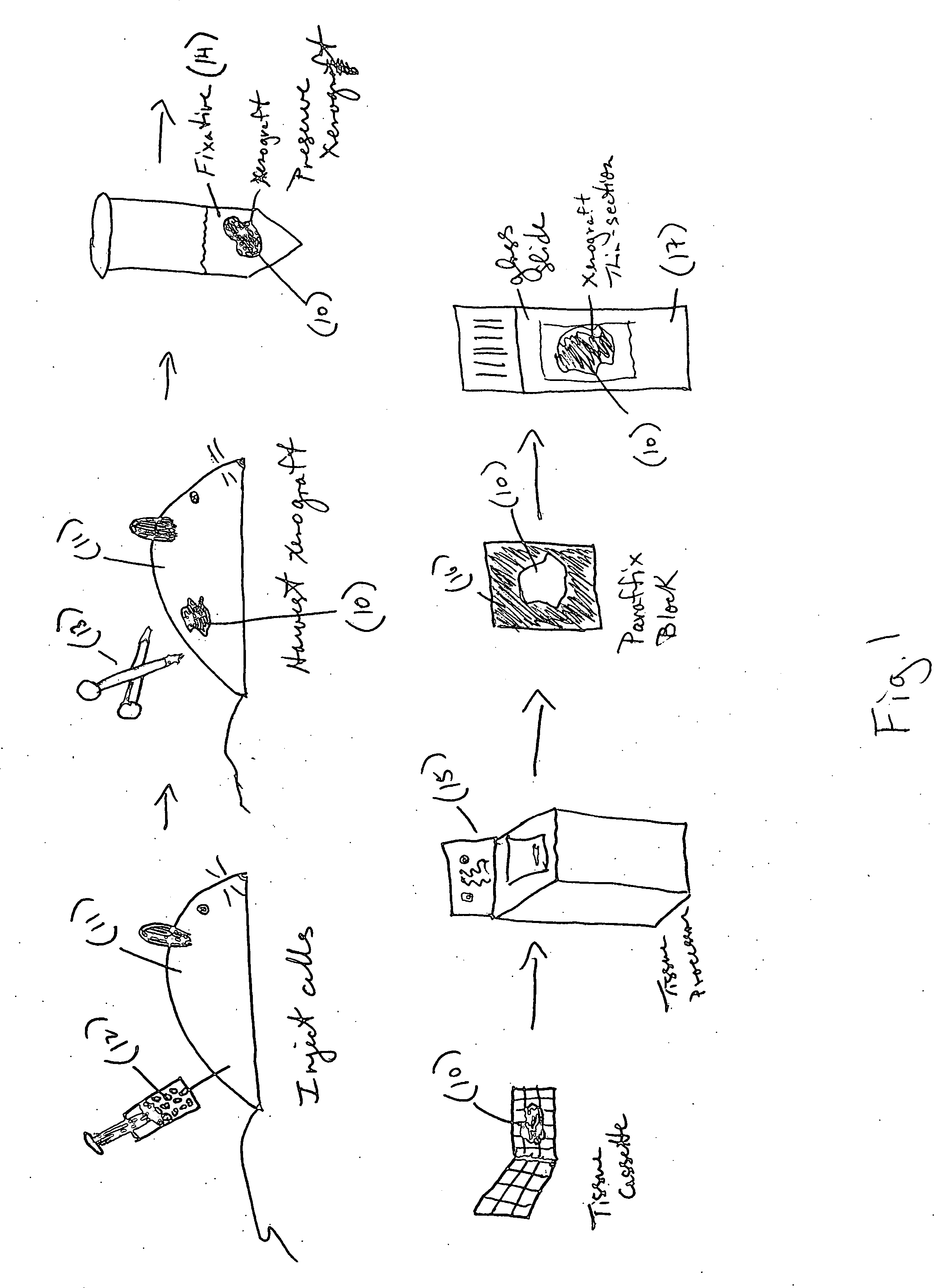
Image

Examples
example 1
- Growing and Characterization of HPV Xenograft Tissue Controls
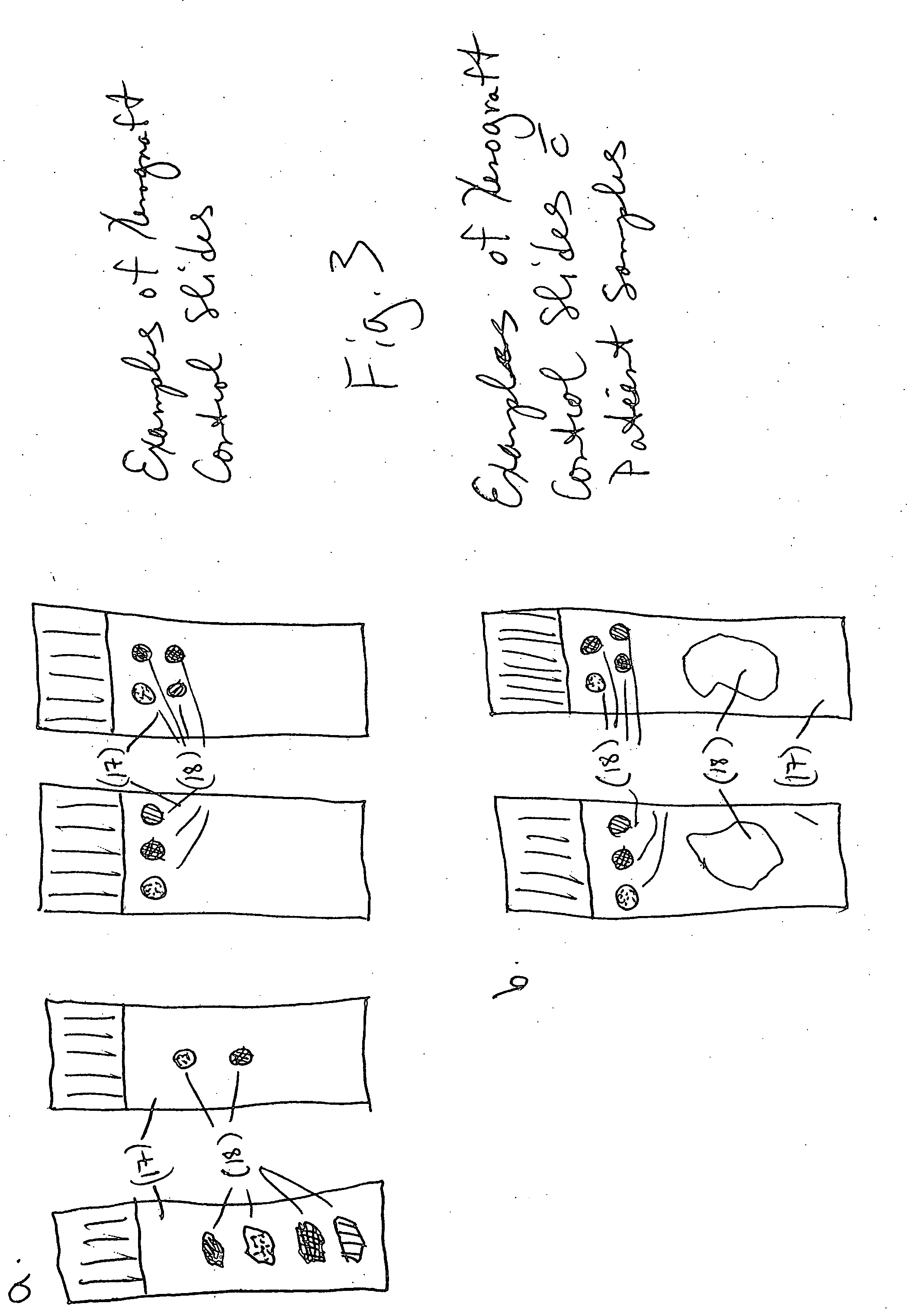
[0062] An example of a xenograft control slide for an in situ HPV assay is a slide containing four cores of xenograft tissue which contain four different amounts of integrated HPV DNA: CaSki (250-500 copies of HPV Type 16), HeLa (25-50 copies, HPV Type 18), SiHa (1-2 copies, HPV Type 16) and T-24 (0 copies). The xenograft control slides are made by growing transformed cervical (CaSki, HeLa and SiHa) and bladder carcinoma (T24) cell lines using standard laboratory practices (as described in R. I. Freshney, Culture of Animal Cells, 4th ed., Wiley-Liss, New York, 2000). The CaSki cells were grown in RPMI-1640 medium plus L-glutamine (MediaTech, Cat.# 10-040-CV) supplemented with 10% Fetal Bovine Serum (FBS) (Gibco, Cat.# 16000-044), 10 mM HEPES (Hyclone, Cat.# SH30237.01), 1 mM sodium pyruvate (MediaTech, Cat.#25-000-CI), 1.5 g / L sodium bicarbonate (Gibco, Cat.#25-035-CI) and 1% penicillin streptomycin (Gibco, Cat.# 15140-...
example 2
Multiblock Construction
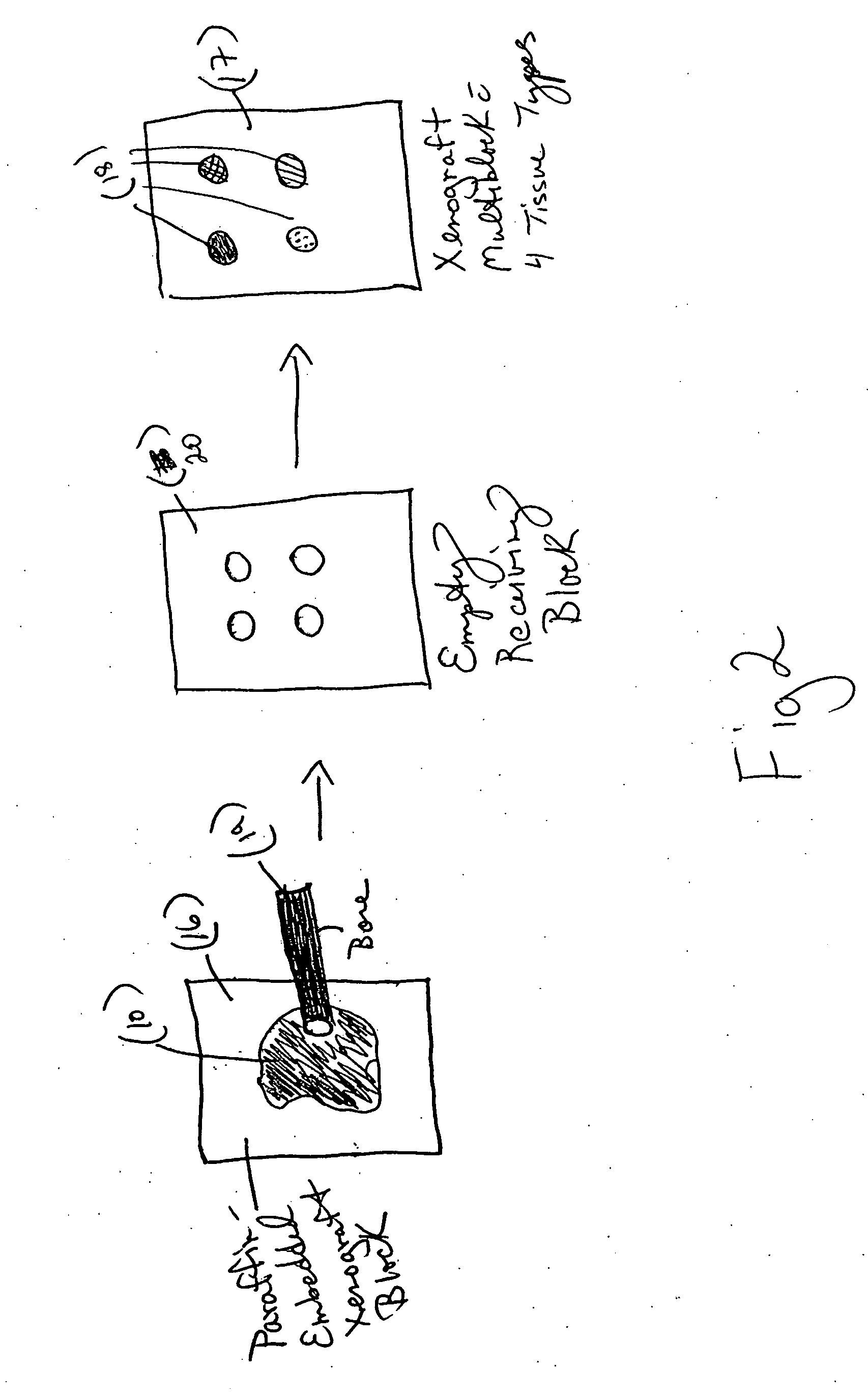
[0065] To create a xenograft control multiblock, a recipient block is first made using a microarrayer instrument. The recipient block was placed face up into the recipient block holder. Using the tissue microarrayor and a 2 mm core, the punch was positioned in the precise area where the first piece of tissue is to be embedded. A core was removed from the receiving block. The coring process was repeated until four paraffin cores were removed from the recipient bock. The recipient block was removed from the microarrayor and replaced with the xenograft tissue block to be cored. Using the H&E slide, the xenograft tissue was lined up with the punch to the precise spot on the xenograft tissue block to be cored. The xenograft tissue block was cored and released from the punch. The xenograft tissue block was replaced with the recipient block, and the recipient block hole lined up to the punch with the xenograft tissue core. The xenograft tissue core was ejected into ...
example 3
Xenograft Controls Run with Ventana's HPV Assay
[0066] FIGS. 4A-C are microphotographs of three HPV xenograft control slides tested with an ISH assay using an HPV DNA probe. Using a BENCHMARK® series autostainer, (Ventana Medical Systems, Tucson, Ariz.) slides containing HPV xenograft controls were stained using INFORM® HPV Family 16 DNA Probe (PN 780-2838), detected with ISH iView Blue Detection Kit (PN 760-092) and counterstained with ISH Red Counterstain. The HPV Family 16 probe is a cocktail of DNA probes with specificity for high-risk HPV types 16, 18, 31, 33, 35, 39, 45, 51, 52, 56, 58 and 66. CaSki (FIG. 4a) and HeLa (FIG. 4b) xenograft tissues exhibit positive staining (blue punctuate pattern) due to nucleus-integrated HPV DNA. Negative staining (no blue punctuate pattern) is seen in the T24 (FIG. 4c) xenograft
PUM
 Abstract
Description
Claims
Application Information