Novel druggable regions in the dengue virus envelope glycoprotein and methods of using the same
a dengue virus and envelope glycoprotein technology, applied in the field of new druggable regions in the dengue virus envelope glycoprotein, can solve the problems of unfavorable fusion of class ii viral fusion proteins, inability to fusion immature particles, and inability to control dengue virus by vaccination
- Summary
- Abstract
- Description
- Claims
- Application Information
AI Technical Summary
Benefits of technology
Problems solved by technology
Method used
Image
Examples
example 1
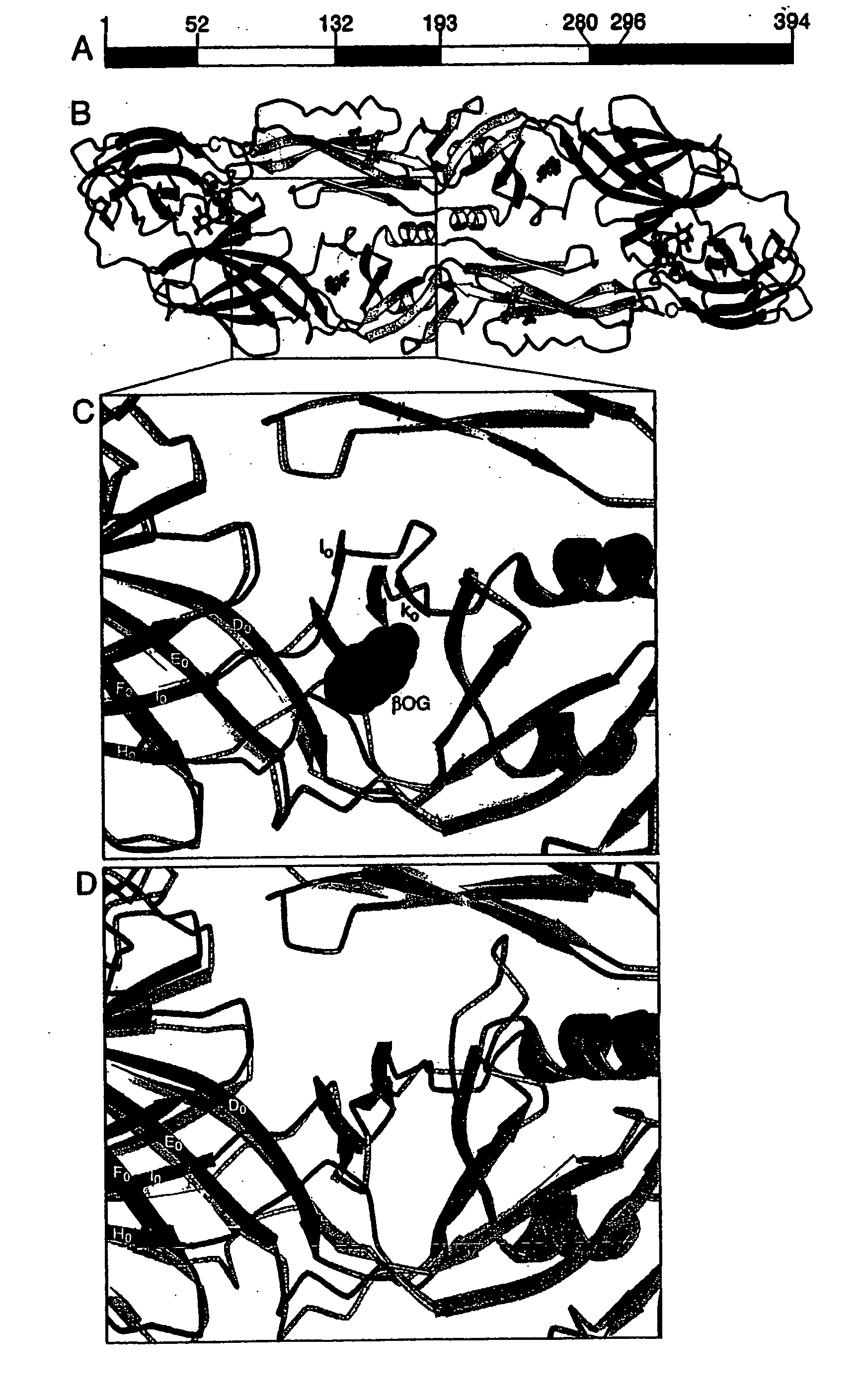
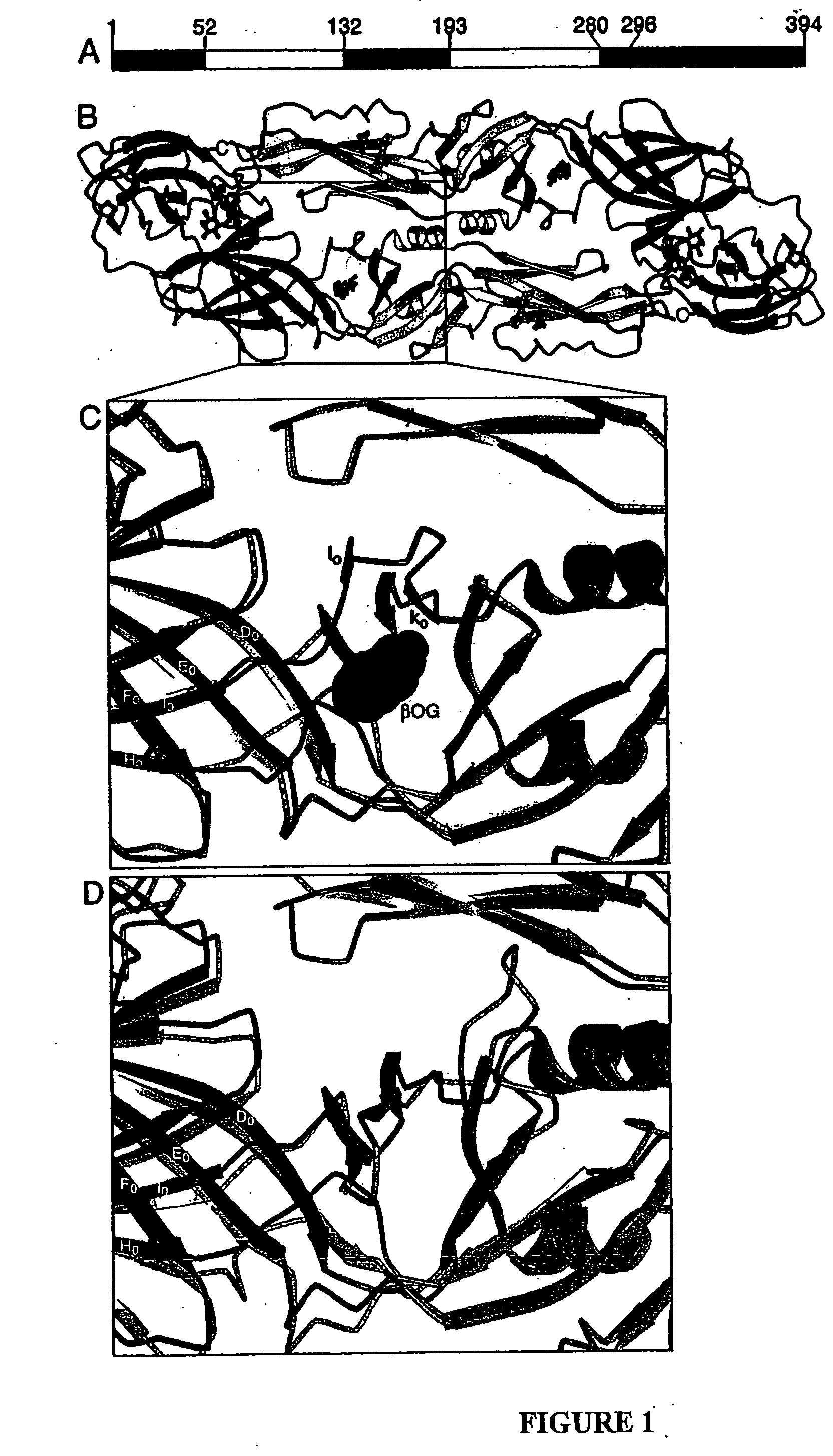
Determination of the Pre-Fusion Structure of E Protein from Dengue Virus Type S1
[0267] A. Expression, Purification and Crystallization of E Protein from Dengue Virus Type 2 S1
[0268] E protein from dengue virus type 2 S1 strain (Hahn et al., 1988) was supplied by Hawaii Biotechnology Group, Inc. (Aiea, Hi.). The construct that encoded the E protein sequence (SEQ ID NO 2) spans nucleotides 937-2118 of GenBank Accession Number M19197 and is described in detail in Hahn, Y. S. et al (1988), Virology 162, p 167-180. The primers that were used to amplify the dengue sE sequence by PCR:primer D2E937p2 5′-cttctagatctcgagtacccgggacc ATG CGC TGC ATA GGA ATA TC-3′ and primer D2E2121m 5′-gctctagagtcga cta tta TCC TTT CTT GAA CCA G-3′. Nucleotides corresponding to dengue cDNA are in upper case; non-dengue sequence is in lower case. The protein was expressed in Drosophila melanogaster Schneider 2 cells (ATCC, Rockville, Md.) from a pMtt vector (SmithKline Beecham) containing the dengue 2 prM and ...
example 2
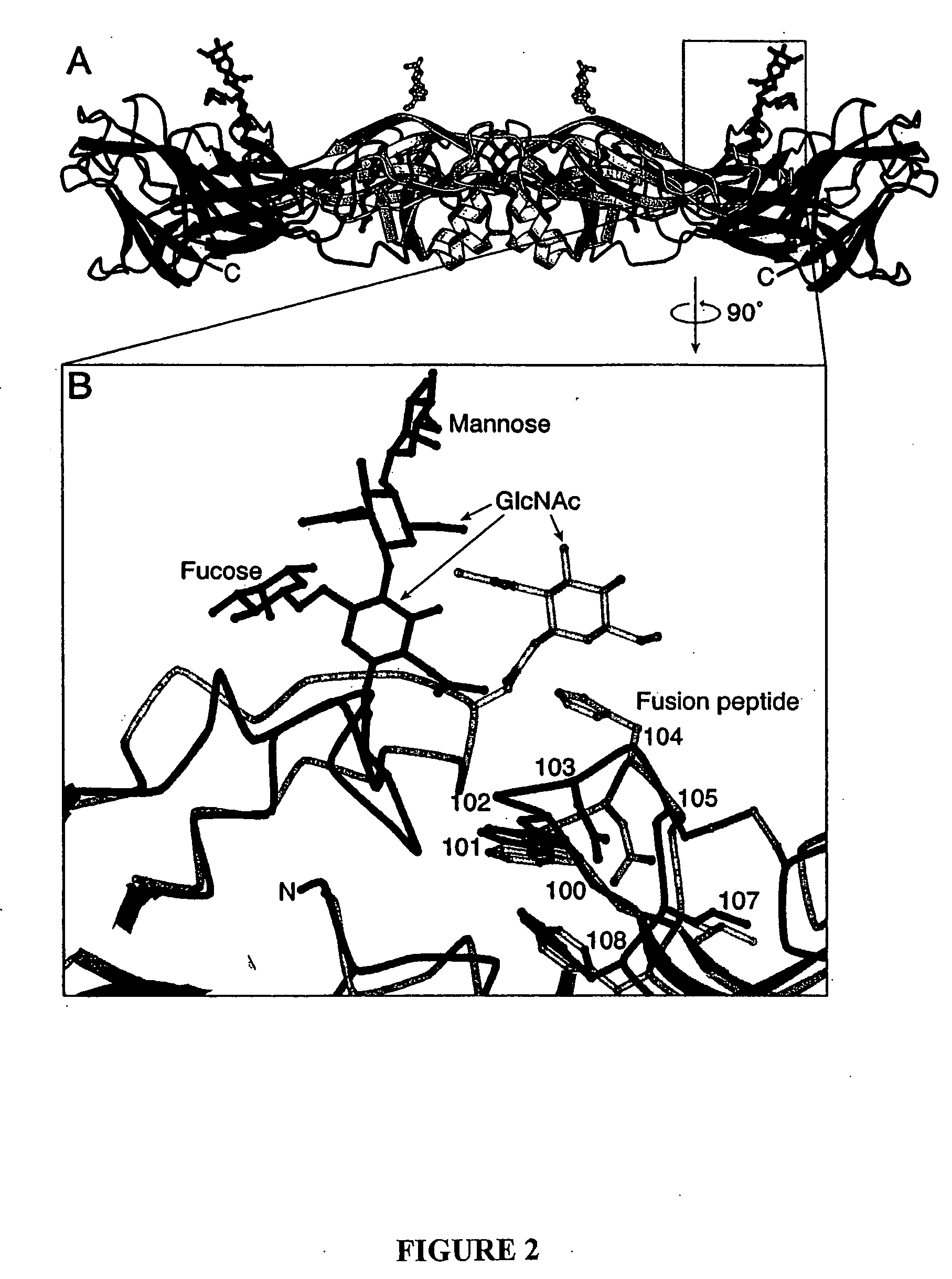
Determination of the Post-Fusion Structure of E Protein from Dengue Virus Type S1
[0290] A. Expression, Purification and Crystallization of E Protein from Dengue Virus Type 2 S1
[0291] Soluble E protein (sE) from dengue virus type 2 S1 strain was supplied by Hawaii Biotech. The protein was expressed in Drosophila melanogaster Schneider 2 cells (obtained from ATCC) using a pMtt vector (GlaxoSmithKline) containing the dengue 2 prM and E genes (nucleotides 539-2121 of the sequence) as described in Section A of Example 1. The resulting prM-E preprotein is processed during secretion to yield sE, which was purified from the cell culture medium by immunoaffinity chromatography.
[0292] Dengue sE trimers were obtained as follows, based on a method developed for tick-borne encephalitis virus sE (Stiasny, K., et al. J. Virol. 76, 3784-3790 (2002)). 1-palmitoyl-2-oleoyl-sn-glycero-3-phosphocholine, 1-palmitoyl-2-oleoyl-sn-glycero-3-phosphoethanolamine (Avanti Polar Lipids) and 1-cholesterol (Si...
example 3
Inhibitors of Flavivirus Entry
[0337] The discovery of a hydrophobic ligand-binding pocket beneath the k1 loop in the pre-fusion structure of dengue sE has suggested one possible strategy for inhibiting flavivirus entry by interfering with the fusion transition. The rationale for that proposal is enhanced by our new structure, which shows that significant rearrangements do occur around the k1 loop during the conformational change. The trimer structure also suggests a second strategy for interfering with fusion, related to an approach successful in developing an HIV antiviral compound. Peptides corresponding to the C-terminal region of the gp41 ectodomain inhibit HIV-1 entry, probably by binding to the trimeric, N-terminal “inner core” of the protein and interfering with the folding back of the C-terminal “outer-layer” against it. An analogous strategy may be possible with some class II viral fusion proteins, such as those of dengue and hepatitis C. The way in which the stem is likel...
PUM
| Property | Measurement | Unit |
|---|---|---|
| pH | aaaaa | aaaaa |
| conformational changes | aaaaa | aaaaa |
| pH | aaaaa | aaaaa |
Abstract
Description
Claims
Application Information
 - R&D
- Intellectual Property
- Life Sciences
- Materials
- Tech Scout
- Unparalleled Data Quality
- Higher Quality Content
- 60% Fewer Hallucinations
Browse by: Latest US Patents, China's latest patents, Technical Efficacy Thesaurus, Application Domain, Technology Topic, Popular Technical Reports.
© 2025 PatSnap. All rights reserved.Legal|Privacy policy|Modern Slavery Act Transparency Statement|Sitemap|About US| Contact US: help@patsnap.com


