[0014] Because the
RF field is also generated or excited only where the image should be acquired, or where image information should be read out, in the inventive magnetic resonance apparatus as a consequence of the defined, locally-delimited excitation field generation, the use of the
saturation pulse required in the prior art is no longer necessary. The sequence
workflow consequently can be accelerated and the
specific absorption rate (SAR) is inevitably reduced because the
saturation pulse is not emitted. The occurrence of artifacts that can also result as described from a rephasing in the prior art is advantageously avoided.
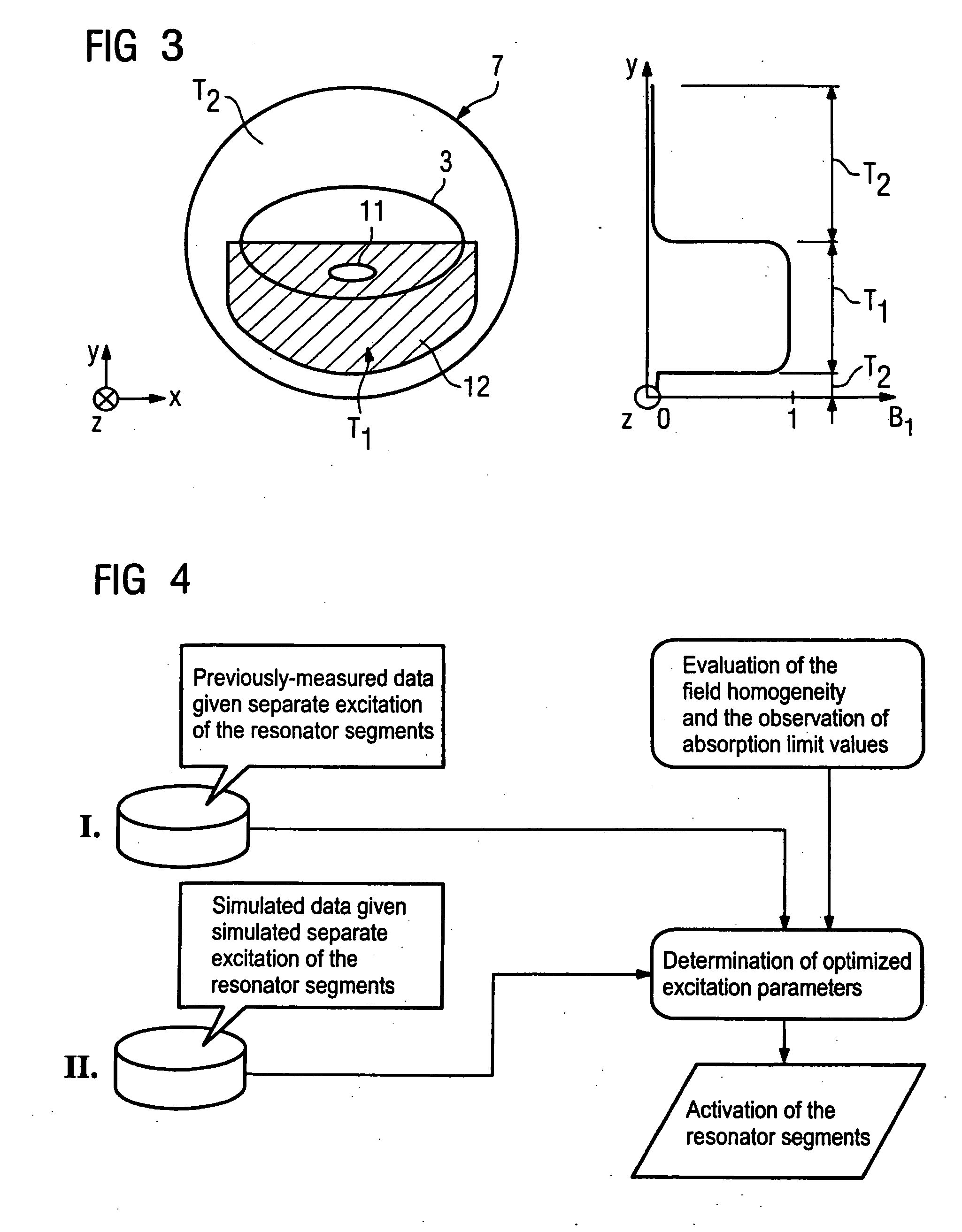
[0019] On the basis of such a procedure it is possible (if applicable, with repetition of the steps of the computational
superimposition, the homogeneity evaluation and the determination of the parameters (iterative optimization)) to fashion or to shape the
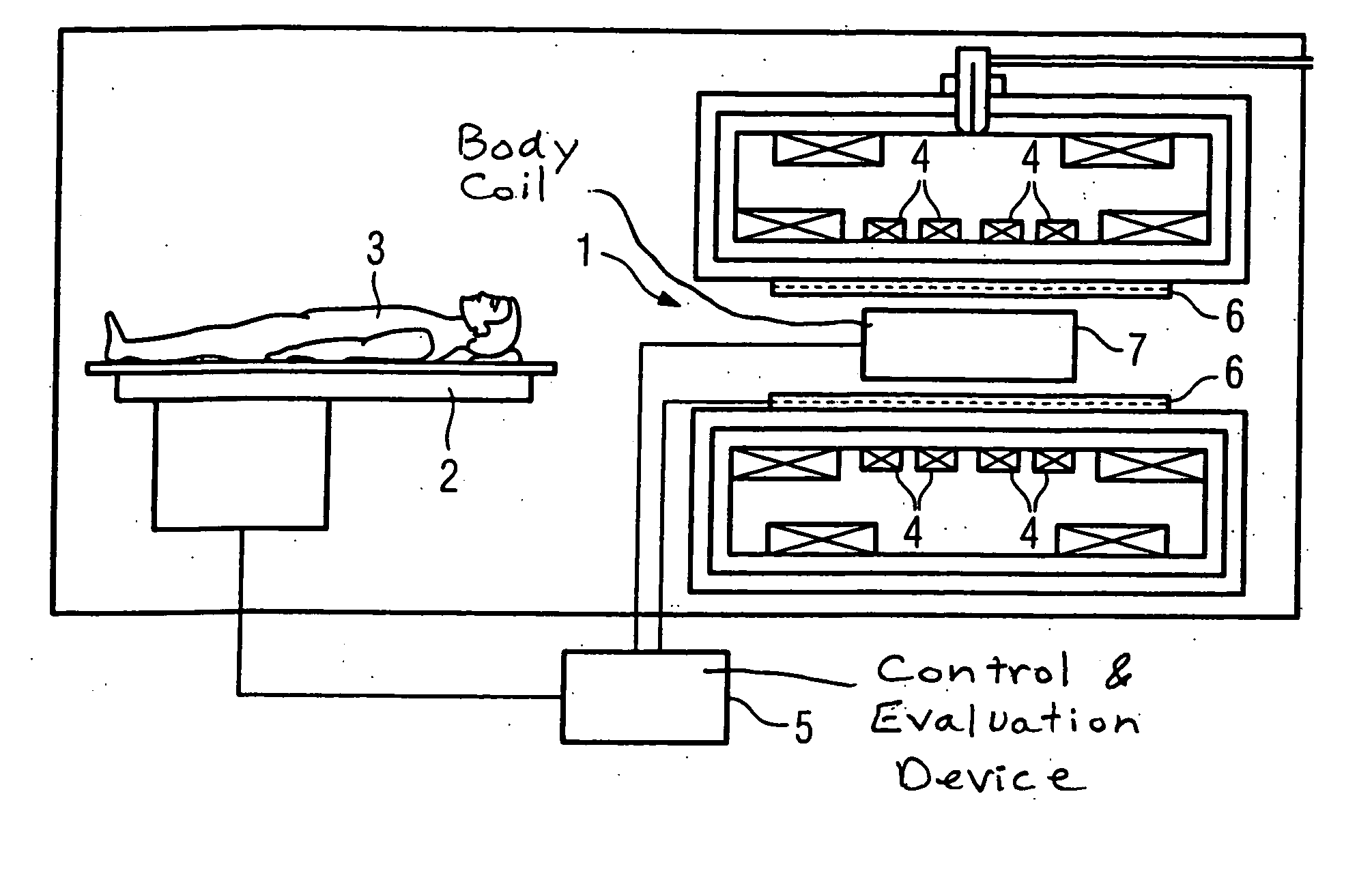
RF field corresponding to the clinical question, thus corresponding to the actual examination volume to be acquired, and to simultaneously minimize the field formation in the second sub-volume. For example, if the excitation field for a
spinal column acquisition should be generated only in the lower region of the coil volume, such that the
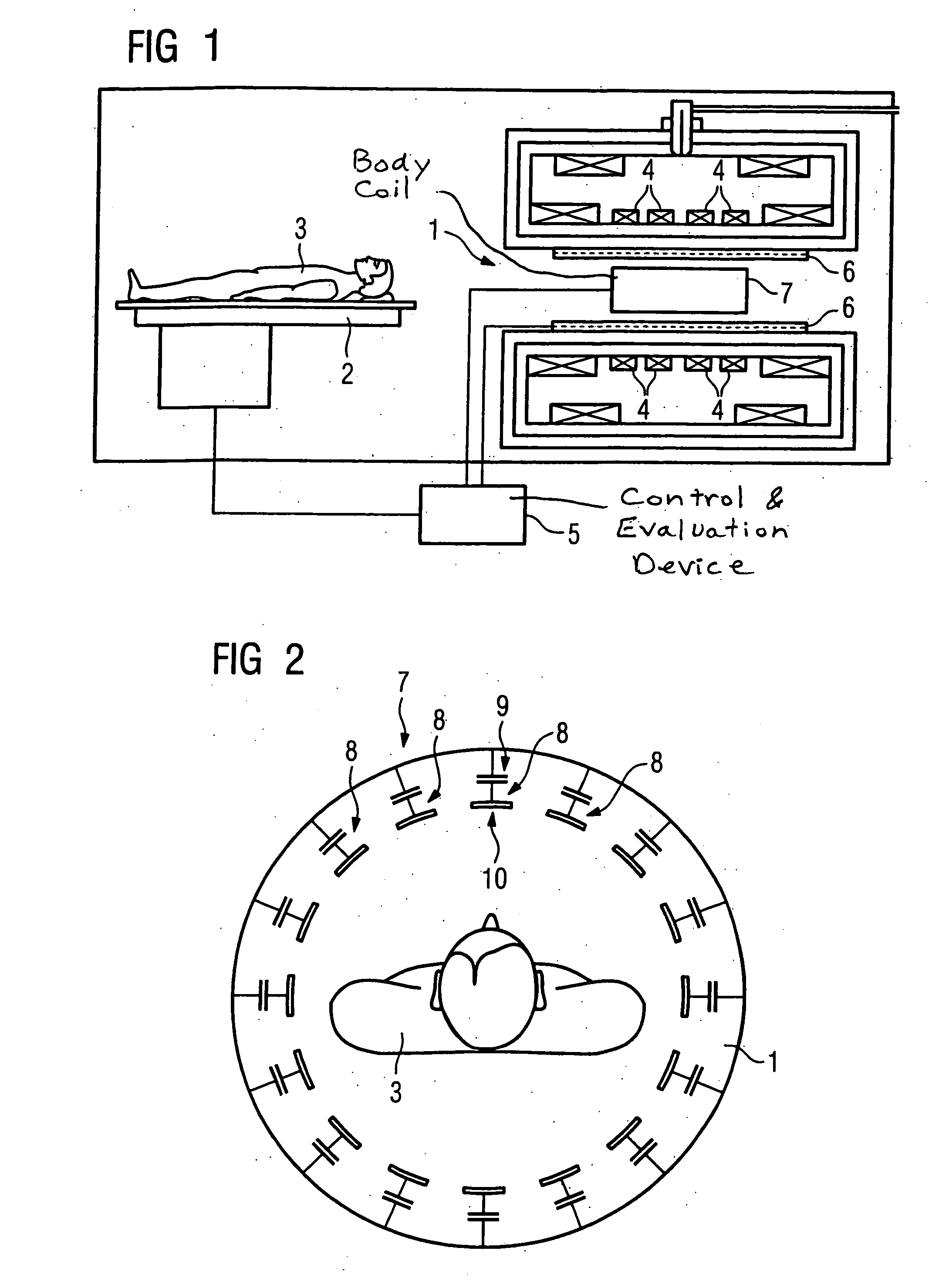
abdominal wall is not situated in the excitation field, the described parameter determination ensues such that just this field geometry is set, by excitation of a few or all resonator segments. For each resonator segment, the corresponding excitation parameters (starting from a normal excitation as a basis of, for example, the iterative parameter prioritization) are determined that cause the individual fields to be generated such that the overall field distribution is adjusted, such that only the desired sub-volume is excited, or the excitation field is generated only in the desired sub-volume, while the other volume region is essentially field-free. Using a changed, optimized set of excitation parameters, a subject-specific excitation of the resonator segments that is thus dependent on the examination subject can ensue for generation of a locally-optimized, geometrically optimally-shaped, polarized
magnetic field with maximum homogeneity. Using such a procedure, the inventive magnetic resonance apparatus and method allow a simple and fast generation of an excitation field with maximum homogeneity in the examination volume, defined by the excitation field sub-volume. Complete scanning by simultaneous or time-offset grouped or individualized activation of only the resonator segments necessary to acquire the individual fields is required for this purpose. A diagnostic
patient exposure is not needed. The parameters required for the subsequent diagnostic
image acquisition can be determined in a simple and fast manner. Since the
evaluation algorithm serves for the optimization of the excitation field with regard to its homogeneity, the
image acquisition can ensue on the basis of an optimally homogeneous excitation field, such that the maximum information yield is possible from the limited examination volume without inhomogeneities of the
image quality resulting from the non-excitation of other volume regions that are not of interest.

