
Method and Apparatus for Performing a Surgical Procedure

a surgical procedure and surgical method technology, applied in the field of methods, can solve the problems of affecting the surgical procedure, and affecting the surgical effect, and achieve the effect of preventing premature tissue resection

Inactive Publication Date: 2006-11-16
ARAGON SURGICAL INC
View PDF19 Cites 43 Cited by
  • Summary
  • Abstract
  • Description
  • Claims
  • Application Information

AI Technical Summary

Benefits of technology

[0010] The invention provides, inter alia, improved methods and devices for performing such procedures as vaginal hysterectomies, and that reduce procedure time and complexity, resulting in improved patient outcomes and potentially increased cost savings to the healthcare system. In one embodiment, the invention offers most advantages when performing a procedure, such as a hysterectomy, through a vaginal approach as described herein, yet is easier for the average surgeon to perform. It will be appreciated, however, that the presently disclosed devices may be modified to allow, for example, the removal of the uterus via open abdominal hysterectomy, which is also within the scope of the invention. Additionally, laparoscopic visualization may be used to guide the procedures of the invention. Those skilled in the art will appreciate that, while the invention is discussed in detail in connection with procedures performed on the uterus, i.e. a hysterectomy, other procedures are equally suited for application of the invention thereto. Accordingly, the invention applies equally to such other procedures and is not limited to the examples provided herein.
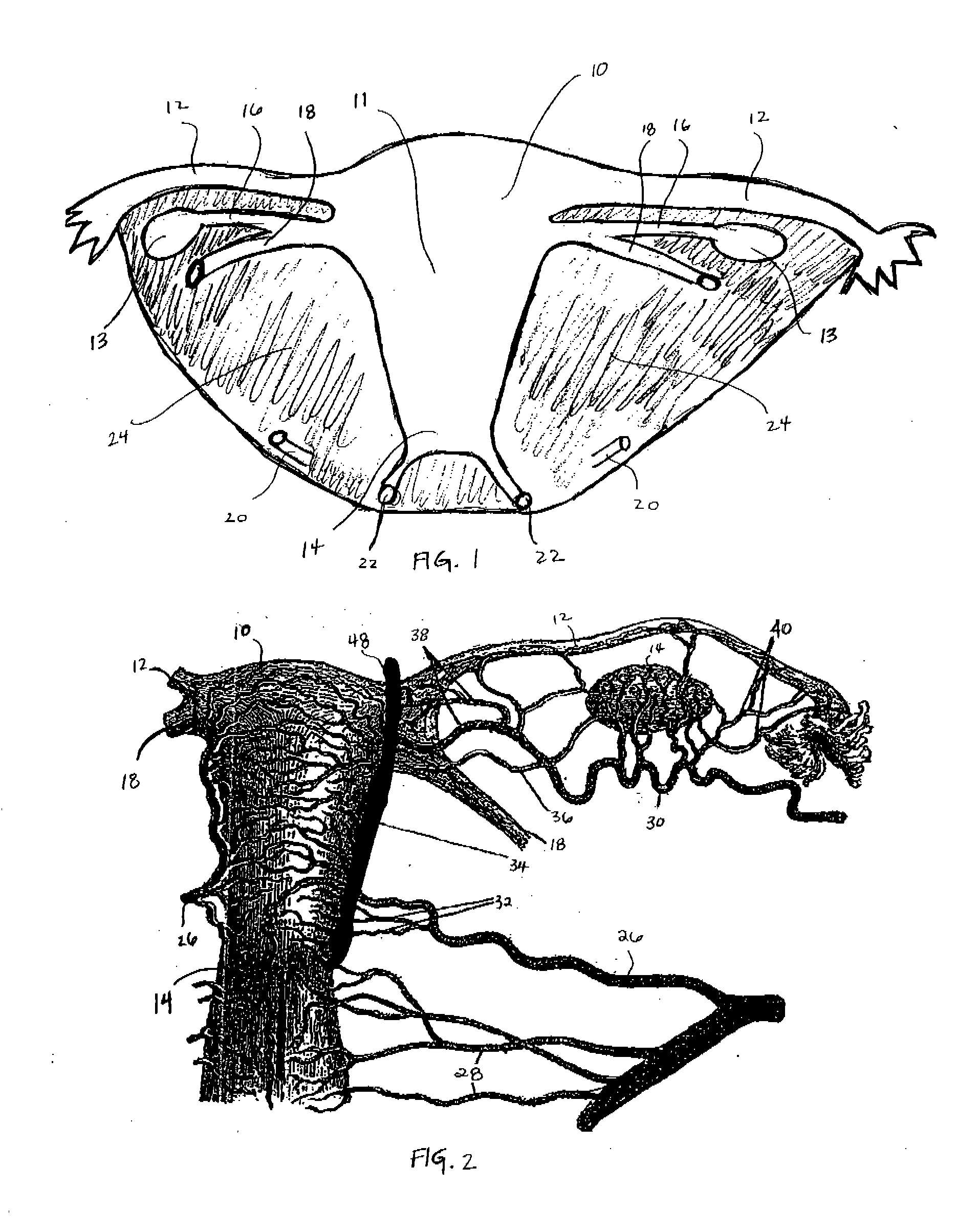
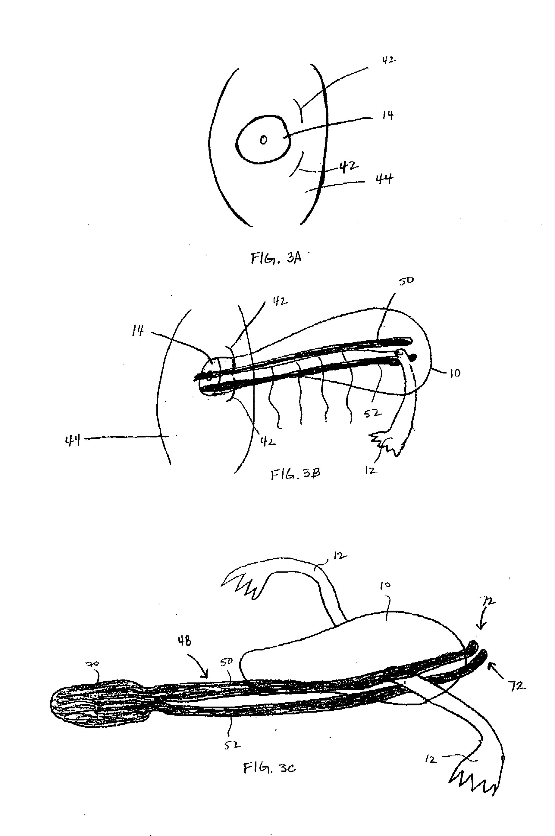
[0012] In this embodiment, the invention avoids heating or ablation of the entire uterus. Instead, the invention focuses on surgically dividing, ligating, and severing the blood vessels, associated ligaments that support the uterus, and optionally the fallopian tube(s) and ovary(ies). This coagulates and seals off the entire blood supply to the uterus to effectively achieve hemostasis, i.e. cessation of bleeding, which is of major concern in removal of an organ or tissue, such as the uterus. This frees up the uterus for subsequent removal through the vaginal opening, as described in more detail below.
[0016] Again, the introduction and engagement of the third and fourth energy transmitting elements may be viewed and guided with a laparoscope. Additionally, a centering post may be inserted into the uterus and located parallel to and between the first and second jaws to allow the surgeon to maneuver the uterus externally. This, in turn, ensures proper viewing and positioning of the first and second jaws along lateral sides of the uterus, wherein all connective tissues and blood vessels are entrapped.
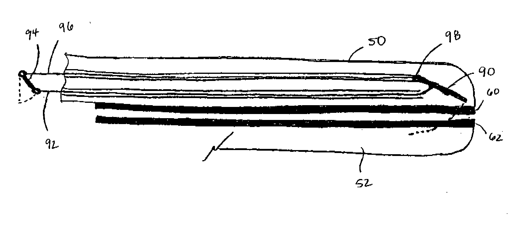
[0022] The surgical tool may also comprise at least one cutting blade recessed within at least one jaw element to allow for tissue resection. The blade may movably traverse a longitudinal channel defined by pairs of electrode elements, as discussed above. The blade may comprise a variety of configurations, including a flexible blade, a cutting wheel, a v-shaped cutter, or a linkage blade, as will be described in more detail below. For safety purposes, a blade guide stop or blade interlock may be coupled to the blade so that the blade is not inadvertently released during the procedure, particularly prior to tissue desiccation. The surgical tool may also comprise at least one trigger mechanism coupled to the handle. For example, actuation of a first trigger clamps the first and second jaw elements together, which triggers the initiation of radio frequency power application. Actuation of a second trigger allows for tissue resection once complete tissue mass coagulation and sealing is verified. In such an embodiment, a change in impedance, current, or voltage is measured to verify that tissue mass coagulation and sealing is completed to prevent premature tissue resection. Further, an audible alarm may be sounded or a visual alarm displayed indicating complete tissue mass coagulation and sealing.

Problems solved by technology

The costs related to performing hysterectomies has burdened the United States healthcare system on the order of billions of dollars annually.
However, open abdominal hysterectomy also suffers from several drawbacks.
For example, the surgical procedure is often lengthy and complicated, requiring longer anesthesia periods and the increased risk of postoperative complications.
Patients also suffer from prolonged recovery periods, pain and discomfort, and large visible scarring on the abdomen.
Further, increased costs are associated with an open abdominal approach, such as prolonged hospital stays.
Unfortunately, less than a third of all hysterectomies are performed vaginally due to a lack of surgeon training, limited access of the uterus and surrounding tissue, and unsuitability of a patient's anatomy, for example a large uterus size, limited vaginal access, severe endometriosis, pelvic adhesions, and the like.

Method used


Image

  • Method and Apparatus for Performing a Surgical Procedure
  • Method and Apparatus for Performing a Surgical Procedure
  • Method and Apparatus for Performing a Surgical Procedure

Examples


Embodiment Construction

[0033] The invention provides methods and devices for performing such procedures as vaginal hysterectomies. It will be appreciated however that application of the invention is not limited to removal of the uterus, but may also be applied for ligation of nearby structures such as the ovaries (oophorectomy), ovaries and fallopian tubes (salpingo-oophorectomy), fallopian tubes, uterine artery, and the like. It will further be appreciated that the invention is not limited to a vaginal approach, but may also allow for removal of the uterus via open abdominal hysterectomy, which is also within the scope of the invention. Additionally, laparoscopic visualization may be used to guide the procedures of the invention. Finally, the invention is likewise applied to other parts of the body in connection with other surgical procedures.

[0034]FIG. 1 illustrates a simplified frontal view of a uterus 10 comprising a body 11 and a cervix 14. Attaching structures of the uterus 10 include fallopian (ut...


PUM


Abstract

One method for performing procedures, such as vaginal hysterectomies, comprises engaging first and second energy transmitting elements against a lateral side of a uterus. The first and second energy transmitting elements are positioned against opposed surfaces of a tissue mass extending from and including a fallopian tube or round ligament to a tip of a cervix. Third and fourth energy transmitting elements are positioned against another lateral side of the uterus and against opposed surfaces of another tissue mass extending from and including another fallopian tube or round ligament to the tip of the cervix. Radio frequency or other high energy power is applied through the energy transmitting elements to the tissue masses. The power is applied for a time and in an amount sufficient to coagulate and seal the tissue masses within the energy transmitting elements. The coagulated tissue masses are then resected and the entire uterus removed.

Description

CROSS REFERENCE TO RELATED APPLICATIONS [0001] This application derives its priority from U.S. Provisional Patent Application Ser. Nos. 60 / 680,937, filed May 12, 2005 and 60 / 725,720, filed Oct. 11, 2005, each of which is incorporated herein in its entirely by this reference thereto.BACKGROUND OF THE INVENTION [0002] 1. Field of the Invention [0003] The invention relates generally to organ resection, and more particularly to methods and devices, for example, for surgical removal of the female uterus or hysterectomy. [0004] 2. Description of the Background Art [0005] Hysterectomy may involve total or partial removal of the body and cervix of the uterus. Hysterectomy next to the caesarian section procedure is the most common surgical procedure performed in the United States. By the age of sixty, nearly one in three American women will have undergone hysterectomy. It is estimated that over a half million women undergo hysterectomy each year in the United States alone. The costs related ...

Claims


Application Information

Patent Timeline
Patent Type & Authority Applications(United States)
IPC IPC(8): A61B18/14
CPCA61B18/1442A61B2018/00559A61B2017/4216A61B17/00A61B18/14
Inventor NEZHAT, CAMRANSTERN, ROGER A.EDER, JOSEPHEDELSTEIN, PETER SETH
Owner ARAGON SURGICAL INC
Learn More