

Non-in situ hybridization method for detecting chromosomal abnormalities
- Summary
- Abstract
- Description
- Claims
- Application Information
AI Technical Summary
Benefits of technology
Problems solved by technology
Method used
Image

Examples
example 1
Preparation of Labeled Nucleic Acid Probes
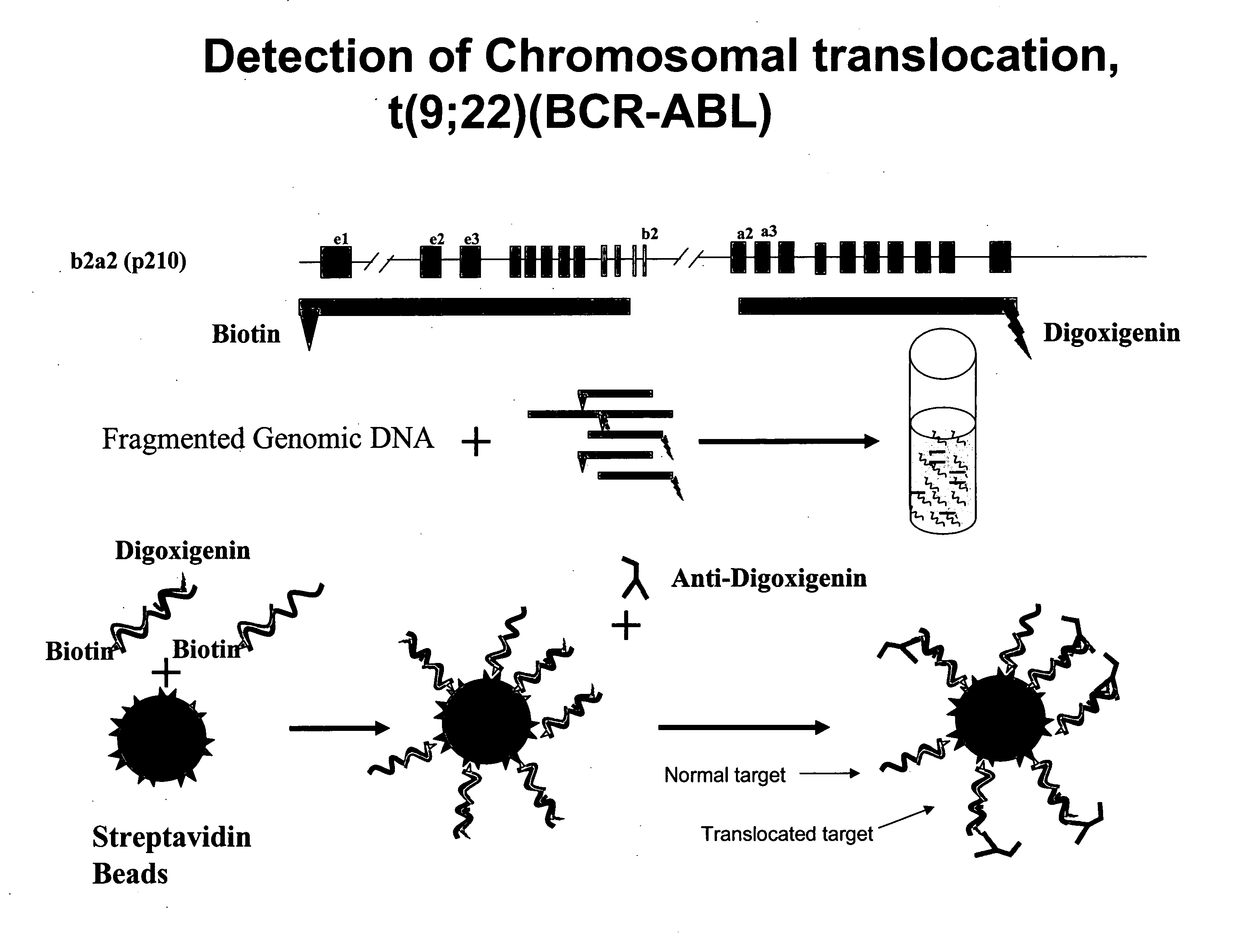
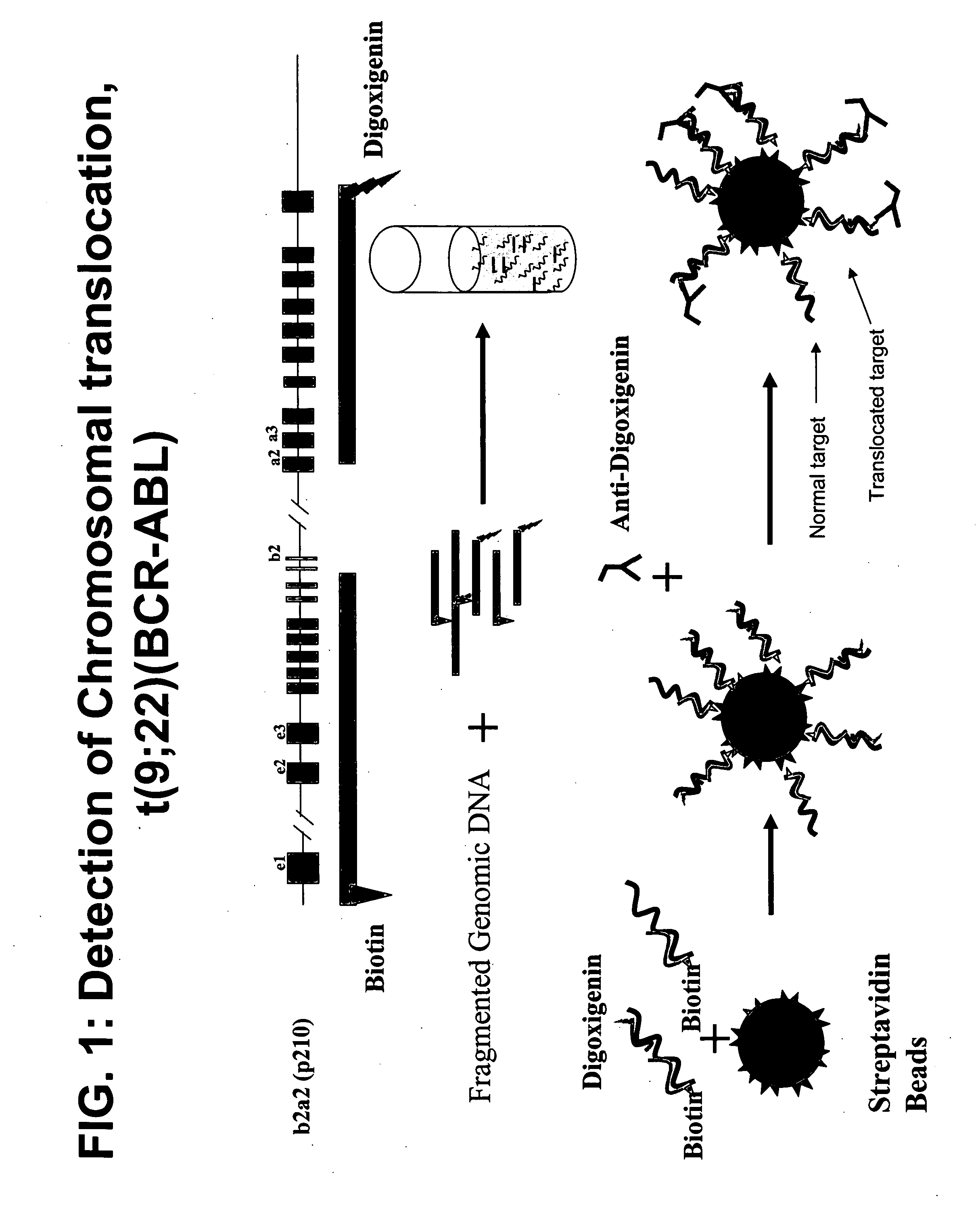
[0197] Bacterial artificial chromosomes (BACs) containing the BCR locus (BCR-BAC) and BACs containing the ABL locus (ABL-BAC) were used to generate probes to detect the Philadelphia chromosome translocation. These BACs were purchased commercially (Invitrogen). The BACs were grown and isolated using standard methods.
[0198] Biotinylation of BACs
[0199] The isolated ABL-BAC was biotinylated using a standard nick translation (NT) protocol. 10 μl of ABL-BAC was mixed with NT enzyme, buffer, and biotin-16-dUTP incubated at 65° C. for 1.5 hours. 0.5 M EDTA was added and the mixture incubated at 65° C. for 10 minutes.
[0200] Detection Labeling of BACs
[0201] The isolated BCR-BAC DNA was digested in aqueous solution with DNAse I for 10 minutes at 37° C. The digestion reaction was stopped with a 10 minute incubation at 65° C. 1 μg of the digested BCR-BAC was ethanol precipitated out of solution using 1 / 10 volume 3M NaOAc and 2 volumes 100% ethanol a...
example 2
Hybridization of Labeled BAC Probes and Genomic DNA
[0203] Labeled probes were hybridized to a test sample of genomic DNA. The biotinylated probe and digoxigenin-labeled probe were mixed and centrifuged at maximum speed for 30 minutes. The resulting pellet was resuspended in hybridization buffer (50% Formamide, 10% dextran sulfate, 2×SSC, 40 mM sodium phosphate buffer and 1× Denhardt's Solution), incubated at 37° C. for 30 minutes and denatured at 73° C. for 10 minutes. The probe mixture was then cooled on ice for 5 minutes and incubated for 30 hour at 37° C. Denaturation solution (70% deionized Formamide, 0.2×SSC) was then added to the probe mixture.
[0204] Genomic DNA was digested with DpnII for 1 hour at 37° C. The digestion was stopped by heat inactivation at 65° C. for 10 minutes. Digested genomic DNA (1 μg) was denatured in denaturation solution (70% deionized Formamide, 0.2×SSC) by incubation at 73° C. for 7 minutes then incubated on ice for 5 minutes.
[0205] The denatured pr...
example 3
Capture of Hybridization Complex on Solid Support
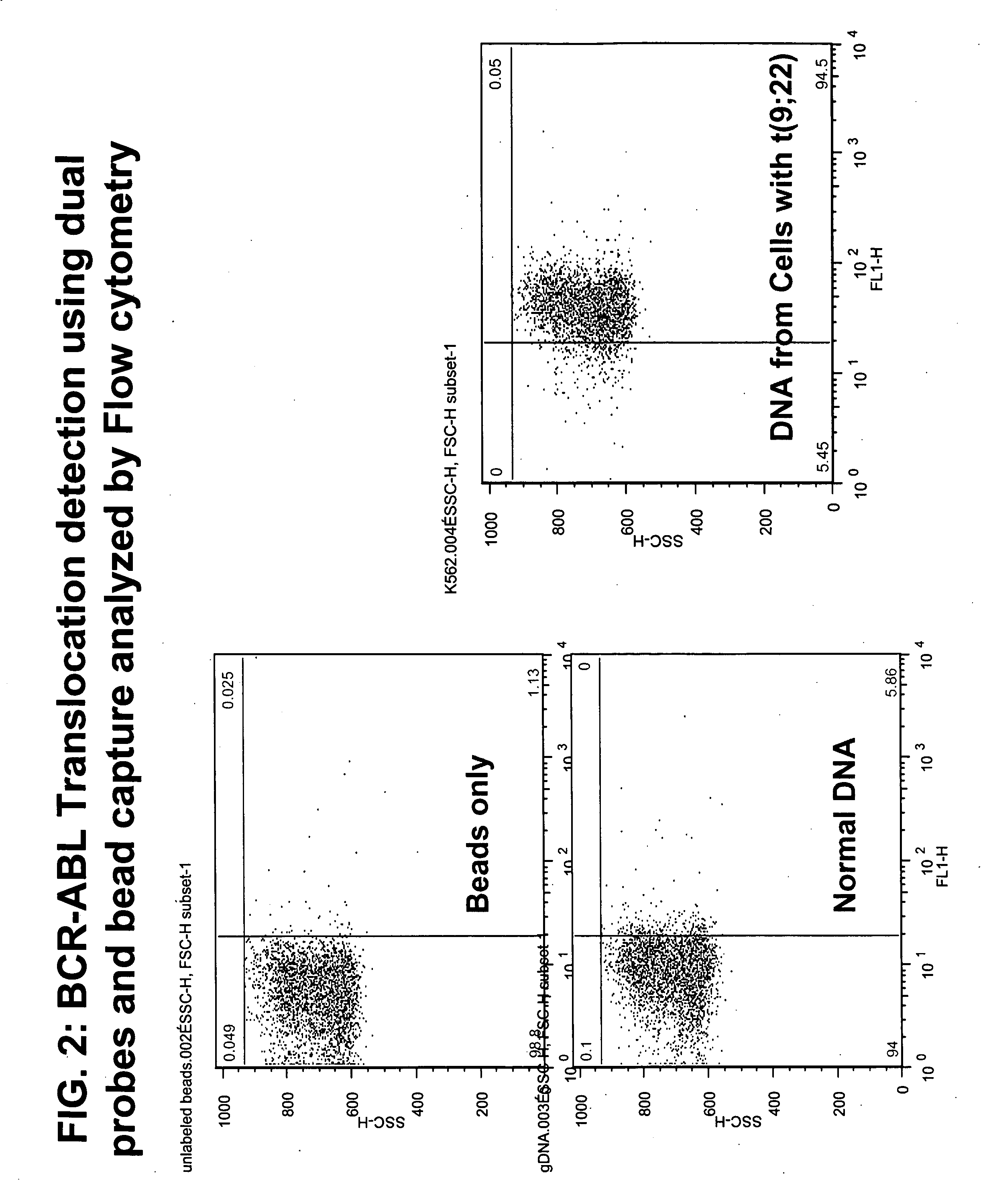
[0206] Hybridization complexes incorporating a biotin-labeled probe were captured on streptavidin-coated beads. 5 μl of streptavidin beads (Bangs Lab, Fishers, Ind.) were washed once with 100 μl TTL solution (100 mM Tris-HCL; pH 8.0, 0.1% Tween 20; and 1 M LiCl) and resuspended in 20 μl TTL. 5 μl probe-DNA complex was added to the beads and the mixture incubated while shaking at room temperature for 30 minutes to form a bead-DNA complex. The bead complex was then washed three times with 2% BSA in phosphate buffered saline (PBS), resuspended in 4% blocking milk, and washed once with 2% BSA in PBS. The bead complex was then resuspended in FITC-labeled anti-digoxigenin antibody at a dilution of 1:500 and rotated for 30 minutes at room temperature in the dark. The bead complex was then washed once with 2% BSA in PBS using a Sorvall CW-2 Cell washer to wash and pellet the beads.
PUM
| Property | Measurement | Unit |
|---|---|---|
| length | aaaaa | aaaaa |
| binding affinity | aaaaa | aaaaa |
| acid | aaaaa | aaaaa |
Abstract
Description
Claims
Application Information