Engineered osteochondral construct for treatment of articular cartilage defects

a technology of articular cartilage and constructs, which is applied in the field of treatment of articular cartilage defects, can solve the problems of less load-bearing tissue, complex autologous chondrocyte transplantation technique, and difficulty in restoring normal function, and achieves the effect of restoring normal function and being easily placed in the defect area

Active Publication Date: 2007-01-11
MUSCULOSKELETAL TRANSPLANT FOUND INC
View PDF99 Cites 25 Cited by
  • Summary
  • Abstract
  • Description
  • Claims
  • Application Information

AI Technical Summary

Benefits of technology

[0018] It is an object of the invention to provide an autograft and / or allograft implant for joints which provides pain relief, restores normal function and will postpone or alleviate the need for prosthetic replacement.
[0019] It is also an object of the invention to provide a cartilage repair implant which is easily placed in a defect area by the surgeon using an arthroscopic, minimally invasive technique.
[0020] It is further an object of the invention to provide an allograft implant procedure which is applicable for both partial and full thickness lesions.

Problems solved by technology

The technique of autologous chondrocyte transplantation is very complex and requires postoperative immobilization of the patient.
This method shows good clinical results including a good load capacity of the operated knee-joint, but is only applicable to small cartilage defects due to the limited availability of autologous osteochondral cylinders.
Classical operation techniques, which do not use cartilage or chondrocyte transplantation (abrasion, debridement, Pridie-drilling), result only in a defect repair with fibrous, less load-bearing tissue unfortunately.
Reports of results of osteochondral plug autografts in a small numbers of patients indicate that they decrease pain and improve joint function, however, long-term results have not been reported.
Factors that can compromise the results include donor site morbidity, effects of joint incongruity on the opposing surface of the donor site, damage to the chondrocytes at the articular margins of the donor and recipient sites during preparation and implantation, and collapse or settling of the graft over time.
The limited availability of sites for harvest of osteochondral autografts restricts the use of this approach to treatment of relatively small articular defects and the healing of the chondral portion of the autograft to the adjacent articular cartilage remains a concern.
However, long term studies of this procedure in rabbits and dogs showed limited success and showed degradation at the implant site.
The original study report has been criticized for not being a prospective controlled randomized study and for lack of quantitative or mechanical data.
As with the perichondrial graft, patient / donor age may compromise the success of this procedure as the chondrocyte population decreases with increasing age.
Disadvantages to this procedure include the need for two separate surgical procedures, potential damage to surrounding cartilage when the periosteal patch is sutured in place, the requirement of demanding microsurgical techniques, and the expensive cost of the procedure which is currently not covered by insurance.
The use of implants for cartilage defects is much more limited than that for bone defects.
Concerns associated with this method are harvest site morbidity and availability, similar to the mosaicplasty method.

Method used


Image

  • Engineered osteochondral construct for treatment of articular cartilage defects
  • Engineered osteochondral construct for treatment of articular cartilage defects

Examples


Embodiment Construction

[0026] The present invention is susceptible of embodiment in various forms as will hereinafter be described with the understanding that the present disclosure is to be considered as an exemplification of the invention, and is not intended to limit the invention to the specific embodiments disclosed herein.

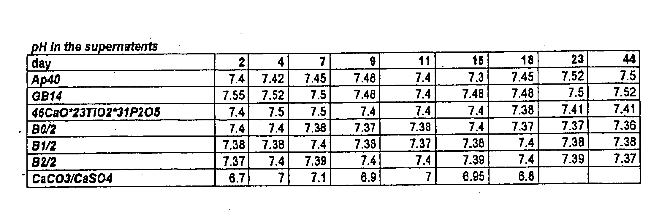
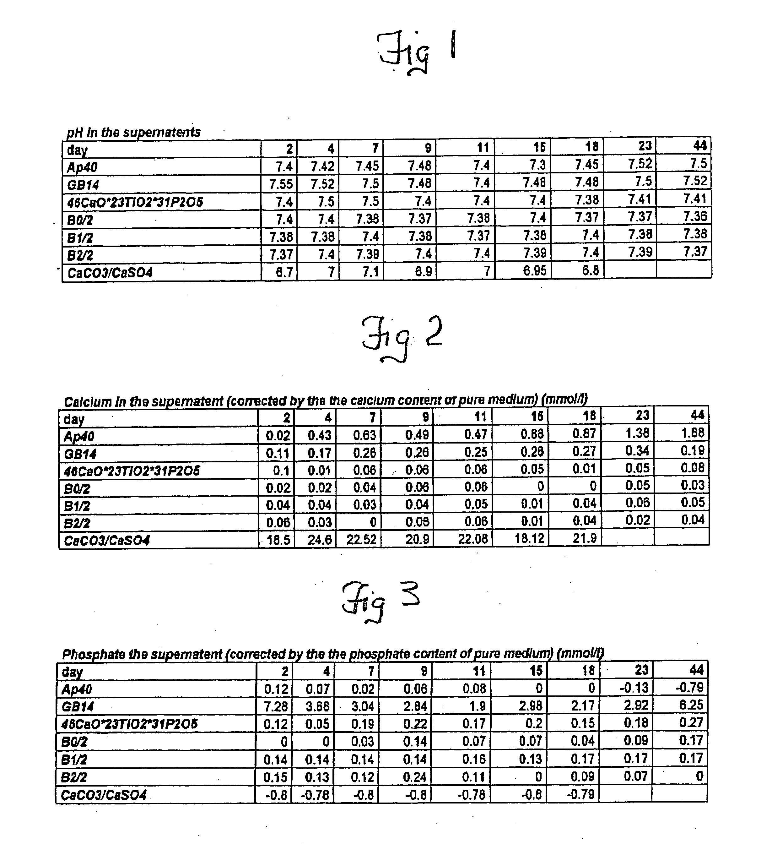
[0027] Sterile cancellous bone replacement structures were utilized for the in vitro grown cartilage replacements, which allow the fabrication of load-bearing constructs. BMP's from the cancellous bone plugs have a positive effect on chondrocyte differentiation in vitro by stimulating the formation of a native, chondrocyte-phenotype and proper matrix production by the cells. The highest stimulation effect of BMP's on chondrocytes can be observed, if BMP's are immobilized onto a carrier or retained in a biological matrix. In these carriers the natural BMP's of the bone are released by the demineralization but retained in the carrier matrix. For evaluating the effect of the biologic...


PUM


Abstract

A method of growing chondrocytes on an allograft cancellous bone structure which had been previously treated by demineralization to form a cartilage repair implant comprises isolating allograft chondrocytes from articular cartilage of a donor other than the patient on which the implant is to be used, cultivating the isolated chondrocytes in a medium to obtain a dedifferentiated fibroblast-like phenotype, placing an at least partially demineralized allograft cancellous bone structure carrier having at least one surface prepared for cell seeding in a culture vessel; adding the cultivated dedifferentiated chondrocytes to a demineralized surface of the cancellous bone structure carrier, and incubating the cancerous bone structure carrier for a period of time of at least 40 days to obtain an implant with a cartilage layer which ranges from about 2 mm to 5 mm n thickness.

Description

RELATED APPLICATIONS [0001] This application claims the benefit of Provisional Application No. 60 / 697,563, filed Jul. 11, 2005.STATEMENT REGARDING FEDERALLY SPONSORED RESEARCH OR DEVELOPMENT [0002] Not applicable. REFERENCE TO SEQUENCE LISTING, A TABLE OR A COMPUTER PROGRAM LISTING COMPACT DISC APPENDIX [0003] None. FIELD OF INVENTION [0004] The present invention is generally directed towards the treatment of articular cartilage defects using an allograft bone scaffold or carrier. In particular, the allograft bone is treated with allograft chondrocytes to form a cartilage surface on the allograft bone. BACKGROUND OF THE INVENTION [0005] Chondrocytes are cells specific to articular cartilage. The isolation and cultivation of chondrocytes is a standard procedure, which has been undertaken for more than 10 years. Under two-dimensional culture conditions in cell culture vessels, chondrocytes dedifferentiate to fibroblast-like cells. During the dedifferentiation they lose their typical p...

Claims


Application Information

Patent Timeline
Patent Type & Authority Applications(United States)
IPC IPC(8): A61K35/34A61K35/12A61K35/32
CPCA61K35/12A61K35/32A61L27/3608A61L27/3654A61L27/3683C12N2533/18A61L27/3852A61L27/3895A61L2430/06C12N5/0655A61L27/3817
Inventor SYRING, CARINA
Owner MUSCULOSKELETAL TRANSPLANT FOUND INC
Why Eureka
Try Eureka