OCT using spectrally resolved bandwidth

a technology of optical coherence and bandwidth, applied in the field of optical coherence tomographic imaging, can solve the problems of myocardial infarction, prone to rupture, no methods are currently available to the cardiologist,

Inactive Publication Date: 2007-01-18
BOARD OF RGT THE UNIV OF TEXAS SYST
View PDF28 Cites 193 Cited by
  • Summary
  • Abstract
  • Description
  • Claims
  • Application Information

AI Technical Summary

Problems solved by technology

Unfortunately, most of us can identify a family member or close friend that has suffered from a myocardial infarction.
Recent evidence from many investigational studies, however, clearly indicate that most infarctions are due to sudden rupture of non-critically stenosed coronary arteries due to sudden plaque rupture.
Unfortunately, no methods are currently available to the cardiologist that may be applied to specify which coronary plaques are vulnerable and thus prone to rupture.
Although treadmill testing has been used for decades to identify patients at greater cardiovascular risk, this approach does not have the specificity to differentiate between stable and vulnerable plaques that are prone to rupture and frequently result in myocardial infarction.
Saline use is limited in duration, however, since myocardial ischemia eventually occurs in the distal myocardium.
Mechanical drag in the rotating shaft can produce significant distortion and artifacts in recorded OCT images of the coronary artery.
Unfortunately, because the catheter will always be forced to make several bends between the entry point in the femoral artery to the coronary artery (e.g., the 180 degree turn around the aortic arch), uneven mechanical drag will result in OCT image artifacts.
As the application of OCT is shifted from imaging gross anatomical structures of the coronary artery to its capability to image at the level of a single cell, non-uniform rotation of the single fiber OCT prototype will become an increasingly problematic source of distortion and image artifact.
Essentially, current endoscope type single channel OCT systems developed by Light Lab Co. suffers by non-constant rotating speed that forms irregular images of a vessel target.
Their approach of a rotary shaft to spin a single mode fiber is prone to produce artifact.
All these bends will cause uneven friction on the rotary shaft, and uneven time distribution of the light on the entire 360 degree arch of the coronary artery.
As the application of OCT is shifted from gross anatomical structures of the coronary artery to its capability to image at the level of a single cell, then non-uniform rotation of the single fiber OCT will become even a greater source of greater artifact.
Many investigators recognize that a single rotating fiber or micro-motor driven mirrors utilized in current designs will not allow imaging at the level of a single cell while the multiphase array approach can provide cellular resolution.

Method used


Image

  • OCT using spectrally resolved bandwidth
  • OCT using spectrally resolved bandwidth
  • OCT using spectrally resolved bandwidth

Examples


Embodiment Construction

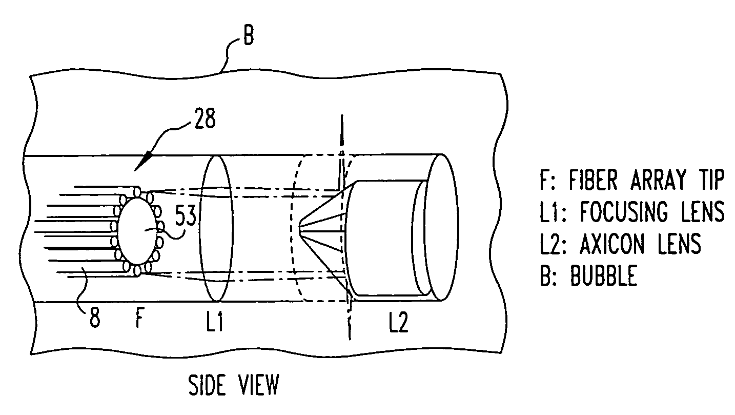
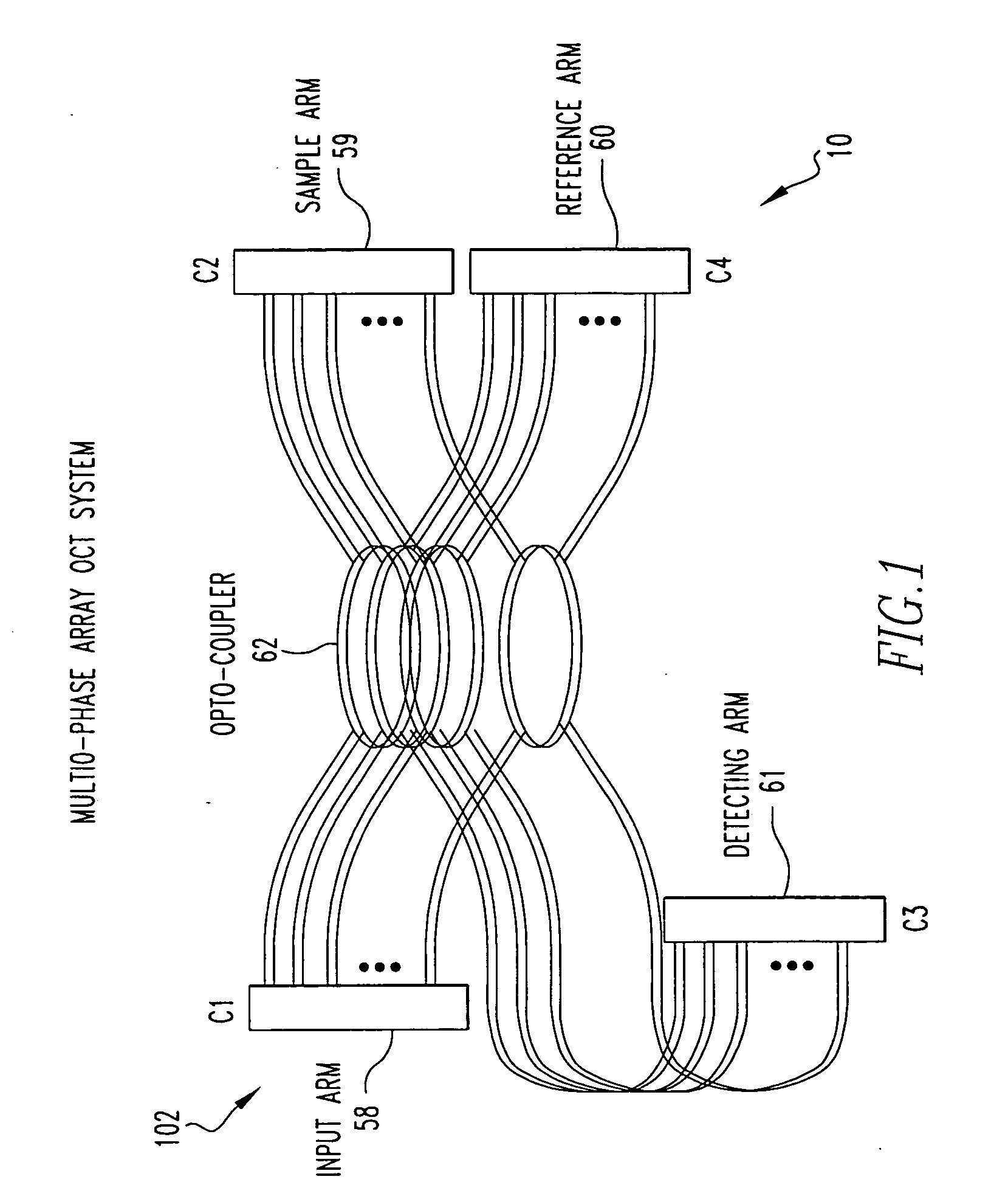
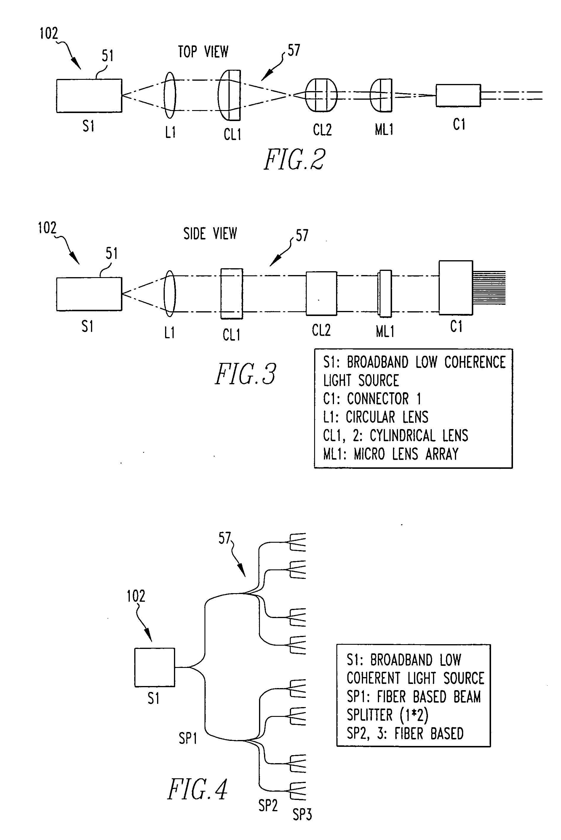
[0059] Referring now to the drawings wherein like reference numerals refer to similar or identical parts throughout the several views, and more specifically to FIGS. 1-5, 15 and 16 thereof, there is shown an endoscope 10 for a patient. The endoscope 10 comprises means 102 for producing light, such as a light source 51. The endoscope 10 comprises an optical fiber array 28 comprising a plurality of optical fibers 8 adapted to be disposed in the patient. The optical fiber array 28 transmits the light from the producing means, preferably including a light source 51, into the patient, and transmits the light reflected by the patient out of the patient. The plurality of the optical fibers 8 of the array 28 is in optical communication with the light producing means 102. The endoscope 10 comprises a detector D for receiving the light from the array 28 and analyzing the light. The plurality of the optical fibers 8 of the array 28 is in optical communication with the detector D.

[0060] Prefer...


PUM


Abstract

The present invention is related to a system for optical coherence tomographic imaging of turbid (i.e., scattering) materials utilizing multiple channels of information. The multiple channels of information may be comprised and encompass spatial, angle, spectral and polarization domains. More specifically, the present invention is related to methods and apparatus for utilizing optical sources, systems or receivers capable of providing (source), processing (system) or recording (receiver) a multiplicity of channels of spectral information for optical coherence tomographic imaging of turbid materials. In these methods and apparatus the multiplicity of channels of spectral information that can be provided by the source, processed by the system, or recorded by the receiver are used to convey simultaneously spatial, spectral or polarimetric information relating to the turbid material being imaged tomographically. The multichannel optical coherence tomographic methods can be incorporated into an endoscopic probe for imaging a patient. The endoscope comprises an optical fiber array and can comprise a plurality of optical fibers adapted to be disposed in the patient. The optical fiber array transmits the light from the light source into the patient, and transmits the light reflected by the patient out of the patient. The plurality of optical fibers in the array are in optical communication with the light source. The multichannel optical coherence tomography system comprises a detector for receiving the light from the array and analyzing the light. The methods and apparatus may be applied for imaging a vessel, biliary, GU and/or GI tract of a patient.

Description

FIELD OF THE INVENTION [0001] The present invention is related to a system for optical coherence tomographic imaging of turbid (i.e., scattering) materials utilizing multiple channels of information. The multiple channels of information may be comprised and encompass spatial, angle, spectral and polarization domains. More specifically, the present invention is related to methods and apparatus utilizing optical sources, systems or receivers capable of providing (source), processing (system) or recording (receiver) a multiplicity of channels of spectral information for optical coherence tomographic imaging of turbid materials. In these methods and apparatus the multiplicity of channels of spectral information that can be provided by the source, processed by the system, or recorded by the receiver are used to convey simultaneously spatial, spectral or polarimetric information relating to the turbid material being imaged tomographically. [0002] The multichannel optical coherence tomogra...

Claims


Application Information

Patent Timeline
Patent Type & Authority Applications(United States)
IPC IPC(8): A61B1/06
CPCA61B1/00096A61B1/00167A61B1/00172A61B5/0073A61B5/0084A61B5/6852A61B5/0066A61B1/06
Inventor FELDMAN, MARC D.MILNER, THOMAS E.OH, JUNG HWANKIM, EUNHAKUMAR, KARTHIKCONDIT, CHRISGRANT, ROBERTKEMP, NATEKIM, JEEHYUNCHEN, SHAOCHENHAN, LI-HSIN
Owner BOARD OF RGT THE UNIV OF TEXAS SYST
Why Eureka
Try Eureka