Device and methods for the detection of locally-weighted tissue ischemia

a tissue ischemia and highly localized measurement technology, applied in the field of highly localized measurement of tissue ischemia, can solve the problems of unreliable clinical detection of ischemia, insufficient oxygen delivery to meet the metabolic needs of tissue, and ischemia is especially difficult to detect, and blood tests are also insensitive to local ischemia

Inactive Publication Date: 2007-01-18
J FITNESS LLC
View PDF98 Cites 42 Cited by
  • Summary
  • Abstract
  • Description
  • Claims
  • Application Information

AI Technical Summary

Benefits of technology

[0013] In view of the foregoing, a general aspect of the present invention is to provide a device and methods for detecting local ischemia in a tissue without regard to site. The present invention teaches that the site at which ischemia occurs is always local, and that local tissue in nearly every case will attempt to compensate for local ischemia, producing a direct effect of early ischemia upon capillary hemoglobin saturation, well in adv...

Problems solved by technology

The clinical detection of ischemia—an insufficient delivery of oxygen to meet a tissue's metabolic needs—is unreliable.
Ischemia is especially difficult to detect when the ischemia is due to a localized reduction of blood flow—such as during a heart attack or a stroke.
Blood tests are also insensitive to local ischemia, since blood tests are not able to reveal the localized nature of the ischemia.
Non-invasive imaging of ischemia also lacks the immediacy that allows for early intervention or real-time feedback to other devices such as pacemakers.
Sensors designed to d...

Method used


Image

  • Device and methods for the detection of locally-weighted tissue ischemia
  • Device and methods for the detection of locally-weighted tissue ischemia
  • Device and methods for the detection of locally-weighted tissue ischemia

Examples


Embodiment Construction

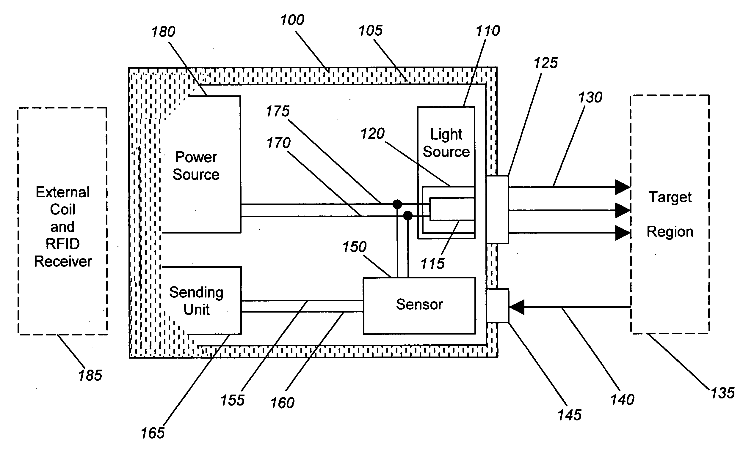
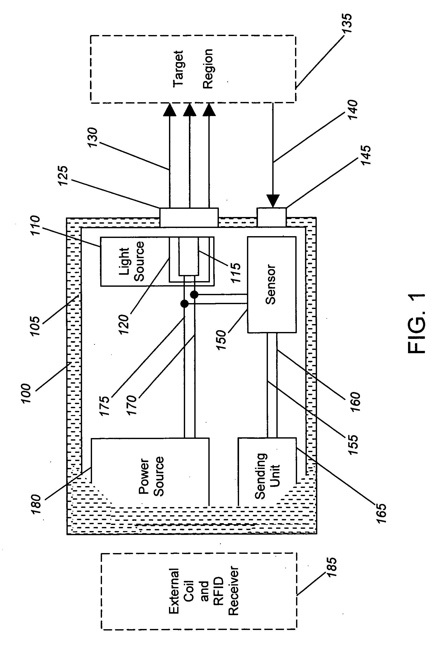
[0026] Generally, the present invention provides devices and methods for detecting local ischemia in a target tissue without regard to tissue site and for producing a target signal generating a direct, quantitative measure or index of ischemia. Tissue, as used herein, may be any material from a living animal, plant, viral, or bacterial subject, with an emphasis on mammals, especially humans. A target tissue may be a tissue material to be detected, imaged, or studied. A target signal may be a detected signal specific to the desired target tissue. This signal may be enhanced through use of known optical techniques, including use of a contrast agent, scattering, absorbance, phosphorescence, fluorescence, Raman effects, or other known spectroscopy and signal processing techniques, provided only that such techniques are applied in a manner to perform substantially locally and capillary weighted ischemia sensing.

[0027] Ischemia, as used herein, may be a condition in which the perfusion o...


PUM


Abstract

An implantable ischemia detection device in which a broadband light source produces a continuous, visible, broadband light illuminating capillary sites within a well localized target tissue is provided. Light backscattered by the target tissue is collected by a sensor, allowing for an index of ischemia to be determined, and subsequently transmitted by a sending unit. Optionally, the device may be provided with an internal power source, the entire device encapsulated by a biocompatible shell to add long-term safety while implanted at a target tissue site.

Description

CROSS-REFERENCE TO RELATED APPLICATIONS [0001] This application is a continuation-in-part of U.S. patent application Ser. No. 10 / 651,541, filed on Aug. 29, 2003, the entire disclosure of which is incorporated herein by reference.FIELD OF THE INVENTION [0002] The present invention relates to a device and methods for providing highly-localized measurements of tissue ischemia. More particularly, in some embodiments the present invention relates to a device comprising a visible light source, a sensor, a power source, and a transmitter embedded into a long-term implantable shell for the purpose of performing a real-time spectroscopic analysis of an in-vivo tissue perfusion that is sensitive to local tissue ischemia and insensitive to regional arterial and venous oxygenation. BACKGROUND INFORMATION [0003] The clinical detection of ischemia—an insufficient delivery of oxygen to meet a tissue's metabolic needs—is unreliable. Ischemia is especially difficult to detect when the ischemia is du...

Claims


Application Information

Patent Timeline
IPC IPC(8): A61B5/00
CPCA61B5/0031A61B5/412A61B5/1459
Inventor BENARON, DAVID A.PARACHIKOV, ILIAN H.
Owner J FITNESS LLC
Why Eureka
Try Eureka