

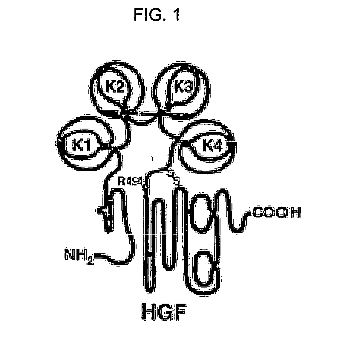
Neutralizable Epitope of HGF and Neutralizing Antibody Binding to the Same
a technology of neutralizing antibody and hgf, which is applied in the field of neutralizing antibody binding to the same and neutralizing epitope of hgf, can solve the problems of not being able to identify the entire tertiary structure of hgf, not yet being clarified, and not having a monoclonal antibody that can neutralize hgf as a single agent, etc., and achieve the effect of inhibiting the binding of hg
- Summary
- Abstract
- Description
- Claims
- Application Information
AI Technical Summary
Benefits of technology
Problems solved by technology
Method used
Image

Examples
example 1
HGF Immunization and Antibody Library Construction
[0096] Over a period of 4 to 5 months, 2 rabbits of the New Zealand White strain were immunized by 5 cutaneous injections of HGF (R&D systems, USA) dispersed in an emulsion of MPL (monophosphoryl lipid A; highly-refined non-toxic lipid A isolated from remutants of S. minnesota)+TDM (synthetic trehalose dicorynomycolate; an analogue of trehalose dimycolate from the cord factor of the tubercle bacillus)+CWS (cell wall skeleton; from deproteinized and delipidated cell walls of mycobacteria) adjuvant (Sigma) at 3-week intervals. Antisera from the immunized animals were analyzed for their binding to recombinant human HGF (R&D systems or Research Diagnostics, Inc.) by ELISA using horseradish peroxidase-conjugated anti-rabbit Fc goat polyclonal antibodies (Pierce). As a result, it was found that while antisera obtained before HGF immunization almost never bind to HGF, antisera obtained after 5 cutaneous injections specifically bound to HGF...
example 2
Amplification of Rabbit-Derived Ab Variable Region and Human-Derived Ab Constant Region
(2-1) Amplification of Rabbit-Derived Ab Variable Region
[0099] In order to amplify variable regions of rabbit VL (Vκ, Vλ) and VH, PCR was performed by using primer combinations described in Table 1.
TABLE 1Variable regionForward primerReverse primerVκSEQ ID NO: 1SEQ ID NO: 4SEQ ID NO: 1SEQ ID NO: 5SEQ ID NO: 1SEQ ID NO: 6SEQ ID NO: 2SEQ ID NO: 4SEQ ID NO: 2SEQ ID NO: 5SEQ ID NO: 2SEQ ID NO: 6SEQ ID NO: 3SEQ ID NO: 4SEQ ID NO: 3SEQ ID NO: 5SEQ ID NO: 3SEQ ID NO: 6VλSEQ ID NO: 7SEQ ID NO: 8VHSEQ ID NO: 8SEQ ID NO: 13SEQ ID NO: 10SEQ ID NO: 13SEQ ID NO: 11SEQ ID NO: 13SEQ ID NO: 12SEQ ID NO: 13
[0100] A PCR reaction solution was prepared by mixing 1 μl of template cDNA (about 0.5 μg) synthesized in Example 1, 60 pmol of each primer, 10 μl of 10× PCR buffer, 8 μl of 2.5 mM DNTP mixture and 0.5 μl of Taq polymerase and adjusted to a final volume of 100 μl. The PCR condition was 30 cycles of 15 sec a...
example 3
Amplification of Light and Heavy Chains of Chimeric Antibody
(3-1) Amplification of Light Chain
[0104] PCR was carried out to amplify the light chain as follows: A PCR reaction solution was prepared by mixing 100 ng each of VL (Vκ, Vλ) PCR product purified in Example (2-1) and Cκ PCR product purified in Example (2-2), 60 pmol each of primers (SEQ ID NOs: 18 and 15), 10 μl of 10× PCR buffer, 8 μl of 2.5 mM DNTP mixture and 0.5 μl of Taq polymerase and adjusted to a final volume of 100 μl. The PCR condition was 20 cycles of 15 sec at 94° C., 30 sec at 56° C. and 120 sec at 72° C. after initial denaturation of 10 mill at 94° C., and final extension of 10 min at 72° C.
[0105] The amplified DNA was subjected to agarose gel electrophoresis and purified from the gel by using Qiaex gel extraction kit (Qiagen).
(3-2) Amplification of Heavy Chain
[0106] Overlap extension PCR was conducted to amplify Fd region (VH and CH1) of a heavy chain as follows: A PCR reaction solution was prepared by ...
PUM
| Property | Measurement | Unit |
|---|---|---|
| volume | aaaaa | aaaaa |
| volume | aaaaa | aaaaa |
| volume | aaaaa | aaaaa |
Abstract
Description
Claims
Application Information