Method for monitoring viability of tissue flaps

a tissue flap and viability technology, applied in the field of optical imaging systems, can solve the problems of transplanted flaps to die, transplanted flaps may sometimes die, transplanted tissue may die, etc., and achieve the effect of preserving the viability of the remainder of the flap

Active Publication Date: 2007-03-08
VIOPTIX
View PDF12 Cites 17 Cited by
  • Summary
  • Abstract
  • Description
  • Claims
  • Application Information

AI Technical Summary

Benefits of technology

[0011] In one embodiment, if the first location is identified as having a poor blood supply, then the tissue at and around the first location may be trimmed to substantially preserve the viability of the remainder of the flap. In another embodiment, the method also includes placing a sensor head of an optical imaging system in contact with the flap tissue at the first location such that the sensor head causes light to be reflected off of the flap tissue to allow the oxygen saturation level to be determined.

Problems solved by technology

A transplanted flap may sometimes die, i.e., transplanted tissue may die, when the blood flow through the transplanted flap is compromised.
For example, a blood clot in the transplanted flap or a pinched vein in the transplanted flap may cause the transplanted flap to die.
Though laser Doppler flap monitoring may be effective for enabling potential flap failures to be detected in some instances, laser Doppler systems are generally able to make measurements on relatively large vessels, and are unable to measure regional perfusion in the micro-vasculature within a skin flap.
Even though flow may be detected in larger vessels when laser Doppler flap monitoring is employed, distal flap tissue may be underperfused and, as a result, may die.
Nicking a transplanted flap is invasive and does not always allow for an accurate determination of the viability of a transplanted flap, as assessing the blood flow in such a manner is highly subjective.
Further, it may be very difficult to determine where in a transplanted flap to make a nick, e.g., a surgeon may inadvertently fail to make a nick near a blood vessel that is pinched.

Method used


Image

  • Method for monitoring viability of tissue flaps
  • Method for monitoring viability of tissue flaps
  • Method for monitoring viability of tissue flaps

Examples


Embodiment Construction

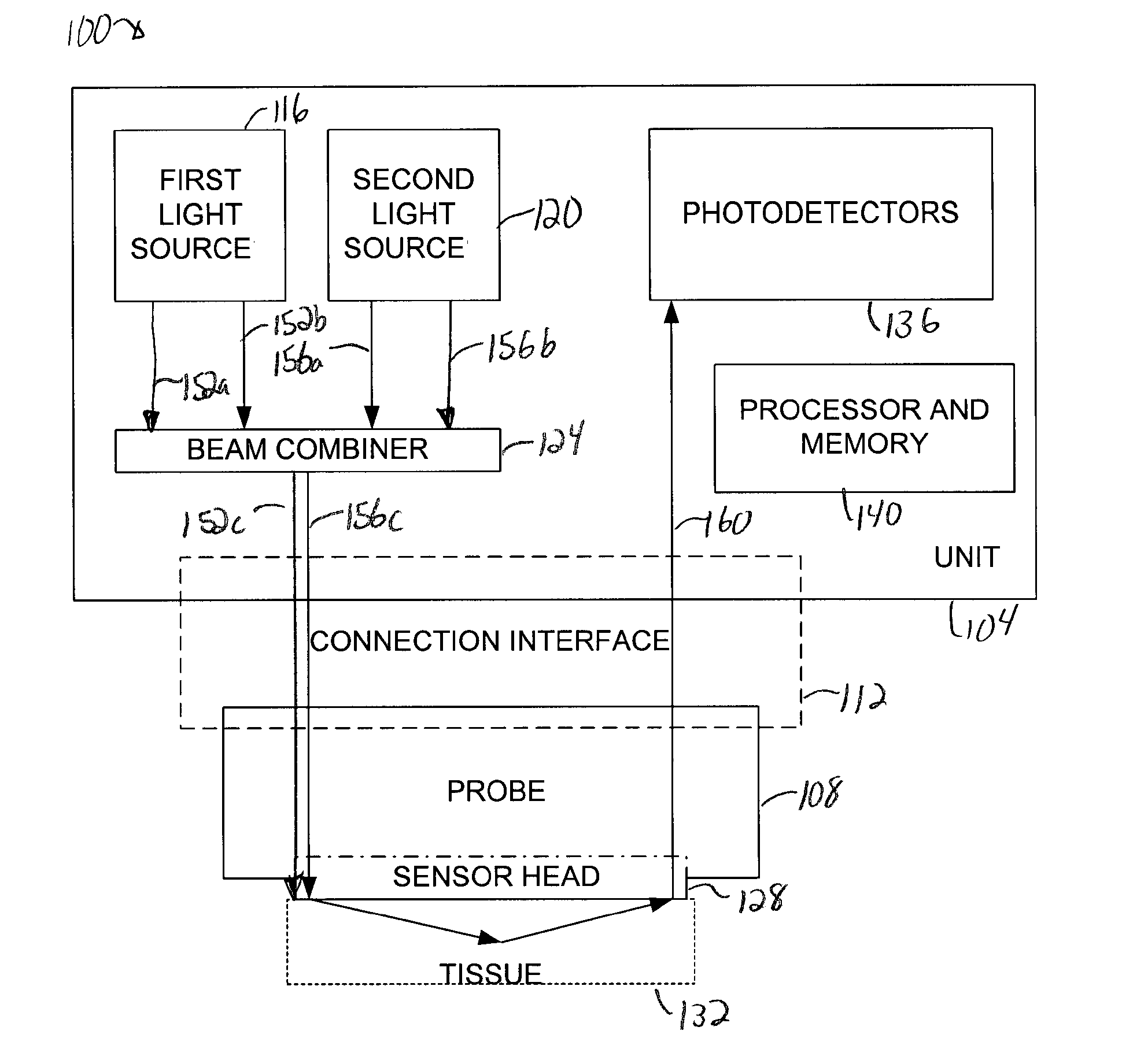
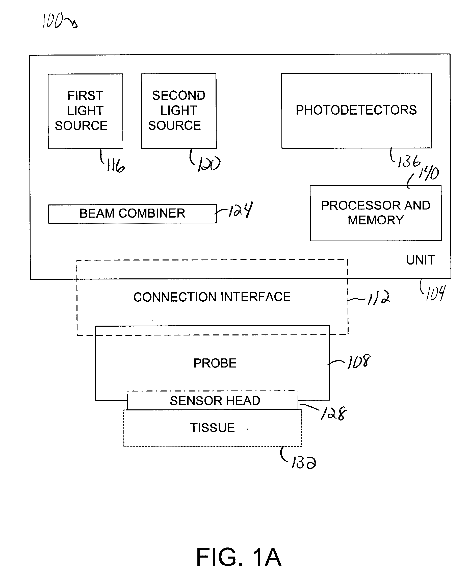
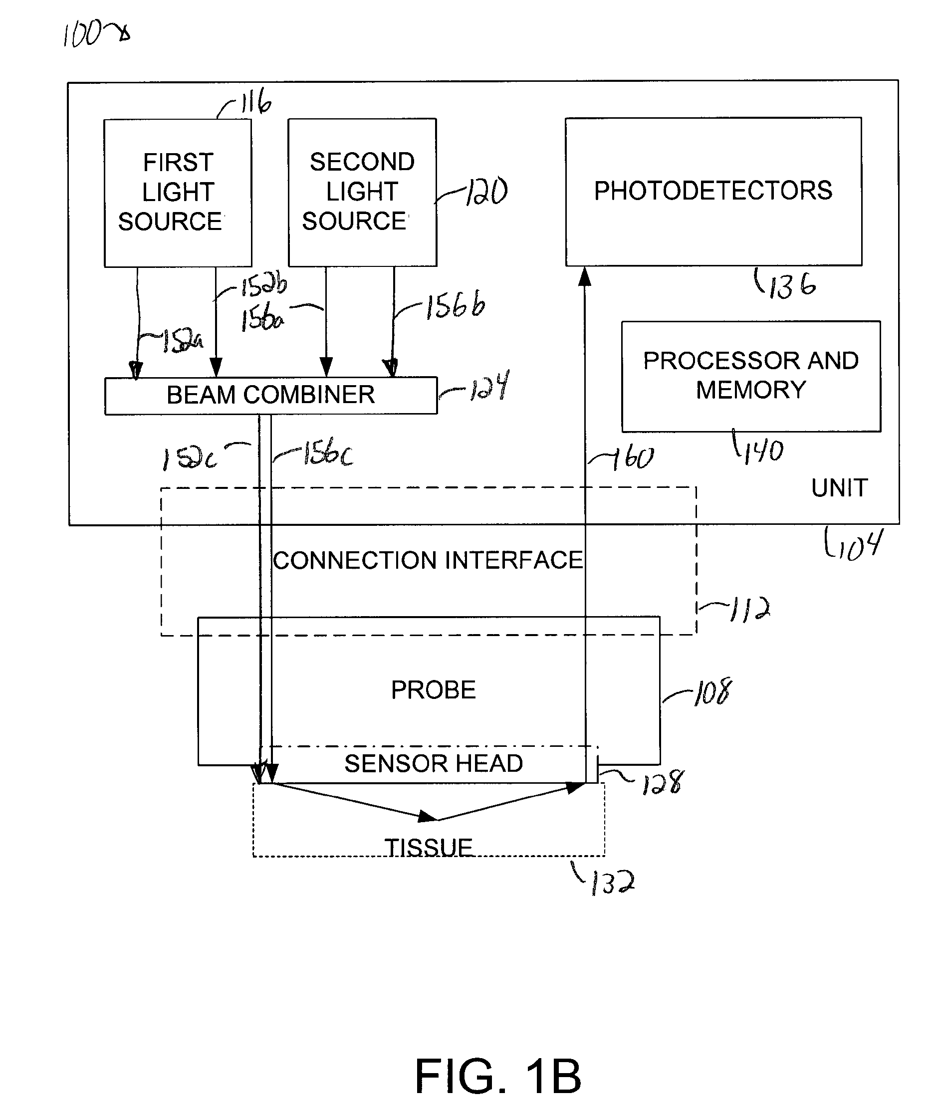
[0029] Assessing the blood supply associated with a flap is crucial to ensure that the flap is viable. By monitoring the oxygen saturation level of an area on flap tissue, the blood flow to at least that area may be determined. Monitoring an oxygen saturation level is generally a non-invasive, non-subject process. Near-infrared spectroscopy has been used for non-invasive measurement of various physiological properties in animal and human subjects. The basic principle underlying the near-infrared spectroscopy is that physiological tissues include various highly-scattering chromophores to the near-infrared waves with relatively low absorption. Many substances in a medium may interact or interfere with the near-infrared light waves propagating therethrough. Human tissues, for example, include numerous chromophores such as oxygenated hemoglobin, deoxygenated hemoglobin, water, lipid, and cytochrome, where the hemoglobins are the dominant chromophores in the spectrum range of approximate...


PUM


Abstract

Methods and apparatus for assessing the viability of tissue such as flap tissue are disclosed. According to one aspect of the present invention, a method for assessing the viability of flap tissue includes obtaining an oxygen saturation level associated with a first location on the flap tissue, determining whether the oxygen saturation level is less than a first level, and identifying the first location as having a poor blood supply if the oxygen saturation level is less than the first level.

Description

BACKGROUND OF THE INVENTION [0001] 1. Field of Invention [0002] The present invention relates generally to optical imaging systems that monitor oxygen levels in tissue. More specifically, the present invention relates to monitoring oxygen levels to determine the viability of flaps before and after a flap transplant. [0003] 2. Description of the Related Art [0004] Flap surgery is a type of plastic or reconstructive procedure that enables tissue from one area of a body to effectively be moved to another area of the body. A flap is a section of living tissue with a blood supply that may be transported from a “donor” area of a body to a new area of the body, i.e., an area onto which the flap is to be transplanted. A flap may be transplanted to an area of the body that has lost, for example, skin, fat, or muscle. Flap surgery generally restores some muscle movement or skeletal support to an area in which muscle movement or skin coverage may have been missing. [0005] There are many differ...

Claims


Application Information

Patent Timeline
Patent Type & Authority Applications(United States)
IPC IPC(8): A61B5/1459
CPCA61B5/413A61B5/14551A61B5/14552A61B5/4312A61B5/4848
Inventor LASH, ROBERT E.MAO, JIAN-MIN
Owner VIOPTIX
Why Eureka
Try Eureka