Catheter mounted automatic vessel occlusion and fluid dispersion devices

a technology of fluid dispersion device and catheter, which is applied in the field of catheter mounted automatic vessel occlusion and fluid dispersion device, can solve the problems of inability to safely or effectively achieve the concentration of the substance required at the target site, and the current oct (optical coherence tomography) imaging system is not able to image much more than, into blood or tissu

Inactive Publication Date: 2007-06-07
ABBOTT CARDIOVASCULAR
View PDF31 Cites 94 Cited by
  • Summary
  • Abstract
  • Description
  • Claims
  • Application Information

AI Technical Summary

Problems solved by technology

This is particularly true when the concentration of the substance required at the target site cannot be safely or effectively achieved by introduction of the substance to a location remote from the target site or the flow of blood washes away or dilutes the substance too rapidly at the target site.
Moreover, the physician may only want to treat the diseased portion of a vessel, organ or tissue and avoid treating the healthy portions.
Also, current OCT (optical coherence tomography) imaging systems are not able to image much more than about 2 mm (typically 1.2 to 1.7 mm) into blood or tissue.
Because vessels for which imaging is desired are generally 2 mm in diameter or greater and the imaging device may be in contact with a vessel wall, it is not practical to expect to image the wall of these vessels over their full 360 degree circumference to any vessel wall depth without clearing the blood from the field of view, eliminating the blood's properties impacting imaging depth and/or controlling the catheter's position relative to the vessel wall.
Use of longer wavelengths to avoid the red blood cell interaction results in a loss of the desired image axial (depth) resolution for vulnerable plaque (VP) detection.
In addition, there are some comparatively minor light energy losses due to absorption and path length changes due to scattering (reflection) by the red cells.
Therefore, such systems will also require a flush for reasons similar to the problems of OCT.
However, a simple flush has quite a few drawbacks and problems:
Thus, the time window for imaging is limited by the ischemia consequences of the solution on the heart muscle.
High velocity jets can be damaging to vessel walls.
Additionally, the pressu

Method used


Image

  • Catheter mounted automatic vessel occlusion and fluid dispersion devices
  • Catheter mounted automatic vessel occlusion and fluid dispersion devices
  • Catheter mounted automatic vessel occlusion and fluid dispersion devices

Examples


Embodiment Construction

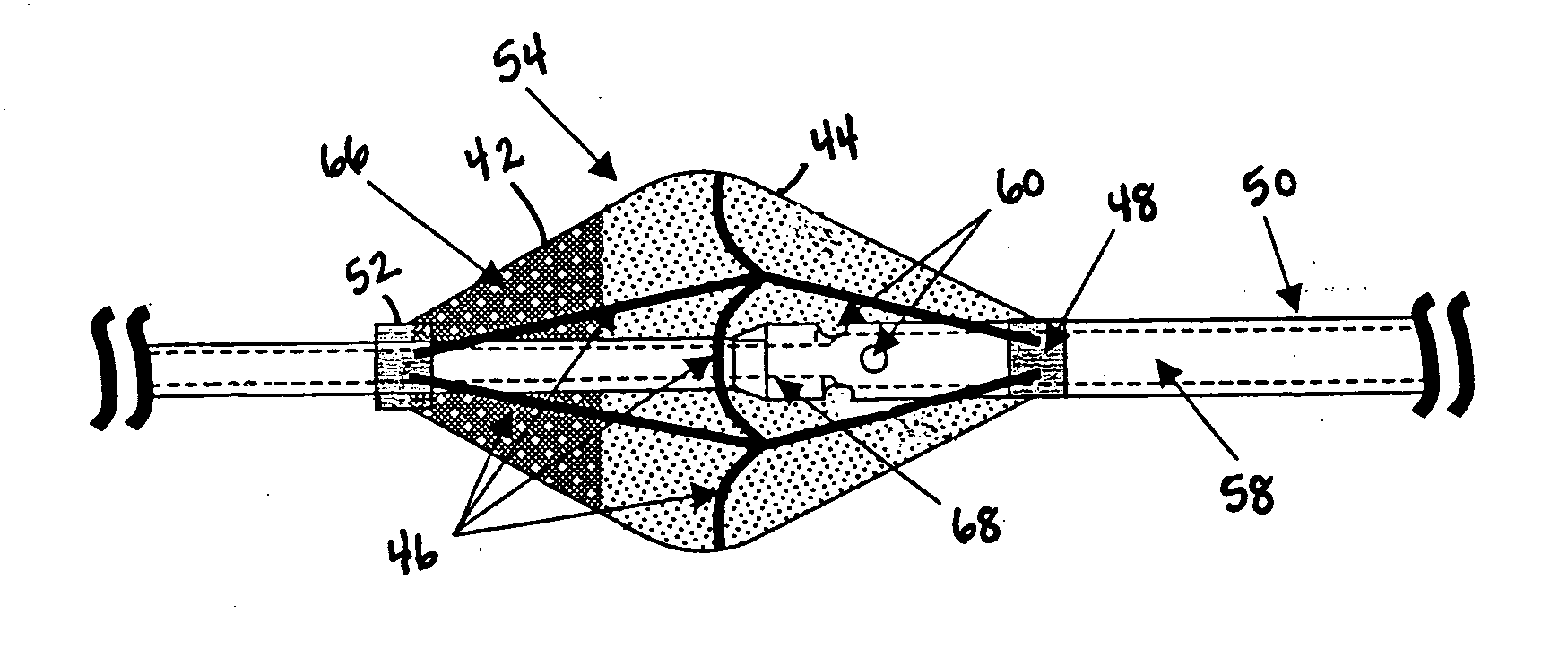
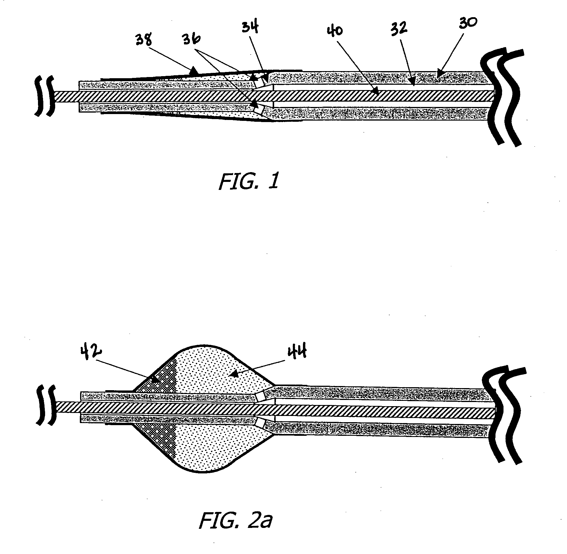
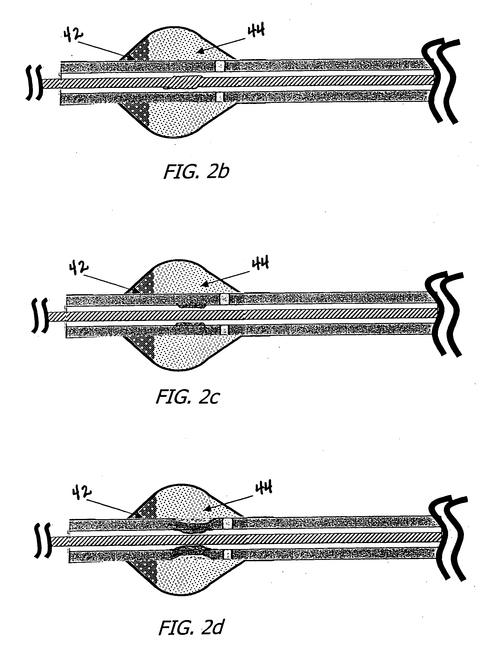
[0037] Embodiments of the present invention comprise automatic occlusion / flush dispersion devices. In these devices, the occlusion / flush dispersion device deploys to occlude or partially occlude the artery (or vein) during the injection of the flushing solution. In fact, the flow or pressure of the flush injection is used to deploy (expand) the occlusion / flush dispersion device. Thus, during the flushing (or infusion), the vessel is at least partially occluded at the device site, so the flush will flow preferentially distal or proximal (depending upon the flush's exit position relative to the occlusion) into the arterial (or venous) tree and clear or dilute the path for the light or ultrasound, allowing one to more effectively introduce an agent (e.g. saline solution, angiographic contrast, ultrasonic contrast) into the region of interest or more effectively expose a therapeutic agent to a vascular region of interest, to tissues adjacent to a vascular region of interest or tissues i...


PUM


Abstract

Catheter or guidewire mounted automatic vessel occlusion and fluid dispersion devices that expand to occlude or partially occlude a vessel when a fluid is flowing in the catheter or guidewire, and that automatically collapse when fluid flow is stopped. Each occlusion device has an elastic skeleton covered with a flexible cover coupled thereto and may have a hole(s) or openings in its distal or proximal end thereof to allow controlled flow through the desired end of the occlusion device. The fluid may be a flush fluid for enabling or improving the performance of imaging devices and image enhancing fluids, of treatment fluids for localized treatment of a vessel or tissues in communication with the vessel and/or of the transmission of energy to the vessel wall and adjacent tissues. Various embodiments are disclosed, including embodiments having provisions for the rapid expansion and collapse of the occlusion device or alternately, for the rapid expansion of the device, followed by much slower collapse of the device.

Description

BACKGROUND OF THE INVENTION [0001] 1. Field of the Invention [0002] The present invention relates to the field of occlusion / infusion and occlusion / imaging devices and systems. [0003] 2. Prior Art [0004] It is increasingly important that a physician or surgeon delivering substances, such as a treatment agent or drug or an image-enhancing agent, is able to efficiently and accurately locate and / or effectively deliver the substance to the desired target tissue. This is particularly true when the concentration of the substance required at the target site cannot be safely or effectively achieved by introduction of the substance to a location remote from the target site or the flow of blood washes away or dilutes the substance too rapidly at the target site. Moreover, the physician may only want to treat the diseased portion of a vessel, organ or tissue and avoid treating the healthy portions. [0005] For example, to achieve localized treatment of tissue, such as tissue in a heart, physicia...

Claims


Application Information

Patent Timeline
IPC IPC(8): A61M29/00A61F2/958
CPCA61F2230/0071A61F2230/0076A61B2017/22067A61F2/013A61M25/0029A61M25/0032A61M25/10A61M2025/0057A61M2025/105A61M2025/1052A61M2025/1086A61F2002/018A61F2230/0006
Inventor WEBLER, WILLIAM E.HAGUE, GARYMANNING, FRANK E.
Owner ABBOTT CARDIOVASCULAR
Why Eureka
Try Eureka