Time-dependent three-dimensional musculo-skeletal modeling based on dynamic surface measurements of bodies
a dynamic surface measurement and musculoskeletal technology, applied in the field of time-dependent three-dimensional musculoskeletal modeling based on dynamic surface measurements of bodies, can solve the problems of large number of people suffering from different musculoskeletal complaints, such as back pain or knee problems, and provide a non-contact technique for obtaining, so as to achieve short recording and analysis time, no preparation time, and the effect of efficient, stable and reliable devices
- Summary
- Abstract
- Description
- Claims
- Application Information
AI Technical Summary
Benefits of technology
Problems solved by technology
Method used
Image
Examples
example 1
Spine
[0094] The internal spine is reconstructed from the external spine using an anatomical formula, estimating the distance of the skin to the centre of a vertebral body, as illustrated on FIGS. 15 and 16. Spinal parameters (e.g. lumbar lordisis angle) can be monitored as a function of time in three dimensions. A detailed deduction of the anatomical formula is described by Drerup et al. in Clinical Biomechanics 9 p 28-36.
example 2
Shoulder
[0095] The shoulder is a complex joints and it is not possible to model it as a 3 degrees-of-freedom spherical joint. The skeletal model 240, as shown in FIG. 20, contains the following bones: the sternum, clavicula, scapula and humerus. Between the bones, three joints have been defined: the sternoclavicular joint, the acromioclavicular joint and the glenohumeral joint. These joints are modelled as three-degrees-of-freedom (DOFs) spherical joints. The scapulothoracic joint is modelled in such a way that the scapula is able to move freely with respect to the thoracic wall, in order to enable winging. The set of measured anatomical features is necessary and sufficient to define all DOF's of the system, and to build a personalised time-dependent three-dimensional model of the shoulder complex.
example 3
[0096] A personalised time-dependent three-dimensional model of the pelvis is reconstructed from the lower back surface and the position of the dimple points.
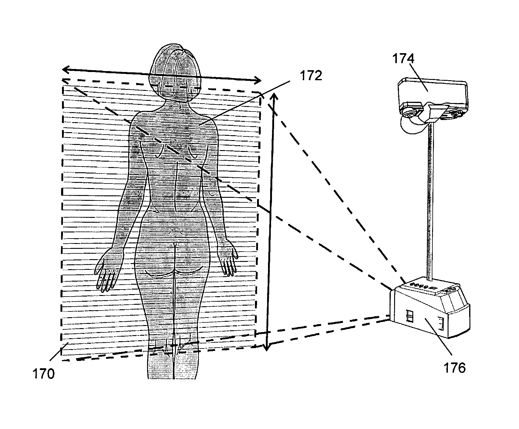
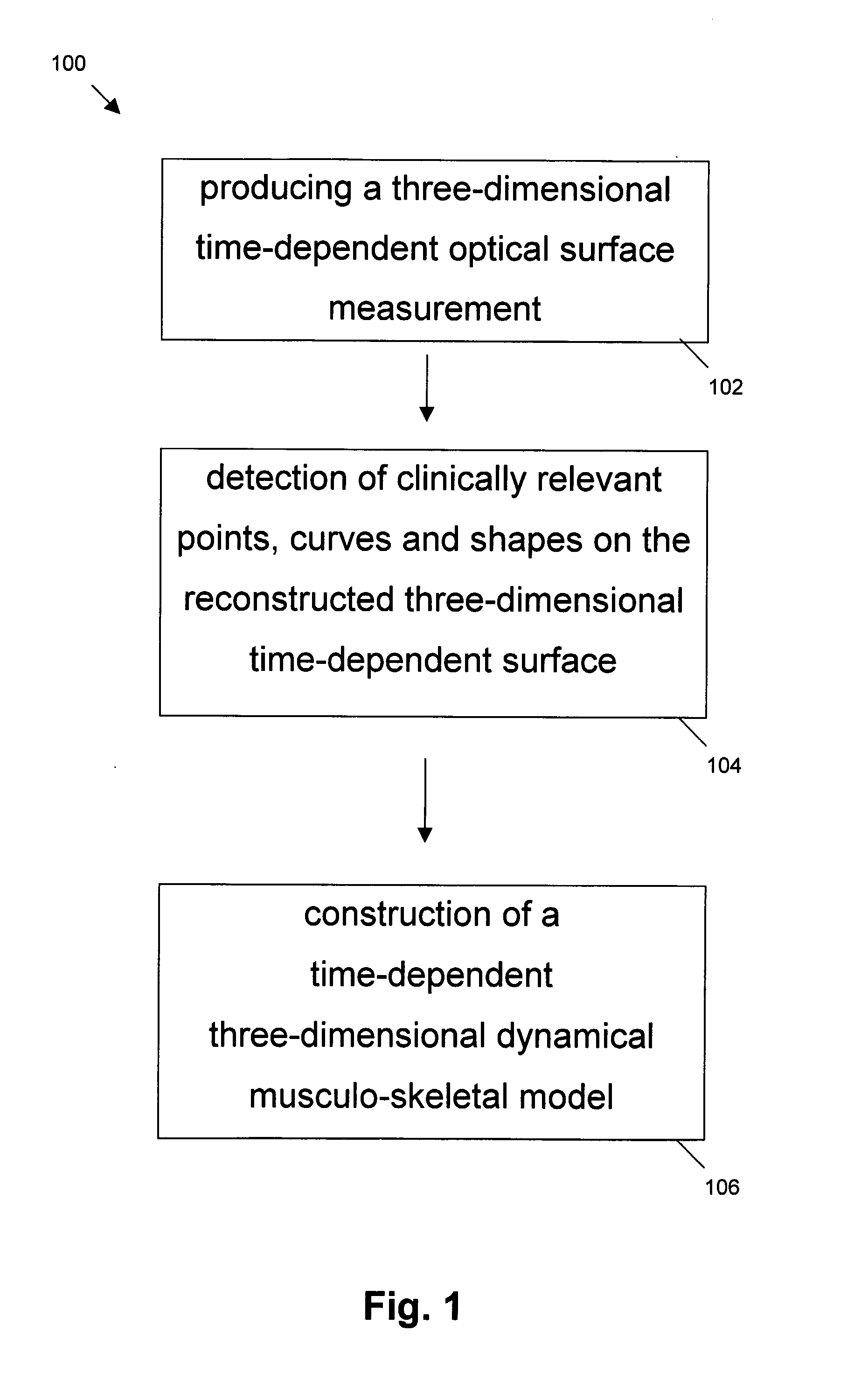
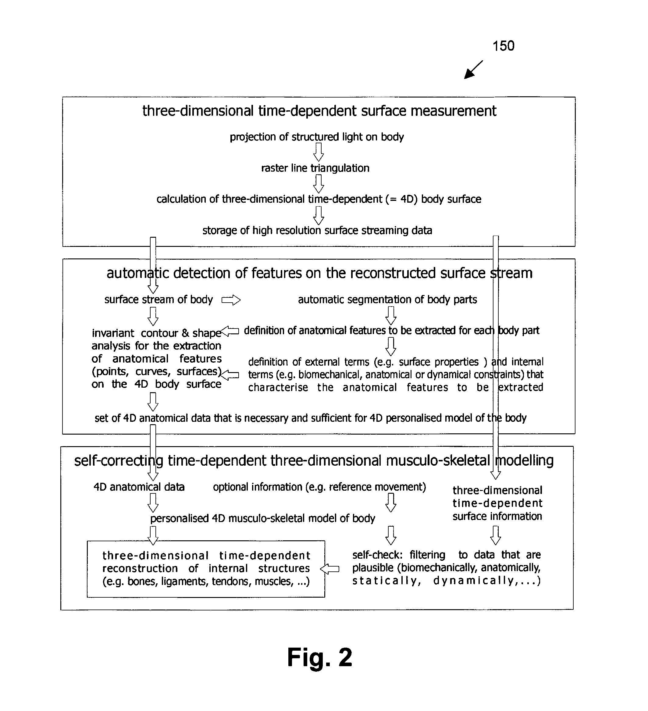
In conclusion, the method of the above described embodiment comprises the provision, e.g. grabbing, of surface images of a moving body by suitable scanning or projection equipment, e.g. optical equipment. Thereby either a one-sided surface (like human back, face, . . . ) or a multi-sided surface (legs, torso, . . . ) is recorded. The method furthermore comprises the reconstruction and mapping on a regular grid of the surface, thus giving a sequence of single static surfaces. On the regular surfaces, invariant features are calculated, like curvature and symmetry and invariant features are used for detecting anatomical landmarks and shapes on each of the static surface, like vertebra prominence, sacrum point (rima ani), left and right dimples, acromium, scapula, spinal symmetry line (processi spinosi). In the next step, ...
PUM
 Abstract
Description
Claims
Application Information
 - R&D
- Intellectual Property
- Life Sciences
- Materials
- Tech Scout
- Unparalleled Data Quality
- Higher Quality Content
- 60% Fewer Hallucinations
Browse by: Latest US Patents, China's latest patents, Technical Efficacy Thesaurus, Application Domain, Technology Topic, Popular Technical Reports.
© 2025 PatSnap. All rights reserved.Legal|Privacy policy|Modern Slavery Act Transparency Statement|Sitemap|About US| Contact US: help@patsnap.com


