Motion monitor system for use with imaging systems

a motion monitor and imaging system technology, applied in the field of medical devices, can solve the problems of difficult continuous ct fluoroscopy, difficult positioning of treatment or biopsy apparatus, and difficult positioning of needles, etc., and achieve the effect of convenient positioning

Active Publication Date: 2007-07-26
MAYO FOUND FOR MEDICAL EDUCATION & RES
View PDF34 Cites 29 Cited by
  • Summary
  • Abstract
  • Description
  • Claims
  • Application Information

AI Technical Summary

Benefits of technology

[0008]One aspect of the invention is the use of wireless communications between the control unit and the display units to enable the display units to be easily positioned within eyesight of the subject to provide real-time position feedback and within eyesight of others performing the medical procedure. This provides flexibility in the positioning of displays to account for variations in patient size and positioning and variations in the arrangement and complexity of equipment in the imaging suite.
[0009]Another aspect of the invention is a manually operable reference switch which enables an operator to establish a desired reference position anywhere within the respiratory cycle from which subject motion is measured. This also establishes the location of the dynamic operating range of the motion detector such that accurate displacement data is produced in the vicinity of the reference position even if the subject motion is greater than the dynamic operating range of the motion detector. This establishes a high resolution signal at the point of importance within the respiratory cycle.

Problems solved by technology

Quick accurate needle advancement or placement of treatment or biopsy apparatus into the body, moreover, is difficult with continuous CT fluoroscopy, even with a needle-holder or other device that may prevent or at least minimize exposure to the primary beam.
The lungs, the diaphragm, and the upper abdomen move during breathing; thus the displacement of the body and its organs during the breath cycle can be a significant problem during certain medical procedures because target structures, such as lesions and tumors also move during breathing.
Inconsistent breath holding by a patient, especially during procedures performed in the area of the thoracic cavity, can cause target structures such as lesions or tumors to move completely out of sight during imaging and intervention.
There is also the potential for decreased diagnostic yield of the biopsy specimen and higher complication rates.
Even healthy patients are unable to reproduce consistent levels of suspended inspiration or expiration without the help of breath-hold monitoring and feedback systems.
The drawback to the systems, however, is that they monitor external changes in body wall girth or position, rather than the actual physical display of the interior portions of the body; although these external changes may be correlated to diaphragm position and internal lesion location, as disclosed in Frolich et al., “A Simple Device For Breath-Level Monitoring During CT” 156 Radiology 235 (1985).
Most of these systems, moreover, linearly correlate the movement of the body to changes in pressure in the transducer, a presumption that is not always accurate.

Method used


Image

  • Motion monitor system for use with imaging systems
  • Motion monitor system for use with imaging systems
  • Motion monitor system for use with imaging systems

Examples


Embodiment Construction

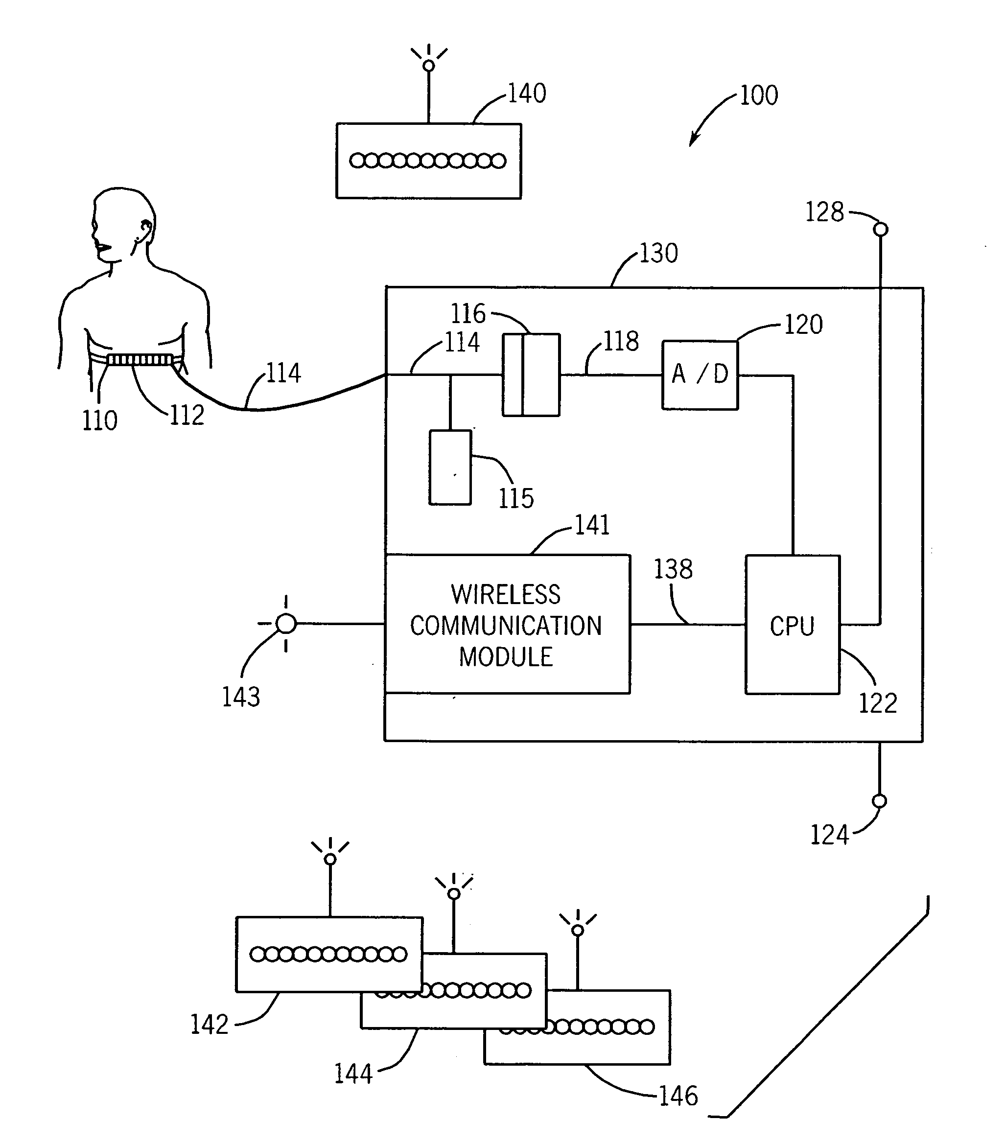
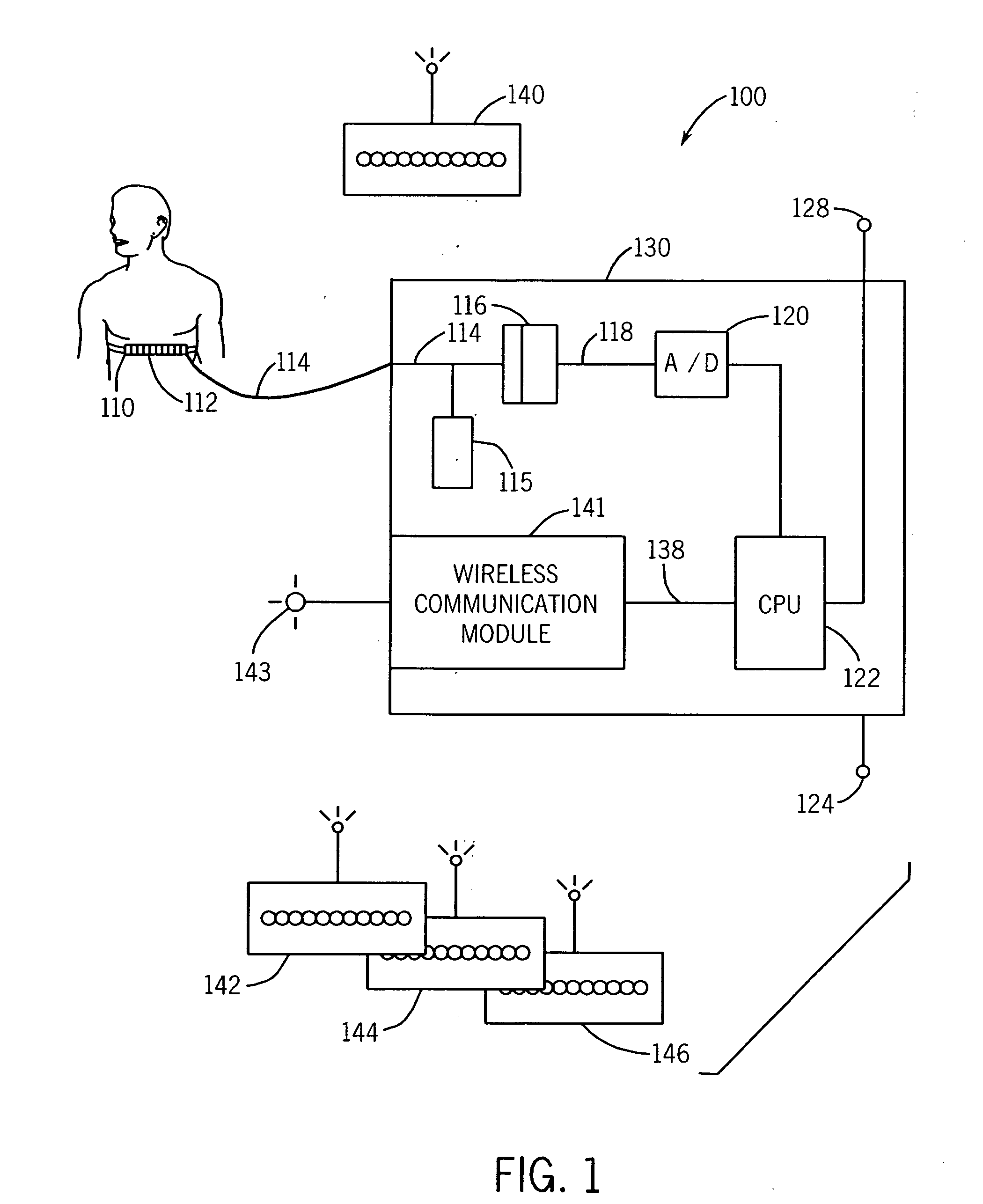
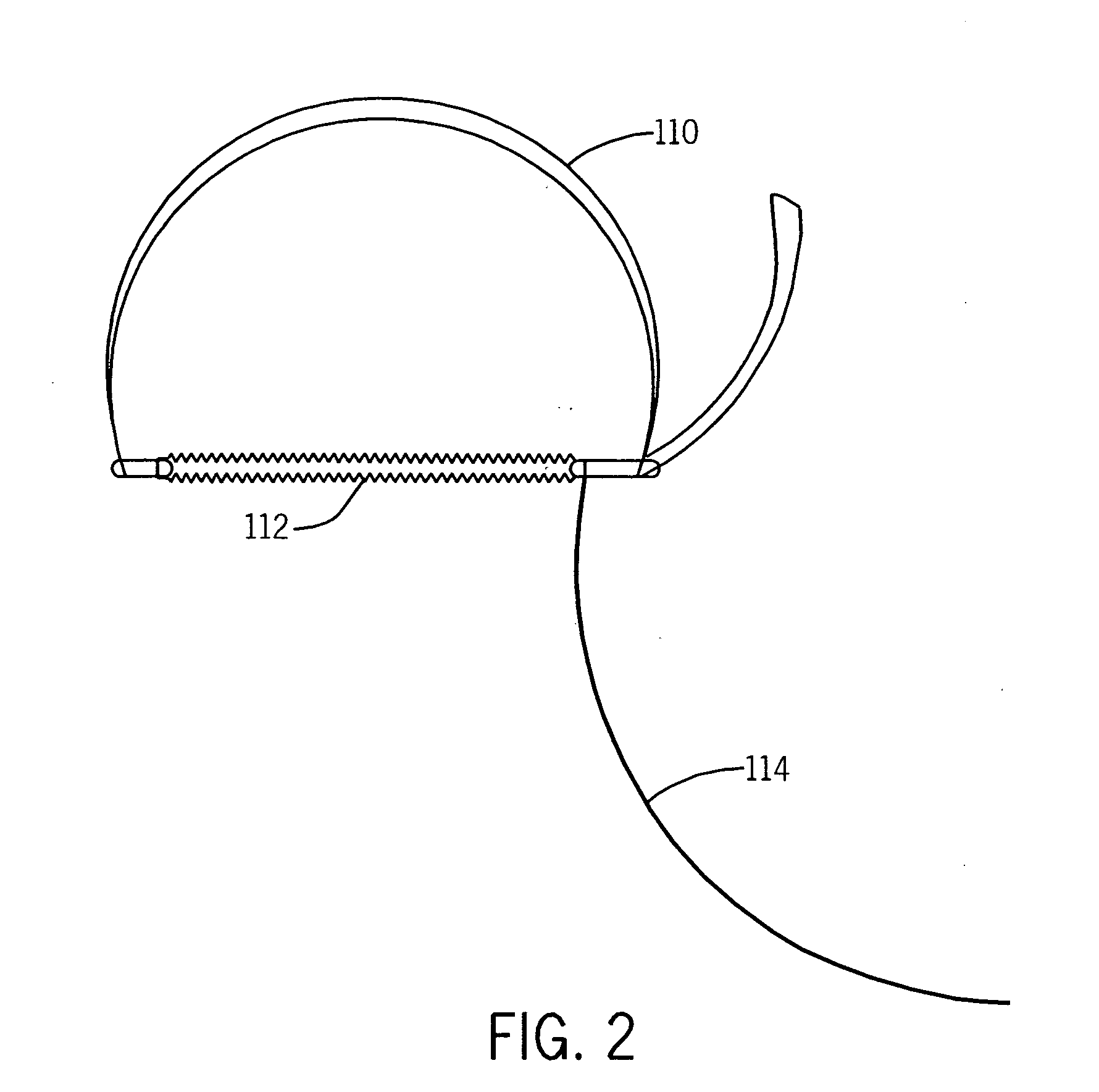
[0016]Referring to FIGS. 1 and 2, a motion detection and monitor system 100 includes an expandable bellows 112 connected on both ends to a strap or a belt 110. The bellows is a motion detector 112 constructed from flexible rubber, silicone, or other expandable material responsive to expanding and contracting chest motion. The strap 110 may be cloth or leather having Velcro or D-rings or other attachment mechanism to circumscribe the body. A hollow tubing 114 containing a gas, preferably ambient air is connected to the interior of the bellows. Changes in patient girth occurs during respiration and this result in expansion and contraction of the tubing 114. Other motion detectors 112 may also be used such as a strain gauge or piezoelectric fabric.

[0017]The belt 110 is positioned around a patient's upper abdomen or lower chest. When the motion to be detected is the respiratory motion, it is preferable to position the motion detector or bellows 112 on the anterior of the body at a locat...


PUM


Abstract

A bellows-based patient breath-hold monitoring and feedback system for use in intermittent mode CT fluoroscopy-guided biopsies of the lung or upper abdomen where respiratory motion is a problem. Breath-hold monitoring and feedback with the bellows system allows a patient to perform consistent breath-holds at a preselected level, which in turn, optimizes intermittent mode CT fluoroscopy-guided biopsies of the lung or upper abdomen by allowing target lesions to be reliably visualized.

Description

BACKGROUND OF THE INVENTION[0001]This invention relates generally to the field of medical apparati and more particularly to an apparatus to provide feedback regarding the movement of the body, for example, the lungs and the internal organs while breathing during imaging procedures, such as computer tomographic fluoroscopy.[0002]Computer-aided tomography (CT) fluoroscopy is a technique used in medical diagnostics wherein x-rays impinge onto and are rotated around a patient to give a detailed real-time three-dimensional image of the interior of the body. CT fluoroscopy is especially useful during medical procedures because its rapid data acquisition and interpretation allow a physician to obtain a tissue sample or administer treatment while viewing the image.[0003]CT fluoroscopy operates in two modes: continuous real-time mode and intermittent “quick-check” mode; and of the two continuous CT fluoroscopy results in a greater radiation exposure to both the patient and others in the vici...

Claims


Application Information

Patent Timeline
Patent Type & Authority Applications(United States)
IPC IPC(8): H05G1/10
CPCA61B5/1135A61B6/541A61B6/504A61B5/7285
Inventor FELMLEE, JOEL P.BRUHNKE, RUSSELL E.ROSSMAN, PHILLIPPOTTER, JAMES P.
Owner MAYO FOUND FOR MEDICAL EDUCATION & RES
Why Eureka
Try Eureka