Apparatus and Method for Monitoring Tissue Vitality Parameters

a technology of vitality and apparatus, applied in the field of apparatus and method for monitoring tissue vitality parameters, can solve the problems of limiting the availability of this process, reducing the form of the molecule, and unable to meet the demand in the tissue or cell, so as to avoid photo-bleaching of the measured, monitor the nadh level, and correct efficiently for the haemodynamic artifact

Inactive Publication Date: 2007-08-02
CRITISENSE
View PDF19 Cites 63 Cited by
  • Summary
  • Abstract
  • Description
  • Claims
  • Application Information

AI Technical Summary

Benefits of technology

[0132] Thus—this being a great advantage of the present invention—the blood flow rate measurement is conducted totally independently from the monitoring of the second set of tissue viability parameters, providing a great deal of flexibility in terms of configuration and design of the monitoring apparatus, as well as in the method of use, as will be evident from the following description.
[0133] The wavelength of the illumination radiation that is required for any of the second set of blood viability parameters—NADH, blood volume and oxy-deoxy state—is determined by the absorption and fluorescence characteristics of the NADH in the tissue, as exemplified in FIG. 1 for human brain tissue (the absorption and fluorescence characteristics of NADH are similar for other tissues). The fluorescence wavelength band is typically between about 400 nm and about 550 nm, and an excitation wavelength between about 300 nm and about 400 nm needs to be provided.
[0134] Preferably, an excitation wavelength is chosen such as to simplify correction for the haemodynamic artifact. The haemodynamic artifact arises from the absorption of the NADH fluorescence emission and excitation light by the blood haemoglobin. A change in blood volume cause misleading changes in apparent NADH fluorescence. Since blood haemoglobin has two oxygenation states namely oxy-haemoglobin and deoxy-haemoglobin each one with its distinct absorption spectrum, as shown in FIG. 10, the precise correction of the haemodynamic effect can become extremely complex. The problem is considerably simplified when the wavelength chosen for NADH fluorescence excitation corresponds to one of the isosbestic points, since at these wavelengths the absorption of both haemoglobin species is identical. At these isosbestic excitation points, fluorescence changes are due substantially to changes in total blood volume, and, of course, to changes in NADH concentration only. Thus, by suitably choosing the excitation wavelength, correction for the haemodynamic artifact is significantly simplified. Even at wavelengths within the isosbestic range of from about 300 nm to about 340 nm, or at the isosbestic point of 390 nm, the haemodynamic artifact requires correction. Suitable algorithms for this purpose are described on the prior art (Koyabashi et al., 1971; Renault G., et al. American Journal of Physiology, 246, H491-H499, 1984; Mayevsky A. and Chance B., Brain Res. 65, 529-533, 1974; Harbig et al., J. Appl. Physiol. 41, 480-488, 1976; U.S. Pat. No. 4,449,535). The most widely used correction algorithm (Jobsis et al. Neurophysiology 3465, 735-749, 1971) utilizes the value of the reflection at the NADH excitation wavelength as an indicator for blood changes. The corrected NADH fluorescence values are calculated by subtraction of the reflection signal from the fluorescence signal.
[0135] At the same time, the wavelength of the illumination radiation that is required for the monitoring of the first tissue viability parameter—i.e., the blood flow rate—may be independently chosen from that required for the other parameters. For the various soft body tissues, there are ranges of wavelengths over which the penetration depths of incident light is relatively independent. This is illustrated by way of example for brain tissue in FIG. 2. Here the penetration depth into tissue is relatively independent of the incident radiation wavelength for wavelengths between 360 nm to about 440 nm, while above this range, the penetration depth increases dramatically with wavelength.
[0136] The Optical Penetration Depth (PD) may be defined as the depth where the total optical power of incident light is reduced to 37% of the incident power. The PD parameter is useful for estimation of light depth penetration into tissues. If two different wavelengths are used in the same instrument it is important to compare the penetration depth for both wavelengths in order to estimate differences in collected light intensities. In general the PD is proportional to 1 / μ1 / 2, where μ is the total absorption coefficient, given by:—μ=μa+μs where
[0137]μa is the apparent absorption coefficient due to light absorption; and μs is the apparent absorption coefficient due to light scattering.

Problems solved by technology

In most pathological states, the limiting factor for this process is O2 availability.
The concentration of the reduced form of the molecule (NADH) rises when the rate of ATP production is low, and is unable to meet the demand in the tissue or cells.
Fp concentration drops when the rate of ATP production is reduced, and is unable to meet the demand in the tissue or cells.
An increase in the level of NADH with respect to NAD and the resulting increase in fluorescence intensity indicate that insufficient Oxygen is being supplied to the tissue.
These devices are relatively complicated and susceptible to interference from ambient light, as well as various electronic and optic drifts.
For the monitoring of different parameters to have maximum utility however, the information regarding all parameters is required to originate from substantially the same layer of tissue, and preferably the same volume of tissue, otherwise misleading results can be obtained.
These devices suffer from a major drawback however.
The complex biochemical mechanisms that determine tissue viability are such that short time deviations between measurement at short distances between points of measurement can provide inaccurate or even misleading information.
Another drawback encountered in NADH measurements is the Haemodynamic Artifact.
This refers to an artifact in which NADH fluorescence measurements in-vivo are underestimated or overestimated due to the haemoglobin present in blood circulation, which absorbs radiation at the same wavelengths as NADH, and therefore interferes with the ability of the light to reach the NADH molecules.
However, U.S. Pat. No. 4,449,535 has at least two major drawbacks; firstly, and as acknowledged therein, using a single optical fiber to illuminate the organ, as well as to receive emissions therefrom causes interference between the outgoing and incoming signals, and certain solutions with different degrees of effectiveness are proposed.
This results in measurements that are incompatible one with the other, the blood volume measurement relating to a greater depth of tissue than the NADH measurement.
Therefore, the device disclosed by this reference does not enable adequate compensation of NADH to be effected using the simultaneous, though inappropriate, blood volume measurement.
Further, there is no indication of how to measure other parameters such as blood flow rate or blood oxygenation level using the claimed apparatus.
Although U.S. Pat. No. 5,916,171 and U.S. Pat. No. 5,685,313 represent an improvement over the prior art, they nevertheless have some drawbacks: (i)The oxidation level of the blood will introduce artifacts, affecting both the Mitochrondrial Redox State measurement (NADH fluorescence) and the microcirculatory blood volume (MBV) since these patents do not specify how to compensate for the oxygenation state of the blood in the tissue, i.e., the relative quantities of oxygenated blood to deoxygenated blood in the tissue.
Using a relatively high intensity UV laser illumination source as proposed raises safety issues, especially for long-term monitoring.
Such lasers at these wavelengths are also not standard components and are indeed quite difficult to come by, which might lead to supply problems.

Method used


Image

  • Apparatus and Method for Monitoring Tissue Vitality Parameters
  • Apparatus and Method for Monitoring Tissue Vitality Parameters
  • Apparatus and Method for Monitoring Tissue Vitality Parameters

Examples


first embodiment

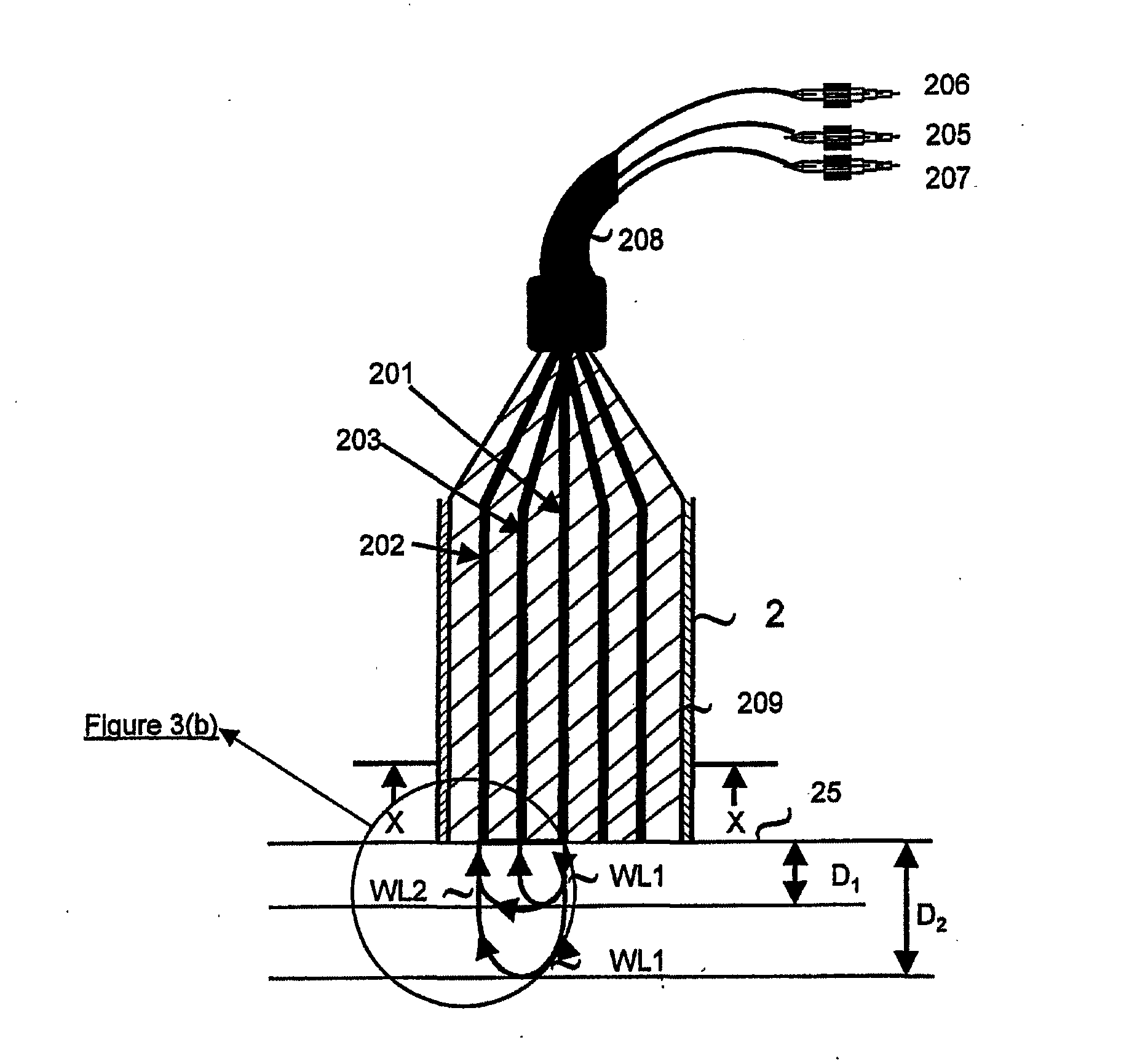
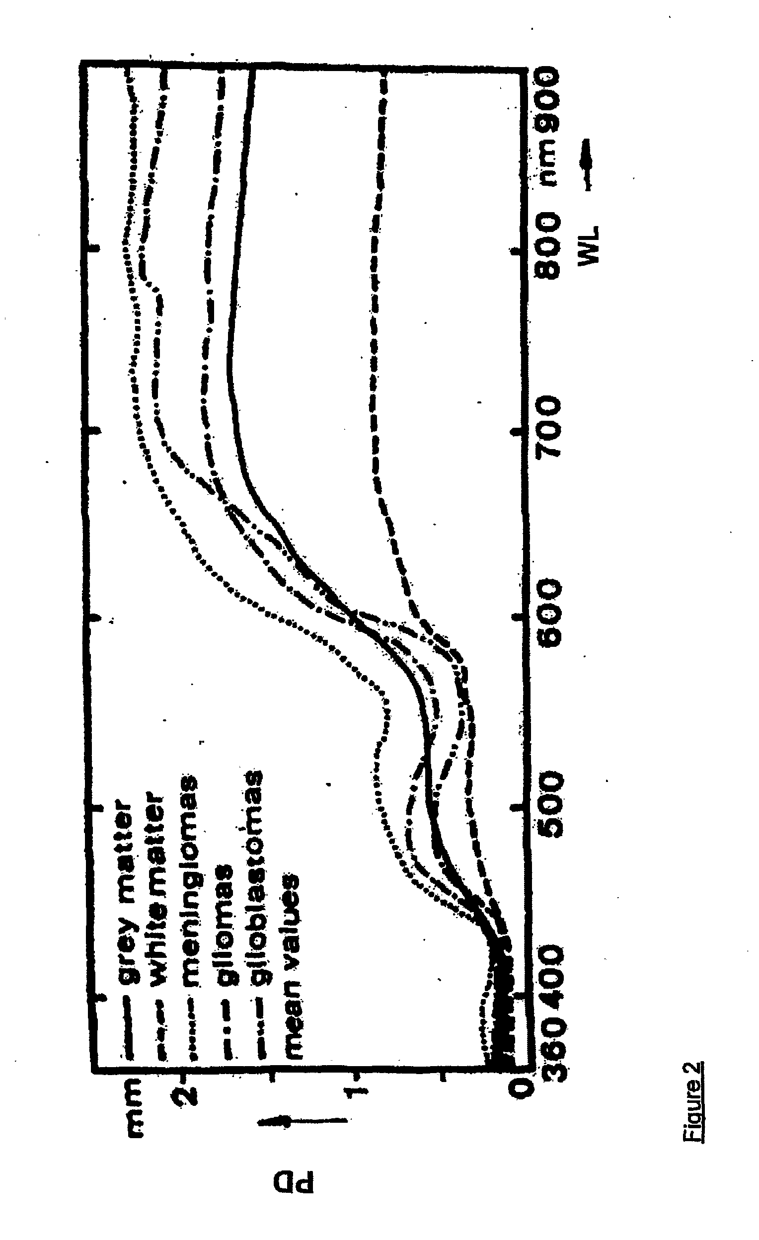
[0145] Thus, in the first aspect of the present invention, and referring to FIGS. 3(a), 3(b), 4(a) and 5, the apparatus generally designated by the numeral (100), is directed to the monitoring of blood flow rate of the first set of tissue viability parameters, and any combination of the three tissue viability parameters of the second set of tissue viability parameters. Thus, the apparatus (100) may be in the form of a probe (2) having at the distal tip thereof contact face (12) for making contact with the surface of the tissue (25) being monitored. In its simplest form, the probe (2) has a single fiber (201) for directing two radiations of different wavelengths to the same point (15) on the tissue (25). Alternatively, a bundle of fibers may replace a single fiber (201). The two radiations may come from first and second sources, (22) and (24) respectively, and are coupled to the fiber (201) by any suitable optical coupler. Referring to FIG. 2, other than at the plateau of wavelength...

second embodiment

[0194] In embodiments where the Doppler excitation wavelength WL1 lies in the range 420 nm to 440 nm, in either the first or second embodiment, the resulting NADH fluorescence will not pass through the dichroic beam splitter (302) designed to split these wavelengths towards detector (301). To overcome this problem at these wavelengths, a simple 1:20 beam-splitter should be substituted for the fore-mentioned dichroic beam splitter. It should be noted, that the strong WL1 reflection will not interfere with the NADH fluorescence, despite the two signals having similar wavelengths, because the signal are separated in time.

[0195] While the appropriate illumination wavelengths for sources (101), (102) referred to hereinbefore are particularly suited for the monitoring of brain tissue in-vivo using the first and second embodiments, corresponding illumination wavelengths may be determined for any other tissue enabling the apparatus (100) to be used with such tissues, mutatis mutandis.

[0196...

fifth embodiment

[0266] The fifth embodiment may be operated in a variety of modes as required by the clinical situation and diagnostic needs to which it is applied. Two particular modes of monitoring for which such multiple probe systems can be usefully applied, are described:

[0267] In the first mode, the mean signal intensities from the multiplicity of probes is calculated and displayed. This results in the parameters detected representing an average response of the multiplicity of tissue volumes probed, and will generally, better reflect the state of the organ layer as a whole. This mode of monitoring could be useful in transplantation surgery when better monitoring of the viability of donated organs are needed.

[0268] In the second mode, by applying one or several of the plurality of probes to each of several locations on the same organ or several different locations of different organs, the quasi-continuous monitoring of these organs over the same time period can be achieved by multiplexing the...


PUM


Abstract

Apparatus for determining the oxygenation state of at least one tissue element, comprising: a) illumination means for illuminating said tissue element with an illuminating radiation at a predetermined wavelength via at least one illumination location with respect to said tissue element, said illuminating radiation being at a wavelength within the NADH excitation spectrum or the Fp excitation spectrum; b) radiation receiving means and detection means for measuring the intensity of the corresponding NADH fluorescence or Fp fluorescence emitted by the tissue element at at least two predetermined wavelengths within the range of wavelengths comprised within the corresponding fluorescence emission spectrum.

Description

RELATED APPLICATIONS [0001] The present application is a divisional application of U.S. application Ser. No. 10 / 381,383, filed on Mar. 25, 2003, which is a U.S. national application of PCT Application No. PCT / IL01 / 00906, filed on Sep. 25, 2001.FIELD OF THE INVENTION [0002] The present invention relates to apparatuses and methods for enabling simultaneous, pseudo-simultaneous, or individual monitoring of a plurality of tissue vitality parameters, particularly in-vivo, with respect to an identical tissue layer element or volume. In particular, such parameters include blood flow rate, NADH concentration, blood volume, blood oxygenation state and flavoprotein concentration. BACKGROUND [0003] Mammalian tissues are dependent upon the continuous supply of oxygen and glucose needed for the energy production. This energy is used for various types of work, including the maintaining of ionic balance and biosynthesis of various cellular components. The ratio or balance, between oxygen supply an...

Claims


Application Information

Patent Timeline
Patent Type & Authority Applications(United States)
IPC IPC(8): A61B5/00A61B5/026
CPCA61B5/0261A61B2562/0242A61B5/14553A61B5/14546
Inventor PEWZNER, ELIAHUMAYEVSKY, AVRAHAM
Owner CRITISENSE
Why Eureka
Try Eureka