Non-invasive method and device to monitor cardiac parameters without use of electrical-mechanical interval

a non-invasive method and cardiac parameter technology, applied in the field of alternative non-invasive methods and devices to monitor cardiac parameters, can solve the problems of e-m intervals that are difficult to standardize, problems such as the normalization of e-m, and the correction of e-m, so as to achieve convenient comparison and easy measurement

Inactive Publication Date: 2007-08-16
HIRSH ROBERT
View PDF14 Cites 49 Cited by
  • Summary
  • Abstract
  • Description
  • Claims
  • Application Information

AI Technical Summary

Benefits of technology

[0023] The present invention frames an alternative noninvasive vector space, N′ consisting of hemodynamic quantities that are easily measured in a non-invasive manner, without requiring the measurement of the E-M interval. The present invention circumvents the problem of standardizing the E-M interval in a way that enables clinicians to easily make comparisons between the non-invasive hemodynamic parameters of one patient with that of another. This alternative vector space is denoted byN′={T, DI, EI, MAP, SV} , where T is the cardiac period measured from the peak of one R-wave on an EKG to the peak of the next R-wave, DI is the diastolic filling interval from the opening to the closing of the mitral valve, EI is the Ejection Interval from the opening to the closing of the aortic valve, MAP is the Mean Arterial Pressure in a peripheral artery such as the radial artery at the wrist, and SV is the Stroke Volume.

Problems solved by technology

A problem with the non-invasive hemodynamic monitoring approach described in the above has to do with the nature of the E-M interval.
While the relation between the E-M interval and the ratio between Stroke Volume and the Ejection Interval (SV / EI) is practically invariant over logarithmic ranges of Systemic Vascular Resistance (SVR) and Left Ventricular End-Diastolic Pressure (LVEDP), the E-M interval is difficult to standardize.
If the anatomical position of the arterial pressure detector is controlled for, say by placing it at the radial artery, there remains the problem of normalizing the E-M by the patient's height, arm-span, or some other feature of the patient's physiognomy.
While this can be easily accomplished, there also remains the problem of correcting the E-M for
Unfortunately, these procedures may need to be performed while the patient is under anesthesia or deep sedation.
The Deltex device simply measures the average blood flow in the descending Aorta and yields a ‘stroke distance’ over the ejection interval that does not correct for variations in the cross sectional area of the aorta.
A second problem with the E-M interval as derived from the EKG signal and the peripheral arterial pressure wave is that the beat-to-beat measurement of the arterial pressure wave has required the use of an invasive indwelling peripheral arterial catheter.
Although the use of such a catheter is routine, it has been associated with patient injury including rare but catastrophic loss of a patient's hand.

Method used


Image

  • Non-invasive method and device to monitor cardiac parameters without use of electrical-mechanical interval
  • Non-invasive method and device to monitor cardiac parameters without use of electrical-mechanical interval
  • Non-invasive method and device to monitor cardiac parameters without use of electrical-mechanical interval

Examples


Embodiment Construction

[0029] The disclosure of U.S. Pat. No. 7,054,679, which is a parent case, has been incorporated herein by external reference.

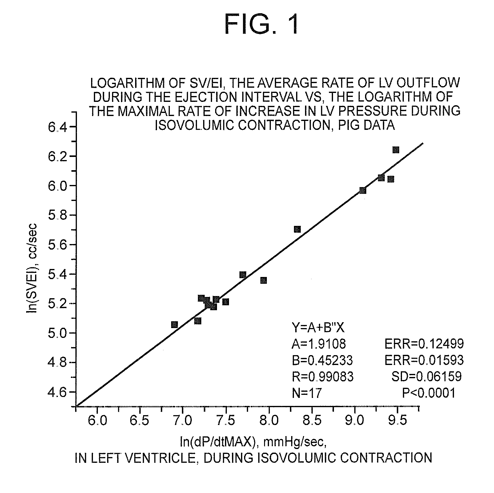
[0030] Equations 2 and 3 are empirical relations. Expressing equations (eqs.) 2 and 3 as a linear proportionality, we have

ln(dP / dtmax)α1 / (E-M)  eq. 2′

exp(1 / (E-M))αSV / EI  eq. 3′

Taking the natural logarithm of both sides of eq. 3′, we get

1 / (E-M)αln(SV / EI)  eq. 4

But by relating to eq. 2′, 1 / (E-M)αln(dP / dtmax)

Since two quantities that are proportional to the same quantity are proportional to each other, we can write

ln(SV / EI)αln(dP / dtmax)  eq. 5

Letting ‘B’ and ‘A’ represent the empirical coefficient and constant of linear proportionality respectively, we have,

ln(SV / EI)=B(ln(dP / dtmax))+A  eq. 6

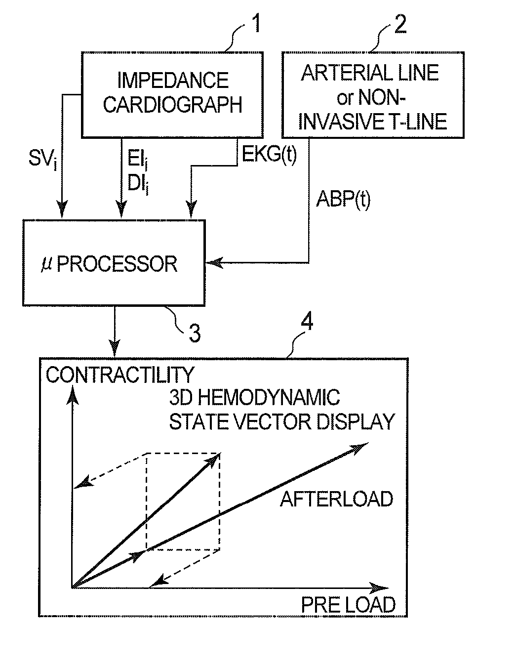
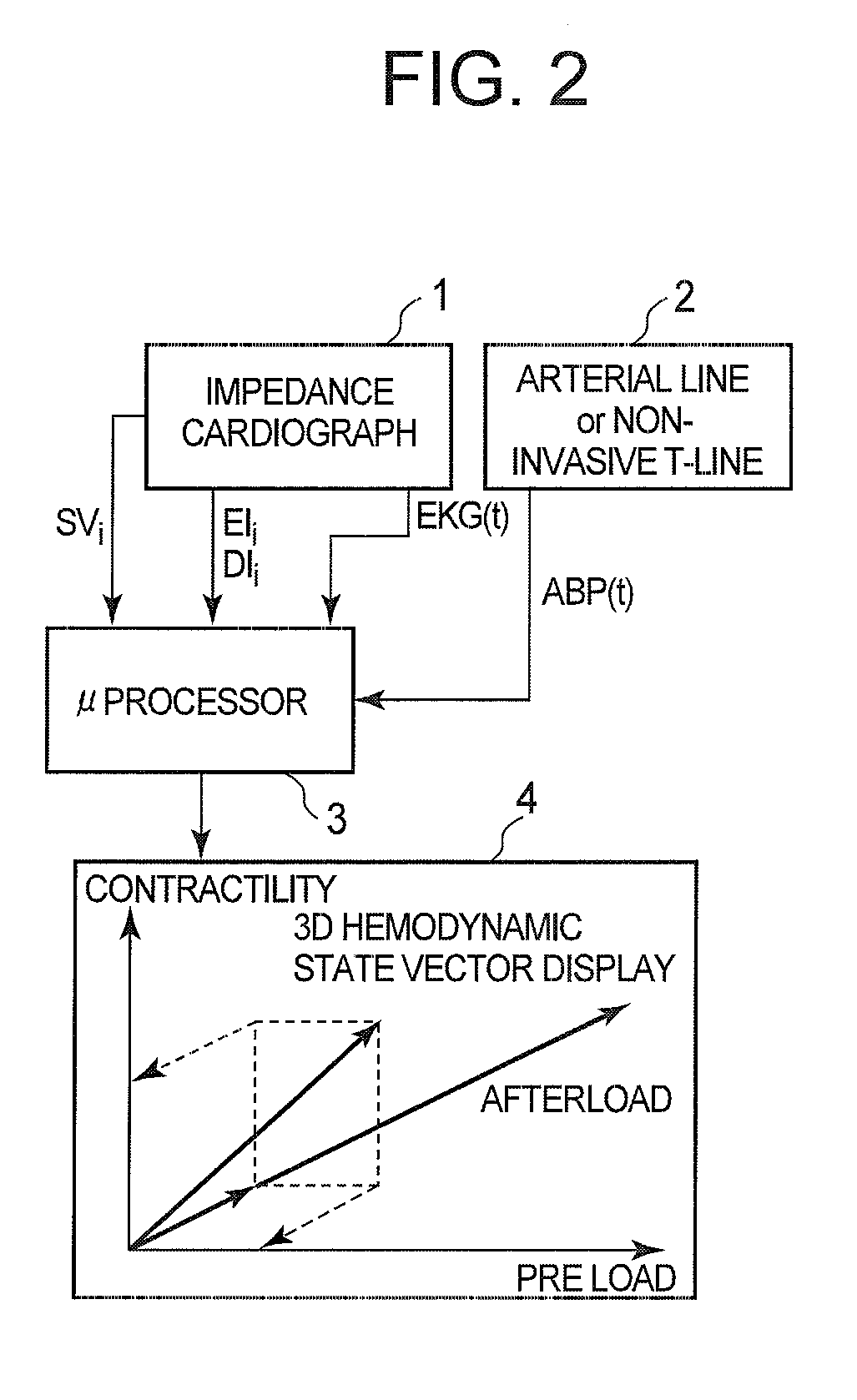
The right hand member of eq. 6 contains a quantity, dP / dtmax, which requires left ventricular catheterization to measure. The left hand side is now easily available with relatively little or no anatomic or physiologic trespass.

[0031] In U.S. Pat. 7,054,679, data from P...


PUM


Abstract

A method of and a device for non-invasively measuring the hemodynamic state of a subject or a human patient involve steps and units of non-invasively or minimally invasively measuring cardiac cycle period, mean arterial pressure, stroke volume, diastolic interval and ejection interval and converting the measured mean arterial pressure, stroke volume, diastolic interval and ejection interval into the cardiac parameters such as Preload, Afterload and Contractility, which are the common cardiac parameters used by an anesthesiologist. In the current invention, the use of electrical-mechanical interval has been eliminated for various advantageous reasons. The converted hemodynamic state of a patient is displayed on a screen as a three-dimensional vector with each of its three coordinates respectively representing Preload, Afterload and Contractility. Therefore, a medical practitioner looks at the screen and—quickly obtains the important and necessary information.

Description

CROSS REFERENCE TO RELATED APPLICATION [0001] This is a continuation-in-part application of the application Ser. No. 11 / 266,122 filed on Nov. 3, 2005, which is a divisional application of the application Ser. No. 09 / 999,125 filed on Oct. 31, 2001 and issued as U.S. Pat. No. 7,054,679 on May 30, 2006.BACKGROUND OF THE INVENTION [0002] 1. Field of the Invention [0003] The present invention relates to an alternative non-invasive method and device to monitor cardiac parameters. [0004] 2. Description of the Prior Art [0005] U.S. Pat. 7,054,679, NON-INVASIVE METHOD AND DEVICE TO MONITOR CARDIAC PARAMETERS issued to Hirsh in 2001, describes a vector space, whose basis vectors are non-invasively obtained in a substantially easy manner. In particular, the vector spaceN={T, MAP, EI, DI, E-M} is described, where T is the Cardiac Period, MAP is the Mean Arterial Pressure in a peripheral artery, preferably the Radial Artery, and E-M is the Electiical-Mechanical Interval. In one embodiment, the E...

Claims


Application Information

Patent Timeline
Patent Type & Authority Applications(United States)
IPC IPC(8): A61B5/04A61B5/352
CPCA61B5/02028A61B5/021A61B5/029A61B2560/0223A61B5/0535A61B8/06A61B8/12A61B5/0456A61B5/352
Inventor HIRSH, ROBERT
Owner HIRSH ROBERT
Why Eureka
Try Eureka