Structure-Based Analysis For Identification Of Protein Signatures: CUSCORE

a structure-based analysis and protein signature technology, applied in the field of bioinformatics, can solve the problems of non-computational methods, antibody generation or compound library screening, and low use value of information

Inactive Publication Date: 2007-10-18
LAWRENCE LIVERMORE NAT SECURITY LLC
View PDF6 Cites 7 Cited by
  • Summary
  • Abstract
  • Description
  • Claims
  • Application Information

AI Technical Summary

Benefits of technology

[0082]Given, the availability and accuracy of computational protein structure prediction, those skilled in the art can easily see the benefit of combining identification of nearest neighbor and target polypeptides using taxonomy and sequence similarity with the preferred

Problems solved by technology

In addition to being costly and time-consuming, non-computational methods are based on the principle of discovery and provide no a priori quantitative characterization of the protein residues forming the signature.
Consequently, traditional methods based on, e.g., antibody generation or compound library screens provide little information that can be used for down-selecting or targeting the possible pool of reagents.
Despite the power of sequence alignment techniques, they suffer from several shortcomings.
For example, the use of sequence alignment as a first step in identifying protein signatures representative of three-dimensional structures is limiting for several reasons.
First, because sequences mutate faster than do overall structures, structural conservation reflective of protein function tends to be incompletely r

Method used


Image

  • Structure-Based Analysis For Identification Of Protein Signatures: CUSCORE
  • Structure-Based Analysis For Identification Of Protein Signatures: CUSCORE
  • Structure-Based Analysis For Identification Of Protein Signatures: CUSCORE

Examples


example 1

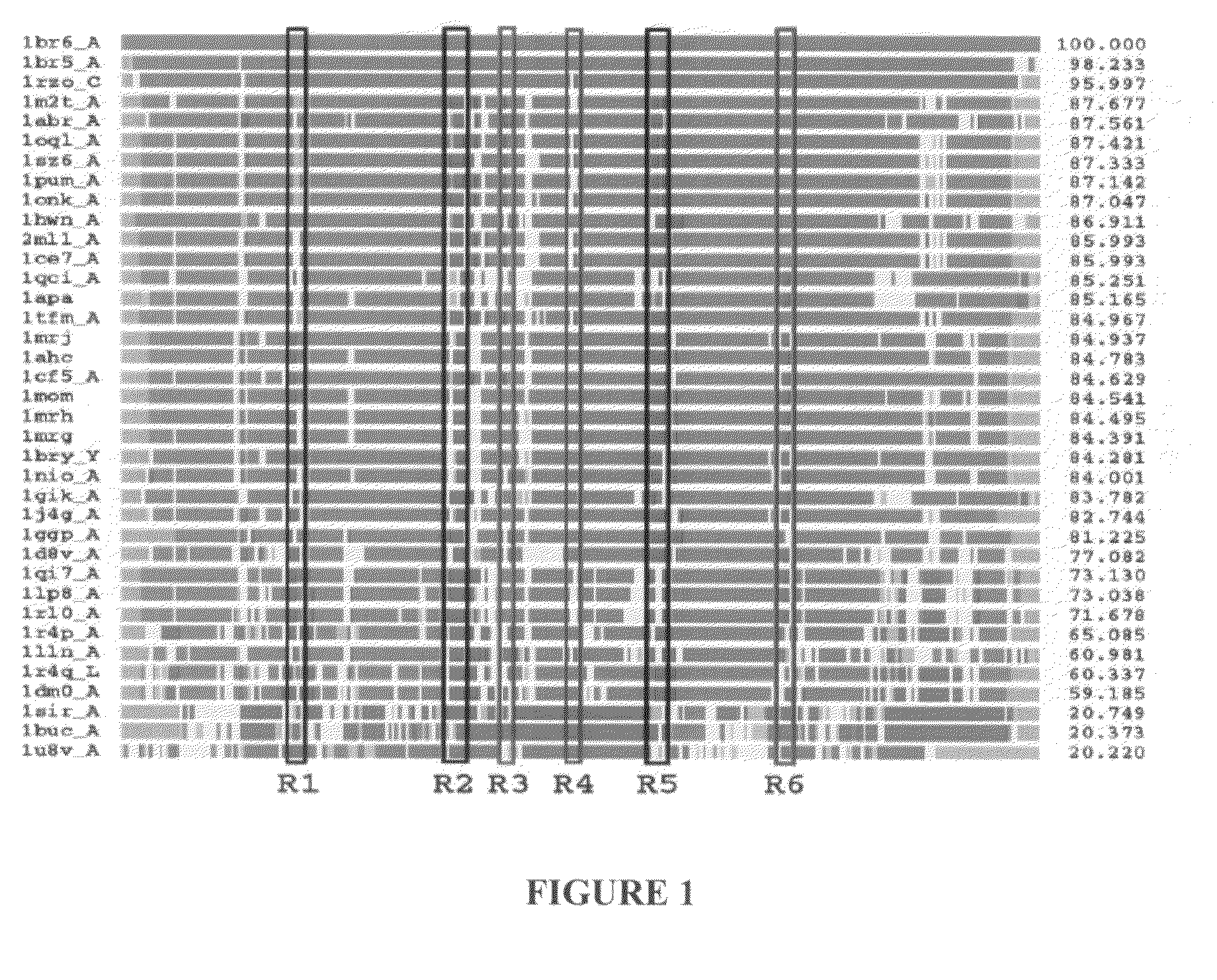
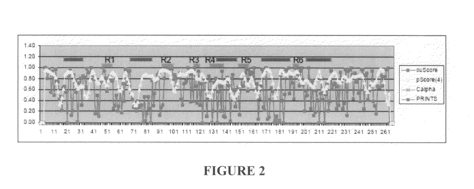
The Identification of Conserved / Unique Signatures in the A chain of Ricin

[0083]An entry (ID=RICI_RICCO, P02879) from the SHIGARICIN family of the PRINTS database of virulence factors as a reference sequence for our analyses of the A chain of ricin. Of the 21 PDB structures of the ricin A chain, the three non-redundant, non-mutant structures that had been solved with highest resolution (PDB entries 1br6, 1br5 and 1rz0) to include in the target set for our structure-based analyses were selected. These structures had sequence similarity between 93 and 100% (and corresponding structure-similarity LGA_S score between 95 and 100%) to the ricin A reference. Using the AS2TS (Zemla et al., 2005) automated homology-based protein structure modeling system, PDB entry lbr6 was selected as the 3D model structure of the ricin A reference sequence, because it had the greatest sequence similarity (100% sequence identity) and structure completeness from among available PDB structures (100% of structu...

example 2

Identification of Conserved / Unique Signatures in the West Nile Virus Envelope Glycoprotein

[0092]The method of the present invention was also used to identify motifs on the envelope glycoprotein of West Nile virus that satisfy conditions of a) conservation / uniqueness with respect to WNV, versus b) potential cross reactivity with other Flaviviruses. Data from the literature was then used to evaluate the success of these predictions. These analyses suggest that PSE correctly predicted conservation / uniqueness and excluded motifs for which targeted detection reagents would likely cross react with other Flaviviruses.

[0093]The envelope glycoprotein of West Nile virus (refseq strain) was selected as the reference sequence and was blasted against the non-redundant (NR) protein database to capture related Flavivirus sequences. The subject and query sequences were then modeled using AS2TS.

[0094]Sequences and models were analyzed using the present method for identification of protein signatures...


PUM


Abstract

Disclosed are computational methods, and associated hardware and software products for scoring polypeptide residues that combine the use of a structure-based alignment with a method of selecting against confounding proteins. The scores can be used to identify protein signatures of interest that are useful, e.g., as targets in developing highly specific ligands for diagnostic or therapeutic uses.

Description

BACKGROUND OF THE INVENTION[0001]1. Field of the Invention[0002]The present invention relates to the field of bioinformatics. More specifically, the invention relates to computational methods for scoring residues based on structural alignment and residue similarity metrics. The invention also relates to methods for identifying protein signatures from the computed scores.[0003]2. Background of the Invention[0004]A motif or signature is a defined region on a target protein that may be used to specifically identify that protein or, indirectly, the organism that produces it. There is increased need to rapidly develop highly specific diagnostic and therapeutic reagents and detection assays for proteins or organisms which cause biological threat. The identification of signatures conserved or unique to proteins or organisms of interest such as pathogens or toxins allows the rapid development of these reagents and detection assays.[0005]Non-computational methods of identifying protein signa...

Claims


Application Information

Patent Timeline
IPC IPC(8): G06F19/00G16B15/30G16B30/10
CPCG06F19/22G06F19/16G16B15/00G16B30/00Y02A90/10G16B30/10G16B15/30
Inventor ZHOU, CAROL E.ZEMLA, ADAM T.
Owner LAWRENCE LIVERMORE NAT SECURITY LLC
Why Eureka
Try Eureka