Method for treating blood coagulation disorders
a blood coagulation disorder and a technology for fviia is applied in the field of blood coagulation disorders to achieve the effect of reducing the immunogenicity of agents
- Summary
- Abstract
- Description
- Claims
- Application Information
AI Technical Summary
Benefits of technology
Problems solved by technology
Method used
Image
Examples
example 1
[0030] This example describes the preparation of the lipidic particles.
[0031] Materials: Albumin free full-length rFVIII (Baxter Health Care Glendale, Calif.) was used as antigen. Advate was provided as a gift from Western New York Hemophilia foundation. Dimyristoyl phosphatidylcholine (DMPC) and soybean phosphatidylinositol (Soy PI) were purchased from Avanti Polar Lipids (Alabaster, Ala.). Cholesterol, IgG-free bovine serum albumin (BSA), and diethanolamine were purchased from Sigma (St. Louis, Mo.). Goat antimouse-Ig and antirat-Ig, alkaline phosphatase conjugates were obtained from Southern Biotechnology Associates, Inc. (Birmingham, Ala.). p-Nitrophenyl phosphate disodium salt was purchased from Pierce (Rockford, Ill.). Monoclonal antibodies ESH4, ESH5, and ESH8 were purchased from American Diagnostica Inc. (Greenwich, Conn.). Monoclonal antibody N77210M was purchased from Biodesign International (Saco, Me.). Normal coagulation control plasma and FVIII-deficient plasma were pu...
example 2
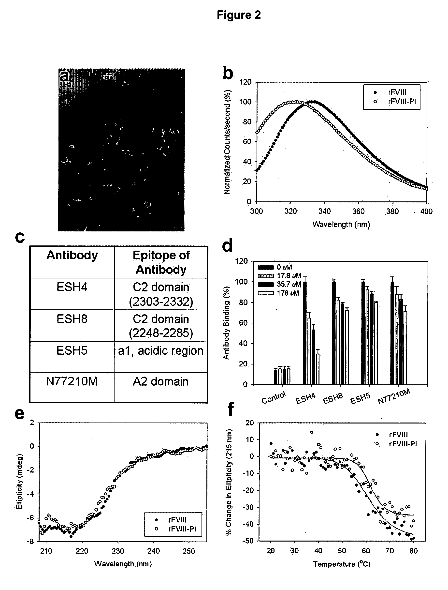
[0033] This example describes characterization studies for the lipid particles prepared in Example 1. The lipid dispersion samples for microscopic analysis were prepared by air-drying on formvar-coated grids and negatively staining them with 2% uranyl acetate for approximately 1 min. The samples were photographed using a Hitachi H500 TEM operating at 75 kV. Negatives were scanned at 300 dpi with an Agfa Duoscan T1200 scanner. The morphology of the particles determined using Transmission electron microscopic studies indicated the following. The particle size was found to be around 100 nm, consistent with dynamic light scattering studies (data not shown). The analysis of the micrograph showed that the donut like structures typical of liposomal lamellarity was not observed instead particles displayed disc like structures (FIG. 2a) and it is possible that unique lipid organization distinct from liposomes are formed that can accommodate higher mol % of FVIII.
example 3
[0034] This example describes fluorescence analysis of rFVIII and rFVIII-PI. The effect of PI on the tertiary structure of rFVIII was determined by exciting the samples either at 280 nm or at 265 nm and the emission was monitored in the wavelength range of 300-400 nm. The spectra were acquired on a PTI-Quantamaster fluorescence spectrophotometer (Photon Technology International, Lawrenceville, NJ). Protein concentration was 5 μμg / ml and slit width was set at 4 nm. The fluorescence emission spectra of FVIII loaded in this particle showed blue shifted emission maxima compared to free protein suggesting that FVIII is located in hydrophobic environment (FIG. 2b). This observation is in contrast to fluorescence spectrum observed for FVIII associated with PS containing liposomes where no change in fluorescence properties was observed (Purohit et al., 2003) and is consistent with molecular model proposed based on crystallographic and biophysical studies. FVIII was associated with PS contai...
PUM
| Property | Measurement | Unit |
|---|---|---|
| size | aaaaa | aaaaa |
| size | aaaaa | aaaaa |
| size | aaaaa | aaaaa |
Abstract
Description
Claims
Application Information
 - R&D
- Intellectual Property
- Life Sciences
- Materials
- Tech Scout
- Unparalleled Data Quality
- Higher Quality Content
- 60% Fewer Hallucinations
Browse by: Latest US Patents, China's latest patents, Technical Efficacy Thesaurus, Application Domain, Technology Topic, Popular Technical Reports.
© 2025 PatSnap. All rights reserved.Legal|Privacy policy|Modern Slavery Act Transparency Statement|Sitemap|About US| Contact US: help@patsnap.com


