Ultrasound diagnosis apparatus

a technology of ultrasound and diagnostic equipment, applied in the field of ultrasound diagnosis equipment, can solve the problems of difficult to recognize the state of the heart or measure the heart function, the plurality of electrodes cannot be brought into direct contact with the fetus, and the inability to perform various measurements using the electrocardiographic signal of the fetus

a technology of ultrasound and diagnostic equipment, applied in the field of ultrasound diagnosis equipment, can solve the problems of difficult to recognize the state of the heart or measure the heart function, the plurality of electrodes cannot be brought into direct contact with the fetus, and the inability to perform various measurements using the electrocardiographic signal of the fetus

US20080009735A1Active Publication Date: 2008-01-10FUJIFILM HEALTHCARE CORP

Image

  • Ultrasound diagnosis apparatus
  • Ultrasound diagnosis apparatus
  • Ultrasound diagnosis apparatus

Examples


Embodiment Construction

[0037]A preferred embodiment of the present invention will be described in detail with reference to the accompanying drawings.

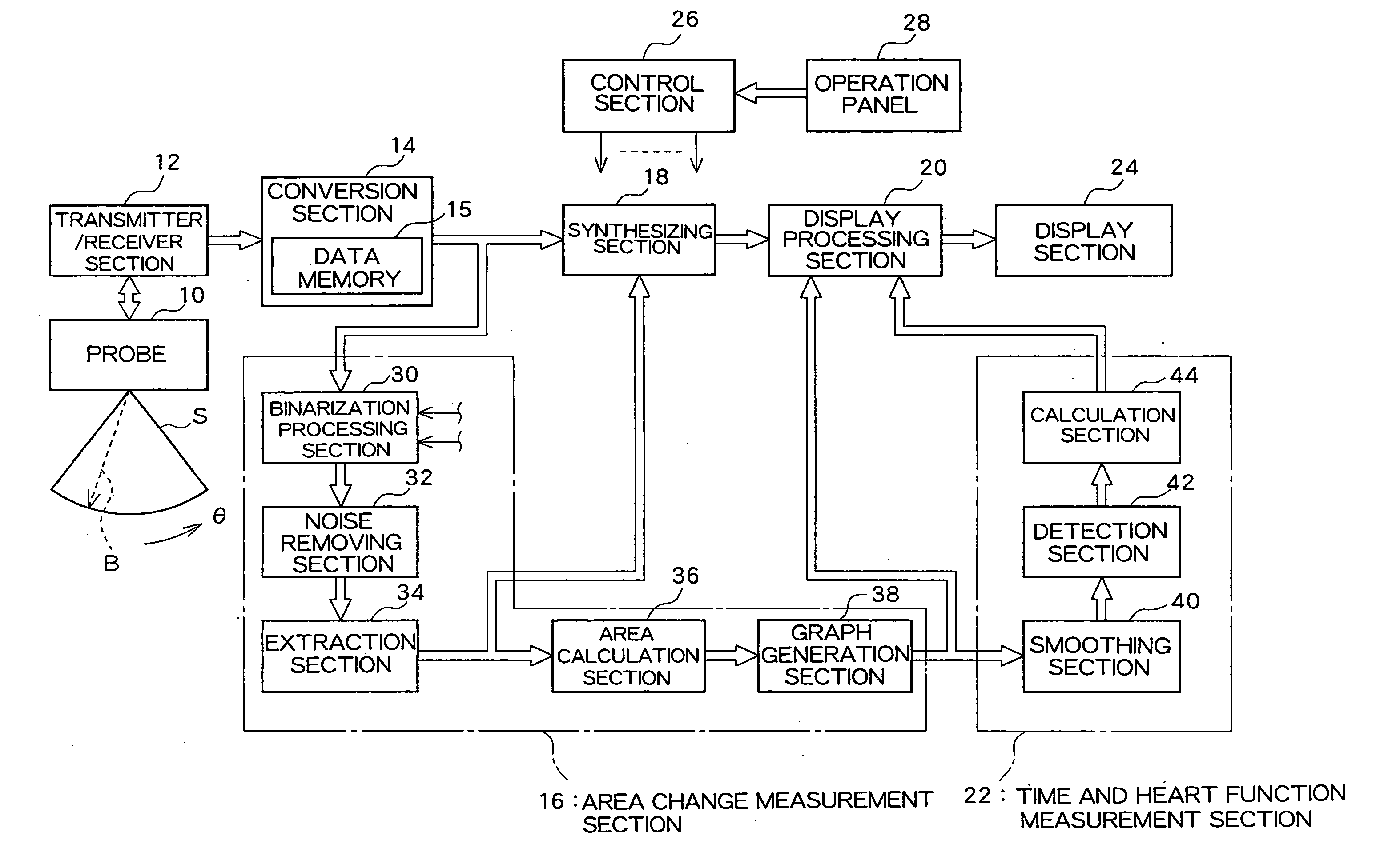
[0038]FIG. 1 shows an ultrasound diagnosis apparatus according to one embodiment of the present invention. Specifically, FIG. 1 is a block diagram showing an overall structure of an ultrasound diagnosis apparatus. The ultrasound diagnosis apparatus according to the present embodiment performs ultrasonic diagnosis concerning the heart in a living body, particularly concerning the heart of a fetus (a fetal heart), although the apparatus of the present embodiment can obviously also be used for ultrasonic diagnosis of a heart in a living body other than a fetus.

[0039]A probe 10 is a transmitter / receiver which transmits ultrasound pulse and receives reflected ultrasound to thereby form an ultrasound beam B. The probe 10 includes an array transducer formed of a plurality of transducer elements. The ultrasound beam B which is formed by the array transducer is electr...


PUM


Abstract

An ultrasound diagnosis apparatus which performs ultrasonic diagnosis with respect of a fetal heart. An area change measurement section calculates an area of a specific heart chamber (left ventricle) in the fetal heart in frame units. Further, the area change measurement section generates a graph representing the area which is calculated in frame units. The maximum value and the minimum value are specified in the graph to thereby specify end-diastole and end-systole. Based on this information, a heart rate and a cardiac cycle with regard to the fetal heart are calculated. Further, an ejection fraction is also calculated from an end-diastolic area and an end-systolic area.

Description

BACKGROUND[0001]1. Technical Field[0002]The present invention relates to an ultrasound diagnosis apparatus, and more particularly to ultrasonic diagnosis technology for a fetal heart.[0003]2. Related Art[0004]Ultrasonic diagnosis of a fetus in it's mother womb has become commonplace in medical practice. One of the purposes of such diagnoses is to diagnose or treat congenital heart diseases at an early stage. If a serious heart disease can be diagnosed prior to delivery, it is often possible to successfully treat the disease using medicine, surgery, or the like immediately after childbirth. In ultrasonic diagnosis of a fetal heart, the shape or size of the heart is displayed on a tomographic image, or the state of blood flow in the heart is displayed on a two-dimensional color Doppler image (or a color flow mapping image). Also, according to the pulse Doppler method, Doppler information obtained from a sample gate (or a sample volume) which is set within the heart is subjected to fre...

Claims


Application Information

Patent Timeline
10 Jan 2008
Publication
US20080009735A1
IPC
A61B8/14
CPC
A61B8/02; A61B8/0866; A61B8/0883; A61B8/483; G06T2207/30048; G06T7/602; G06T2207/10136; G06T2207/30044
Inventors
MURASHITA, MASARU