
Methods of screening based on the EGF receptor crystal structure

a screening method and crystal structure technology, applied in the field of screening based on the crystal structure of the egf receptor, can solve the problems that the cost of humanised mab production is likely to limit the application of this type of therapy, and achieve the effects of enhancing binding, affecting association rate, and increasing affinity of binding

Inactive Publication Date: 2008-01-31
COMMONWEALTH SCI & IND RES ORG +2
View PDF3 Cites 13 Cited by
  • Summary
  • Abstract
  • Description
  • Claims
  • Application Information

AI Technical Summary

Benefits of technology

[0028] The compound may interfere with ligand binding to one or more of the specified residues in a number of ways. For example the compound may bind or interact with the receptor at or near one or more of the specified residues or corresponding regions and by steric overlap and / or electrostatic repulsion prevent natural ligand binding. Alternatively the compound may bind to the receptor so as to interfere allosterically with natural ligand binding. For example the compound may bind to the L1 and L2 domains in manner such as to decrease the “gap” between the L1 and L2 domains thereby preventing access of the ligand to one or more of the specified residues.
[0030] (i) The compound may bind to the L1 and L2 domains in manner such as to decrease the “gap” between the L1 and L2 domains thereby preventing access of the ligand to one or more of the specified residues.
[0041] ErbB2 ErbB2 alone has not been reported to bind any ligand with high affinity but is preferred heterodimerisation partner for the other three EGF receptor family members, enhancing their affinities for their respective ligands and amplifying their signals.
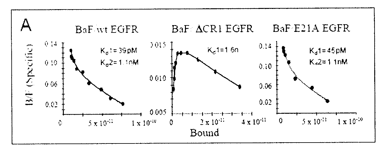
[0091] Additionally, the mutation of residues which are outside of the relevant binding interface may also alter the binding affinity by changes in the long range electrostatic interactions. These changes can affect the rate of association between two interacting proteins without greatly changing the rate of dissociation, and hence change the equilibrium binding constant (Selzer and Schreiber (1999) J Mol Biol. 287: 409-419; Seizer et al., (2000) Nat Struct Biol. 7: 537-541.). In one example of increasing the affinity of binding by mutating residues outside of the protein-protein interface, selected residues of the β-lactamase inhibitory protein that were outside of the interface were mutated so as to change their charge e.g. a basic residue mutated to a neutral residue and then the affinity and rate constants of the mutant binding to TEM1 β-lactamase was measured. In one mutant, the change of four amino acids led to an enhancement of binding by a factor of more 250-fold (Selzer et al., (2000) Nat Struct Biol. 7: 537-541.). In this example, the authors specified a formula which predicted the changes in the association constant upon mutation to within a factor of two (Selzer et al., (2000) Nat Struct Biol. 7: 537-541.). In this way, the structure of the EGFR or a model of one other EGFR family members could be used to predict mutations that would likely lead to an enhancement of the rate of association of the relevant EGFR family extracellular fragment to its interacting protein. Calculation and subsequent visualization of the electrostatic isopotentials (e.g. Smith and Treutlein (1998) Protein Sci. 7: 886-896.) may assist the selection of residues to mutate in order to increase the protein's rate of association. The most likely candidate residues for mutation are those on the periphery of the interface and those outside of the interface but which are within a specified distance of the interacting protein and are not completely buried in the L1 or L2 domain (as judged by visual examination). Cysteine residues, which are needed for the maintenance of the EGFR structure were also excluded from the list. For the EGFR, the preferred residues are:

Problems solved by technology

The large doses required and the cost of production of humanised Mab is likely to limit the application of this type of therapy.

Method used


Image

  • Methods of screening based on the EGF receptor crystal structure
  • Methods of screening based on the EGF receptor crystal structure
  • Methods of screening based on the EGF receptor crystal structure

Examples


example 1

Protein Preparation of sEGFR501

[0223] The derivation of stably transfected Lec8 cells expressing sEGFR501 and the subsequent purification and characterisation of the secreted ectodomain has been described in detail (Elleman et al., 2001, Biochemistry 40:8930-8939.). Purified sEGFR501 was shown, by isoelectric focusing gels to be unstable on storage, the majority of isoforms being transformed into products with less acidic isoelectric points. This change was accompanied by a small mobility increase (estimated at 1-2 kDa) on SDS polyacrylamide gels. N-terminal sequence analysis showed that the new product retained the expressed N-terminus of sEGFR501, suggesting that the apparent 1-2 kDa reduction in mass and increase in positive charge might be due to partial or complete loss of the acidic-residue rich C-terminal tag and enterokinase cleavage site. Prolonged storage led to the majority of protein converting to the least acidic isoform of pI˜6.6, which appeared to remain stable. The ...

example 2

Crystallization and Data Collection

[0224] sEGFR501 obtained from the above procedures appeared nearly homogeneous on SDS and IEF gels and was used in crystallization trials alone and in combination with several ligands. The best diffracting crystals were obtained from mixtures containing a five-fold molar quantity of human TGFα (GroPep receptor grade) compared to sEGFR501. Crystals of sEGFR501 in complex with TGFα were grown in 7% PEG 3350, 20% Trehalose, 10 mM CdCl2 and 100 mM HEPES, pH 7.5, and belonged to the space group P21 (a=51.59, b=198.71, c=78.90 Å, β=102.03°). These crystals were cryo-cooled to −170° C. in the same mother liquor. Data were recorded on a Rigaku RAXIS VI area detector using a Siemens M18XHF X-ray generator with Yale / MSC mirrors or a Rigaku RU300 generator and AXCO capillary optics. Crystals were also derivatised by soaking in mother liquor containing 1-10 mM heavy atom compounds and diffractions data were collected as before and statistics are given in Tabl...

example 3

Phase Determination and Structure Refinement

[0225] Phasing by multiple isomorphic replacement was performed with programs from CCP4 (Collaborative Computational Project Number 4, 1994) and SHARP (De La Fortelle and Bricogne, 1996, Methods Enzymol. 276: 472-494) and the resulting electron density maps were improved by solvent flattening and histogram matching with DM (Cowtan, K. 1994, Joint CCP4 and ESF-EACBM Newslett. Protein Crystallogr. 31:34-38). Details are given in Table 1. Density averaging using noncrystallographic symmetry was not of much value as the proteins corresponded to more than three rigid groups. The polypeptide chains for two receptor and two ligand molecules were fitted manually and refined with CNS (Brunger, et al., 1998, X-PLOR Reference Manual 3.851, Yale Univ., New Haven, Conn.). As the highest resolution data were collected for the PIP derivative these data were use for the final stages of refinement. During the refinement an overall anisotropic temperature ...


PUM

PropertyMeasurementUnit
massaaaaaaaaaa
three-dimensional structuresaaaaaaaaaa
NMRaaaaaaaaaa

Abstract

This invention relates to the structure of members of the epidermal growth factor (EGF) receptor family and to receptor / ligand interactions. In particular, it relates to the field of using the EGF receptor family structure to select and screen for compounds that inhibit the formation of active receptor dimers.

Description

FIELD OF THE INVENTION [0001] This invention relates to the structure of members of the epidermal growth factor (EGF) receptor family and to receptor / ligand interactions. In particular, it relates to the field of using the EGF receptor family structure to select and screen for compounds that inhibit the formation of active receptor dimers. BACKGROUND OF THE INVENTION [0002] Epidermal growth factor is a small polypeptide growth factor that stimulates marked proliferation of epithelial tissues and is a member of a larger family of structurally related growth factors such as transforming growth factor α (TGFα), amphiregulin, betacellulin, heparin-binding EGF and some viral gene products. Abnormal EGF family signalling is a characteristic of certain cancers (Yarden and Sliwkowski, 2001, Nature Reviews Mol Cell Biol. 2, 127-37; Soler and Carpenter, 1994 In Nicola, N. (ed) “Guidebook to Cytokines and their Receptors”, Oxford Univ. Press, Oxford, pp 194-197; Walker and Burgess, 1994, In Ni...

Claims


Application Information

Patent Timeline
Patent Type & Authority Applications(United States)
IPC IPC(8): C07K16/22A61K39/395G01N33/50G16B15/30A61K45/00A61P35/00A61P43/00C07K14/71C07K16/28C07K16/46C07K19/00G01N33/15G01N33/68G01N33/74
CPCC07K14/71C07K2299/00G06F19/16G01N33/74G01N2333/71G01N33/6803G16B15/00G16C20/50A61P35/00A61P43/00G16B15/30
Inventor ADAMS, TIMOTHY EDWARDBURGESS, ANTONY WILKSELLEMAN, THOMAS CHARLESGARRETT, THOMAS PETER JOHNJORISSEN, ROBERT NICHOLASLOU, MEIZHENLOVRECZ, GEORGE OSCARMCKERN, NEIL MORETONNICE, EDOUARD COLLINSWARD, COLIN WESLEY
Owner COMMONWEALTH SCI & IND RES ORG
Learn More