Method Of Cell Isolation

a cell isolation and cell technology, applied in the field of cell isolation, can solve the problems of poor understanding and the inability to evaluate the comparative repopulating capacity of purified cell populations at limiting dilution, and achieve the effect of facilitating autologous cell transplant therapy and tissue regeneration

Inactive Publication Date: 2008-02-14
WALTER & ELIZA HALL INST OF MEDICAL RES
View PDF2 Cites 11 Cited by
  • Summary
  • Abstract
  • Description
  • Claims
  • Application Information

AI Technical Summary

Benefits of technology

[0017] The present invention advantageously provides a method for isolating MaSCs without the need to first maintain the tissue from which the MaSCs are derived in culture. As a result, the MaSCs isolated in accordance with the method of the present invention retain characteristics of MaSCs ii) vivo which may otherwise be modified or lost if the MaSCs underwent a period of culture prior to isolation.
[0018] The isolation of the MaSCs provided by the present invention may be performed using any cell-selection means which facilitates cell selection according to levels of cell surface proteins. Preferably, the cell-selection means comprises contacting the MaSCs to be selected, either sequentially or simultaneously, with molecules capable of interacting with cell surface proteins which are conjugated to a reporter compound which allows cell selection and identification. Most preferably, the molecules are conjugated to a fluorescent reporter compound, thereby facilitating cell-selection according to fluorescence intensity using fluorescence activated cell sorting (FACS).
[0021] The ability to isolate MaSCs according to the method of the present invention provides methods and compositions for use in tissue replacement and / or augmentation therapy, particularly mammary tissue replacement and / or augmentation therapy. In particular, the MaSCs isolated in accordance with the method of the present invention facilitate autologous cell transplant therapies and reduce, therefore, the need for allogenic tissue transplantation and the concomitant use of immunosuppressive agents.
[0022] Furthermore, the ability to isolate MaSCs according to the method of the present invention also enables the identification agents which modulate MaSC survival, self-renewal, proliferation and / or differentiation in both normal and diseased tissue in vitro and / or in vivo. In particular, the identification of agents which regulate the in vivo activity of MaSCs provides a method to induce or otherwise facilitate the regeneration and / or augmentation of tissue, particularly mammary tissue, in situ, that is, without the need for tissue transplantation.

Problems solved by technology

Despite a significant improvement in the management of breast cancer over the last few years, about 25% of women diagnosed will die from the disease, revealing that those tumor cells have intrinsic properties that are refractory to current treatment strategies.
The heterogeneous nature of breast cancer suggests the involvement of multiple genetic factors and cell types but these are poorly understood.
However, in these studies MFP repopulation required large numbers (several thousand) of cells, and the comparative repopulating capacity of purified cell populations was not evaluated at limiting dilution.

Method used


Image

  • Method Of Cell Isolation
  • Method Of Cell Isolation
  • Method Of Cell Isolation

Examples


example 1

General Experimental Procedures

Mammary Cell Preparation

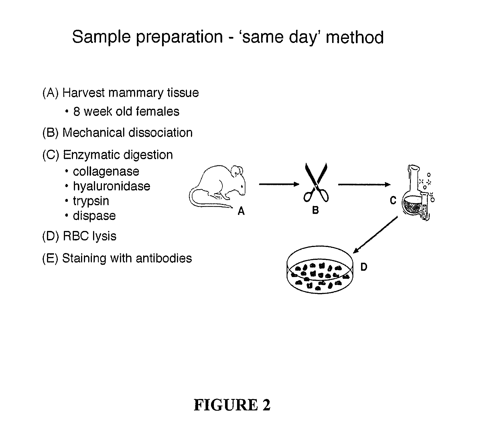
[0106] The nature of mouse mammary epithelial stem cells was evaluated using the in vivo mammary epithelial cell transplantation approach described in Alvi et al., Breast Cancer Res 5:R1-R8, 2003. The protocol for mammary epithelial cell purification was optimised and is summarized in FIG. 2. It initially involved the harvesting of the 3rd, 4th (after first removing the visible lymph node) and 5th mammary glands from eight-week-old mice. The harvested glands were mechanically dissociated using a McIllwain tissue chopper and then enzymatically disrupted with 300 U / ml collagenase and 100 U / ml hyaluronidase in dissociation medium (DME-HAM, 5% v / v FCS, 5 μg / ml insulin, 500 ng / ml hydrocortisone 10 ng / ml EGF and 20 ng / ml cholera toxin) for one hour at 37° C., with forceful titurations every 20 minutes. The resulting organoid suspension was serially treated with 0.25% w / v trypsin / 1 mM EGTA for 1-2 minutes at 37° C. to disrupt cell-c...

example 2

Limiting Dilution Studies

[0115] To establish the frequency of mammary stem cells in a cell population, limiting dilution analysis of mammary repopulating capacity was performed. Limiting dilution analysis is a well-established method for determining the frequency of cells in a specific population that have a certain characteristic (in our case, the ability to form a mammary epithelial structure in vivo). It assumes that the cells in question have this characteristic independent of other cells in the suspension. In our method, decreasing numbers of cells transplanted should produce a progressively smaller proportion of positive outgrowths, such that there is a linear relationship between the log of the number of cells transplanted and the proportion of positive outgrowths. Statistical analysis of our repopulation data was performed using L-Calc software (Stem Cell Technologies, Vancouver, Canada).

[0116] The mammary repopulating cell frequency in the overall cell population was anal...

example 3

Flow Cytometric Analysis of Mammary Cell Preparation Stained with Hoechst33342

[0117] SP cells were identified in our freshly isolated mammary epithelial cell preparation using the Ho dye efflux assay. Prior to antibody staining, Ho dye was added to the cells at a concentration of 3 mg / mL and incubated at 37° C. for one hour. The presence of SP cells was confirmed by treatment of cells with verapamil, which has been shown to inhibit the BCRP1 / ABCG2 membrane transporter pump responsible for the efflux of Hoechst dye. SP cells accounted for approximately 1% of the cells in our mammary cell preparation (FIG. 5).


PUM

PropertyMeasurementUnit
timeaaaaaaaaaa
temperatureaaaaaaaaaa
volumeaaaaaaaaaa

Abstract

The present invention relates generally to a method for the generation of a substantially homogenous population of undifferentiated cells. More particularly, the present invention relates to a method for isolating a substantially homogenous population of stem cells, and in particular, mammary stem cells (MaSCs). The MaSCs of the present invention are isolated on the basis of differential levels of proteins present on their cell surface. The MaSCs of the present invention are particularly useful as targets for identifying agents which modulate MaSC survival, self-renewal, proliferation and / or differentiation in both normal and diseased tissue such as, but not limited to, tumor tissue, and, also as source of tissue for the regeneration, replacement and / or augmentation of tissue damaged and / or lost after disease or injury.

Description

BACKGROUND OF THE INVENTION [0001] 1. Field of the Invention [0002] The present invention relates generally to a method for the generation of a substantially homogenous population of undifferentiated cells. More particularly, the present invention relates to a method for isolating a substantially homogenous population of stem cells, and in particular, mammary stem cells (MaSCs). The MaSCs of the present invention are isolated on the basis of differential levels of proteins present on their cell surface. The MaSCs of the present invention are particularly useful as targets for identifying agents which modulate MaSC survival, self-renewal, proliferation and / or differentiation in both normal and diseased tissue such as, but not limited to, tumor tissue, and, also as source of tissue for the regeneration, replacement and / or augmentation of tissue damaged and / or lost after disease or injury. [0003] 2. Description of the Prior Art [0004] Reference to any prior art in this specification is...

Claims


Application Information

Patent Timeline
Patent Type & Authority Applications(United States)
IPC IPC(8): C12N5/08A61K35/55A61K45/00C12Q1/02C12N5/00C12N5/071C12N5/074G01N33/50
CPCC12N5/0631C12N2501/01G01N33/5073C12N2501/33C12N2501/39C12N2501/11A61P43/00
Inventor LINDEMAN, GEOFFREY JOHNSHACKLETON, MARKVAILLANT, FRANCOISVISVADER, JANE ELLEN
Owner WALTER & ELIZA HALL INST OF MEDICAL RES
Why Eureka
Try Eureka