Fluorescence endoscope system, fluoroscopy apparatus, fluoroscopy method, fluorescence-information processing apparatus, and fluorescence-information processing method

Inactive Publication Date: 2008-02-14
OLYMPUS CORP
View PDF3 Cites 20 Cited by
  • Summary
  • Abstract
  • Description
  • Claims
  • Application Information

AI Technical Summary

Benefits of technology

[0035]Accordingly, the fluorescence endoscope system, the fluoroscopy apparatus, the fluoroscopy method, the fluorescence-information processing apparatus, and the fluorescence-information processing method according to the present invention allow superior fluoroscopy by reducing the effect of autofluorescence of biological tissue by using correction information that can be used to correct for the effect of autofluorescence.

Problems solved by technology

In fluoroscopy using fluorescent probes, however, irradiation of biological tissue under examination with excitation light has the side effect of inducing autofluorescence from the biological tissue.
This autofluorescence causes noise in fluoroscopy using fluorescent probes.

Method used


Image

  • Fluorescence endoscope system, fluoroscopy apparatus, fluoroscopy method, fluorescence-information processing apparatus, and fluorescence-information processing method
  • Fluorescence endoscope system, fluoroscopy apparatus, fluoroscopy method, fluorescence-information processing apparatus, and fluorescence-information processing method
  • Fluorescence endoscope system, fluoroscopy apparatus, fluoroscopy method, fluorescence-information processing apparatus, and fluorescence-information processing method

Examples


first embodiment

[0046]A fluorescence endoscope system 1 according to the present invention will now be described with reference to FIGS. 1 to 5.

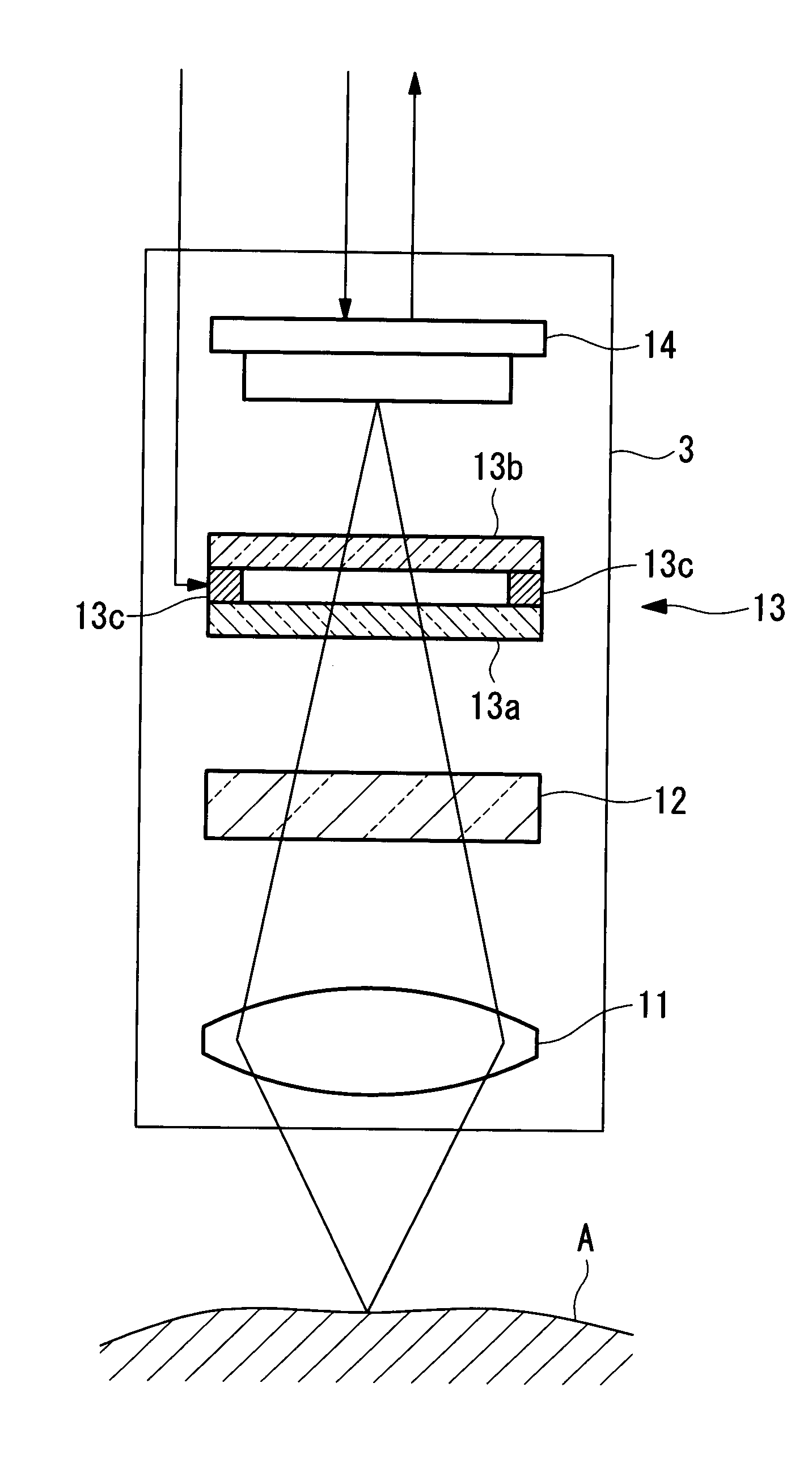
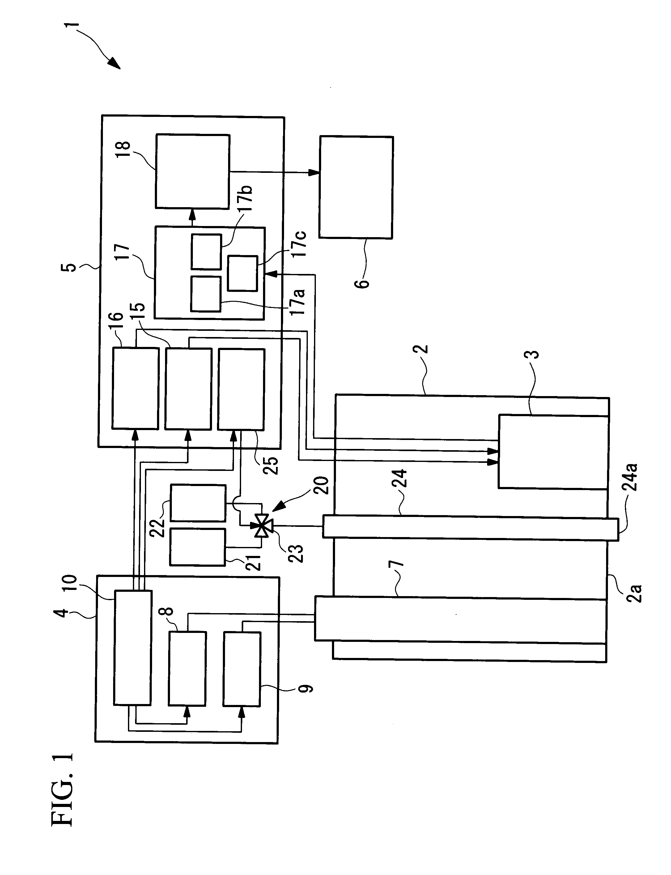
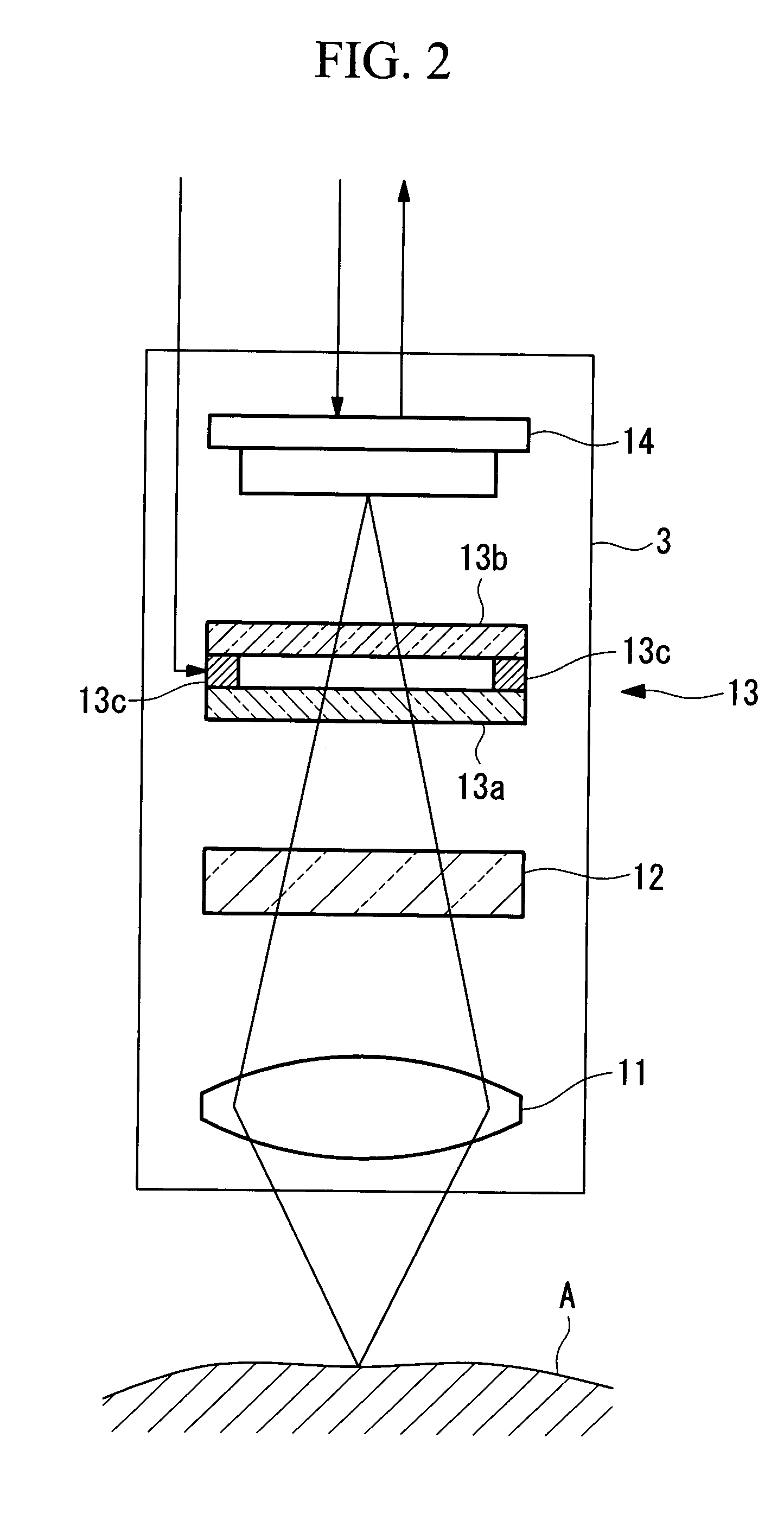
[0047]In FIG. 1, the fluorescence endoscope system 1 according to this embodiment includes an insertion part 2 for insertion into a body cavity of a living organism, an image-acquisition unit (image-acquisition part) 3 disposed in the insertion part 2, a light source unit (light source part) 4 for emitting different types of light, a feed unit 20 for supplying a liquid to be discharged from a front end 2a of the insertion part 2, a control unit (control part) 5 for controlling the image-acquisition unit 3, the light source unit 4, and the feed unit 20, and a display unit (output part) 6 for displaying an image acquired by the image-acquisition unit 3.

[0048]The insertion part 2 has outer dimensions small enough that it can be inserted into the body cavity of the living organism. The insertion part 2 accommodates the image-acquisition unit 3 and a light guide...

second embodiment

[0124]Next, a fluorescence endoscope system 1′ according to the present invention will be described with reference to FIGS. 7 to 9.

[0125]In the description of the second embodiment, the same parts as used in the above fluorescence endoscope system 1 according to the first embodiment are indicated by the same reference numerals, and no description thereof will be given.

[0126]The fluorescence endoscope system 1′ according to this embodiment differs from the fluorescence endoscope system 1 according to the first embodiment in the configuration of a light source unit 4′ and the transmittance characteristics of the tunable-spectrum device 13 and the excitation-light cutting filter 12.

[0127]Referring to FIG. 7, the light source unit 4′ of the fluorescence endoscope system 1′ according to this embodiment includes two excitation light sources 31 and 32.

[0128]The first excitation light source 31 is a semiconductor laser that emits first excitation light with a peak wavelength of 490±5 nm. Th...


PUM


Abstract

The invention provides a fluorescence endoscope system, a fluoroscopy apparatus, a fluoroscopy method, a fluorescence-information processing apparatus, and a fluorescence-information processing method which allow superior fluoroscopy by reducing the effect of autofluorescence. Provided are an excitation-light radiating part for irradiating an object under examination in a body cavity with excitation light having a wavelength included in an absorption spectrum of a fluorescent dye deposited on or absorbed in the object under examination in a state where the fluorescent dye is not deposited on or absorbed in the object under examination; a correction-information acquiring part for acquiring correction information that can be used to correct for the effect of autofluorescence of the object under examination on observation results of fluorescence emitted from the fluorescent dye deposited on or absorbed in the object under examination upon irradiation with the excitation light, based on observation results of the object under examination upon irradiation with the excitation light by the excitation-light radiating part.

Description

BACKGROUND OF THE INVENTION[0001]1. Field of the Invention[0002]The present invention relates to fluorescence endoscope systems for fluoroscopy of an object under examination, fluoroscopy apparatuses, fluoroscopy methods, fluorescence-information processing apparatuses, and fluorescence-information processing methods.[0003]This application is based on Japanese Patent Application No. 2006-221107, the content of which is incorporated herein by reference.[0004]2. Description of Related Art[0005]In endoscopy, the observation of fluorescence images is useful in diagnosis and examination of diseases because the fluorescence images provide different biological information from that provided by reflection images.[0006]For example, the concentration distribution of a disease-derived substance can be observed on a fluorescence image acquired by applying a fluorescent probe that changes from being nonfluorescent to fluorescent in response to the substance.[0007]In addition, changes in a living...

Claims


Application Information

Patent Timeline
IPC IPC(8): A61B1/06
CPCA61B1/043A61B5/0084A61B5/0071A61B1/0638A61B1/0655
Inventor TAKAOKA, HIDEYUKIHASEGAWA, AKIRA
Owner OLYMPUS CORP
Why Eureka
Try Eureka