Method for increasing tumor cell immunogenicity using heat shock protein

a technology of immunogenicity and tumor cells, applied in the field of tumor immunotherapy, can solve the problems of limited vaccine development, ineffective use of hsp for cancer immunotherapy, and inability to introduce a new gene into vaccine development, so as to reduce cancer cell growth, eradicate cancer in patients, and maximize cell delivery

Inactive Publication Date: 2008-03-27
TRAN ANNIE CHEN
View PDF5 Cites 3 Cited by
  • Summary
  • Abstract
  • Description
  • Claims
  • Application Information

AI Technical Summary

Benefits of technology

[0043] Administration of heat shocked tumor cells to a cancer patient can be achieved in various ways known to skilled practitioners. The cells can be injected intratumorly: the tumor, the placement of the needle and release of the contents of the syringe may be visualized either by direct observation (for easily accessible tumors such as surface tumors or tumors easily exposed by surgical techniques), by endoscopic visualization, or by electromagnetic imaging techniques such as ultrasound, magnetic resonance imaging (MRI), CT scans. The cells can also be administered via injection into the bloodstream using a cannula or catheter; the vein or artery is selected to maximize delivery of cells to the tumor or affected tissue. The cells can be injected into cerebro-spinal fluid (i.e., into intracisternal, intraventricular, intrathecal or subarachnoid compartments). In cystic or vesicular tumors or tissues, the cells may be delivered intracystically or intravesicularly.
[0044] It is contemplated that, in such cancer treatment, heat shocked tumor cells will be administered under the guidance of a physician. The concentration and number of cells to be administered at a given time and to a given patient may vary from, for example, about 104 to about 1010 cells per patient. Generally, the number of cells to be administered is the amount necessary to reduce cancer cell growth and / or to destroy cancer cells and / or to eradicate the cancer in the patient. The exact number is a function of the size and compactness (or diffuseness) of the particular transformed cell mass to be treated, and the distance or accessibility of the tissue to be treated from the point of administration of the cells. More than one administration may be necessary. As with any medical treatment, the supervising physician will monitor the progress of the treatment, and will determine whether a given administration is successful and sufficient, or whether subsequent administrations are needed.
[0045] The injected cells and cells junctionally coupled thereto may be destroyed by administration of a pro-drug to the patient that will target for destruction the injected cells and cells that are junctionally coupled to them. For example, the enzyme cytosine deaminase converts 5-fluorocytosine (“5FC”) into the lethal metabolite 5-fluorouracil (“5FU”). Cells that express cytosine deaminase, when exposed to 5FC, will die as a result of the formation of 5FU. Cells that do not express the deaminase but that are junctionally coupled to deaminase-expressing cells will also die. Another gene that confers drug sensitivity upon a host cell is the thymidine kinase gene, which confers sensitivity to GCV. Administration of the pro-drug may be local or systemic and may be achieved by any of the methods for administration of the cells.
[0046] Tumor regression and other parameters of successful treatment may be assessed by methods known to persons of skill in the art. Such methods include, for example, any imaging techniques that are capable of visualizing cancerous tissues (e.g., MRI), biopsies, methods for assessing metabolites produced by the cancer tissue or affected tissue in question, the subjective well-being of the patient.
[0047] The following examples are put forth so as to provide those of ordinary skill in the art with a complete disclosure and description of how to make and use the present invention, and are not intended to limit the scope of what the inventors regard as their invention nor are they intended to represent that the experiments below are all or the only experiments performed. Efforts have been made to ensure accuracy with respect to numbers used (e.g. amounts, temperature, etc.) but some experimental errors and deviations should be accounted for. Unless indicated otherwise, parts are parts by weight, molecular weight is weight average molecular weight, temperature is in degrees Centigrade, and pressure is at or near atmospheric.

Problems solved by technology

The introduction of a new gene into vaccine development is problematic, however, and the effective use of transgene-directed expression of hsp for cancer immunotherapies has not been realized.
However, the required harvesting, isolation and purification of hsp and antigenic peptides for the compositions has limited vaccine development.
Direct hyperthermia dosage to tissues to induce hsp therein has also been evaluated, but has not led to any sustained expression of hsp in tumors or surrounding tissues.

Method used


Image

  • Method for increasing tumor cell immunogenicity using heat shock protein
  • Method for increasing tumor cell immunogenicity using heat shock protein
  • Method for increasing tumor cell immunogenicity using heat shock protein

Examples


example 1

Induction of Endogenous Heat Shock Protein in HelaS3 Human Cervical Carcinoma Cells

[0048] HelaS3 cells used were commercially obtained from Cell Applications, Inc of San Diego, Calif. Cultures of HelaS3 cells (1-2×10⁶ cells / mL) were harvested (trypsinization) and subject to a heat shock condition of approximately 44° C. for approximately 2 hours by immersion in a constant temperature water bath. Cells remained healthy after subjection to heat shock in this manner. The heat shocked cells were fixed (paraformaldehyde), permeabilized (Triton X-100 / BSA in TBS) and stained with FITC-conjugated mouse anti-h ihsp70. The expression level of induced ihsp 70 in the heat shocked cells was evaluated with fluorescence activated cell sorting (FACS) flow cytometry analysis against an isotype-matched control. Flow cytometry analysis for expression of ihsp70 was also performed for log phase HelaS3 cells in culture for more than 48 hours, and harvested HelaS3 cells plated back approximately one day ...

example 2

Induction of Endogenous Heat Shock Protein in PC3 and LNCaP Prostate Cancer Cells

[0056] PC3 cells and LNCaP cells were commercially obtained from BRFF of Ijamville, Md. PC3 prostate cancer cells and LNCaP prostate cancer cells were separately cultured and harvested in the manner described above for Example 1. The PC3 and LNCaP cells and were subject to heat shock treatment at 44° C. for 1 hour via immersion in a constant temperature water bath. Flow cytometry analysis results for harvested and replated cells, irradiated cells (5000 rads) and heat shocked cells (44° C. for 1 hour) are shown in Table 5.

TABLE 5PC3 / gmPC3 / gmPC3 / gmLNCaP / gmLNCaP / gmLNCaP / gmCellHarvestedIrradiatedheat shockHarvestedIrradiatedHeat shock%1308717436TotalGeo.5569587Mean

Constitutive hsp70 expression for PC3 and LNCaP cells were approximately 95% to 99% positive for all conditions. Inducible hsp70 increased approximately 14 fold (geometric mean) in heat shocked PC3 cells. In the case of the above prostate cell...

example 3

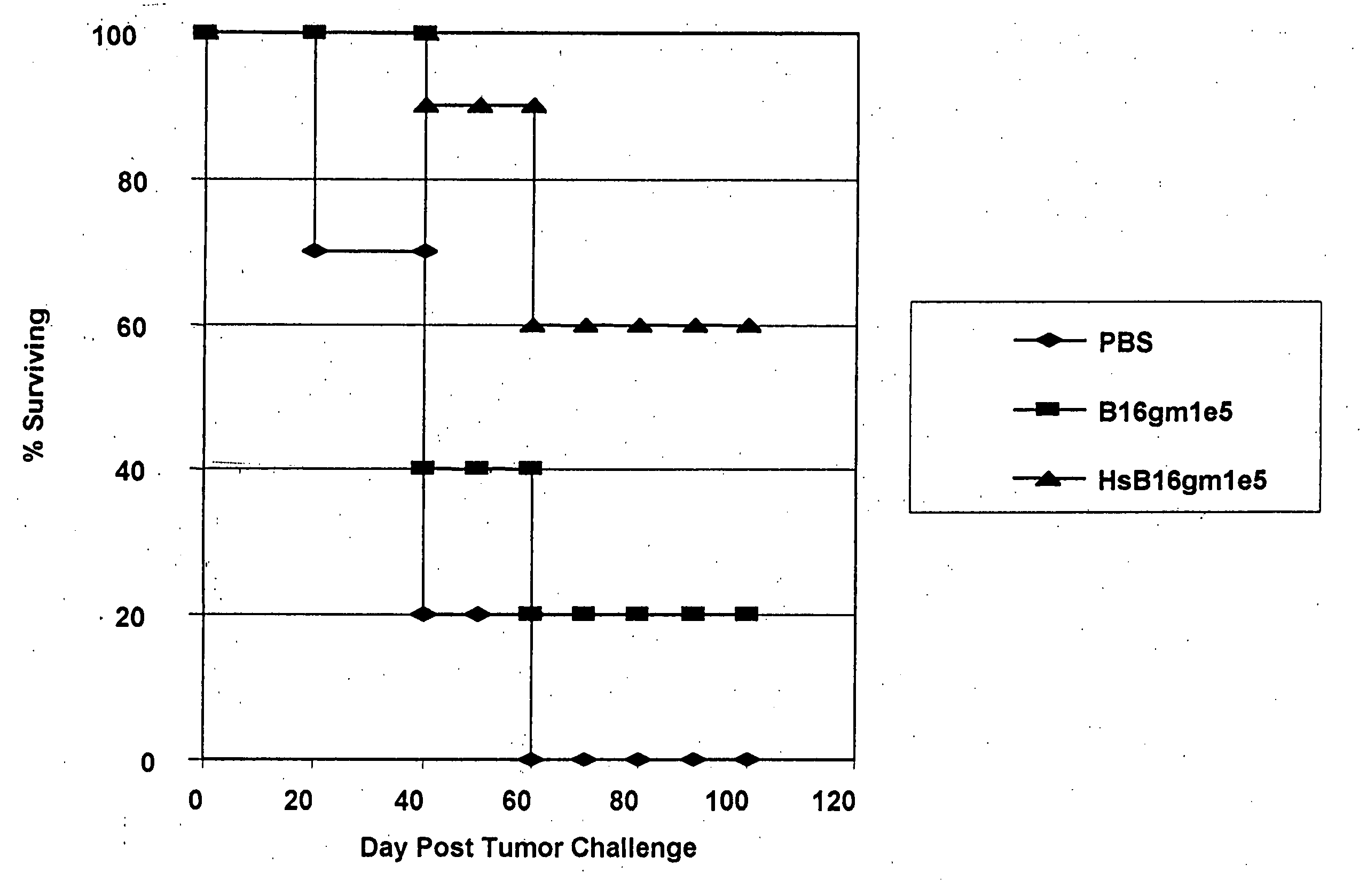
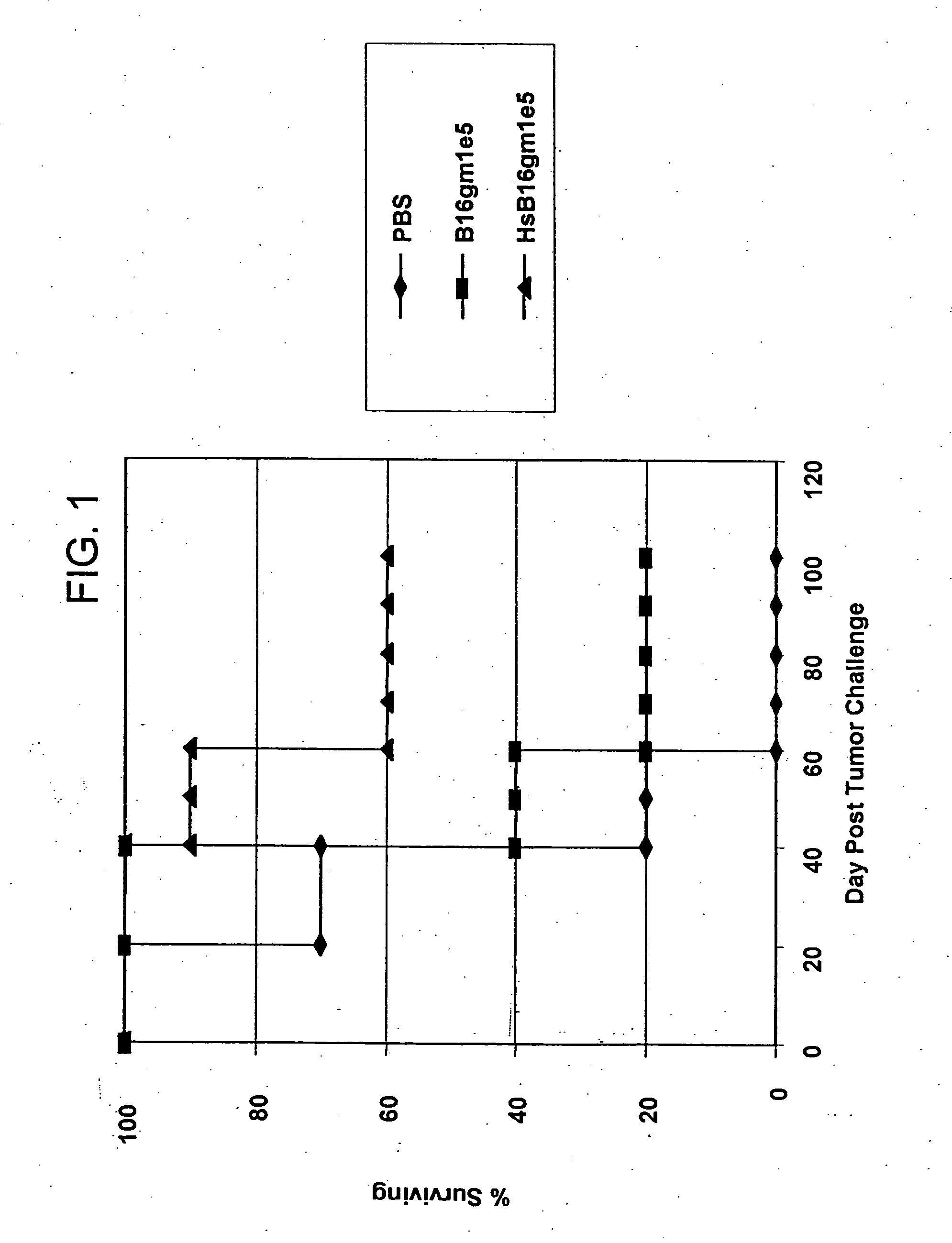
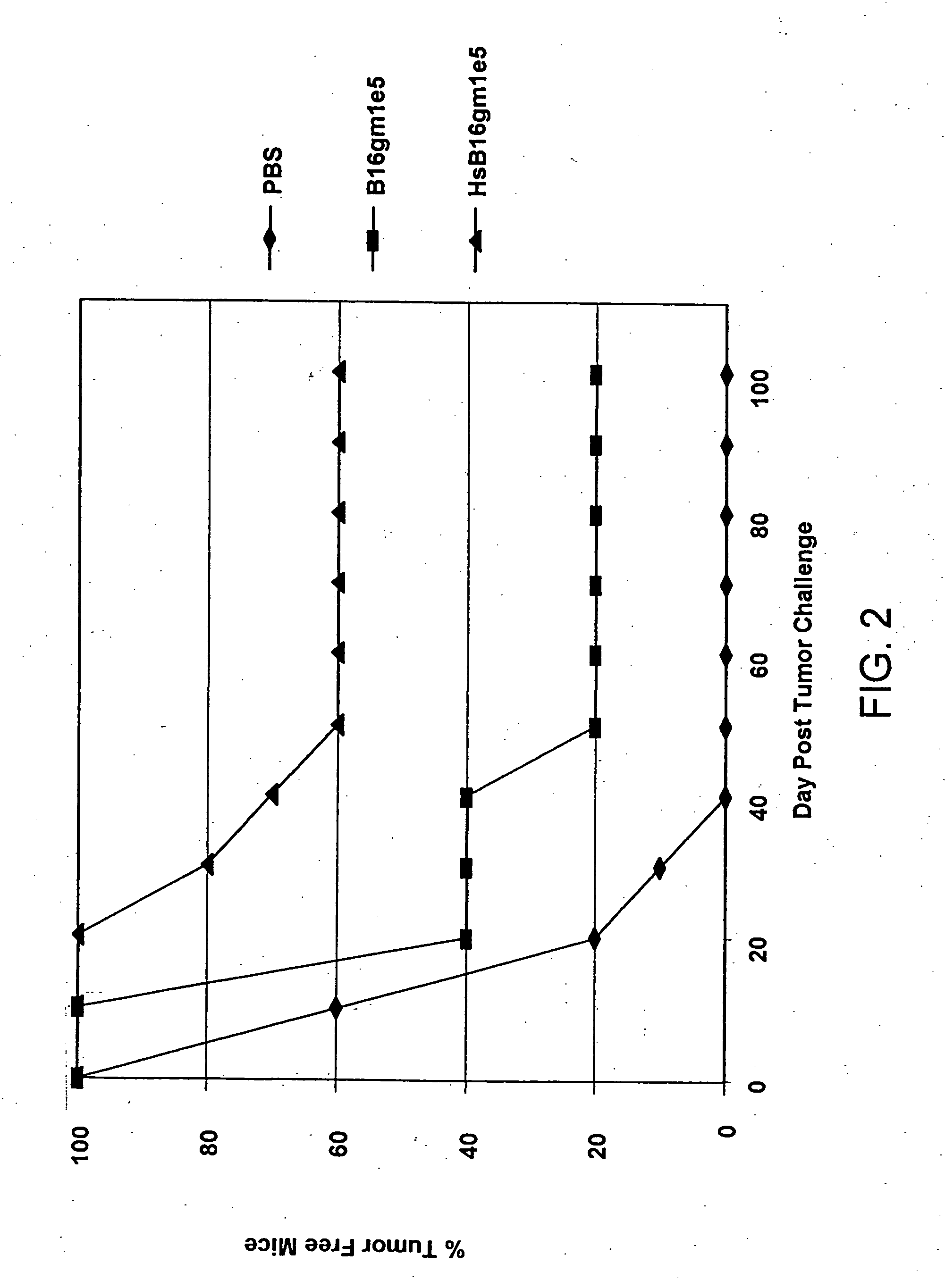
Induction of Endogenous Heat Shock Protein in B16 Melanoma Cells

[0057] B16 melanoma cells were prepared according to Vile et al., Cancer Res. 53:962 (1993). B16 melanoma cells were cultured and harvested in the manner described above for Example 1 and were subject to heat shock treatment at 44° C. for one half hour (30 minutes) by immersion in a constant temperature water bath. Flow cytometry analysis results for harvested and replated cells and heat shocked cells are shown in Table 6.

TABLE 6B16gmB16 / gmCellHarvestedHeat shocked% Total7.0683.56Geo. Mean29.8890.43

Heat shock treatment in this case provided an approximately 3-fold increase in ihsp70 expression in heat shocked cells over harvested cells, with greater than 90% ihsp70 expression in the heat shocked cells.


PUM

PropertyMeasurementUnit
diameteraaaaaaaaaa
temperatureaaaaaaaaaa

Abstract

A method for induction of endogenous heat shock protein in tumor cells using a simple heat shock treatment which provides a simple and inexpensive method for augmenting antitumor vaccine potency. The method comprises administering to a mammal tumor cells which have been subject to a heat shock condition sufficient to cause induction of endogenous heat shock protein therein. Also disclosed is a composition for enhancing tumor cell immunogenicity comprising a therapeutically effective amount of attenuated tumor cells which have been subject to a heat shock condition.

Description

CROSS-REFERENCE [0001] This application claims the benefit of U.S. provisional patent application No. 60 / 606,429, filed Jul. 7, 2001.FIELD OF THE INVENTION [0002] This invention pertains to systems and methods for tumor immunotherapy. More particularly, the invention pertains to a method for increasing tumor cell vaccine immunogenicity wherein tumor cells are subject to heat shock conditions causing induction of endogenous heat shock protein therein prior to administering the tumor cells to a patient. BACKGROUND OF THE INVENTION [0003] Vaccines based on autologous tumor cells have long been of interest for promoting tumor immunogenicity. Unmodified tumor cells are poor stimulators of immunity, and cellular tumor vaccines have generally involved some form of gene transfer modification to stimulate host antitumor responses. Gene transfer of MHC, co-stimulatory, chemokine, and cytokine genes in tumor cells are known. In the case of cytokines, transgene modified tumor cells expressing I...

Claims


Application Information

Patent Timeline
Patent Type & Authority Applications(United States)
IPC IPC(8): A61K35/12A61P35/00A61P37/00A61K39/00A61K41/00C12N5/09
CPCA61K39/0011A61K41/00C12N2501/07A61K2039/5156C12N5/0693A61K2039/5152A61P35/00A61P37/00
Inventor TRAN, ANNIE-CHEN
Owner TRAN ANNIE CHEN
Why Eureka
Try Eureka