Method for providing a 3D X-ray image dataset of a patient's heart

Inactive Publication Date: 2008-06-12
SIEMENS HEALTHCARE GMBH
View PDF9 Cites 8 Cited by
  • Summary
  • Abstract
  • Description
  • Claims
  • Application Information

AI Technical Summary

Benefits of technology

[0011]The object of the present invention is to develop the approaches known from the prior art so as to achieve an as good as possible imaging quality,

Problems solved by technology

The 2D X-ray images can not, therefore, be readily reconstructed from

Method used


Image

  • Method for providing a 3D X-ray image dataset of a patient's heart
  • Method for providing a 3D X-ray image dataset of a patient's heart
  • Method for providing a 3D X-ray image dataset of a patient's heart

Examples


Example

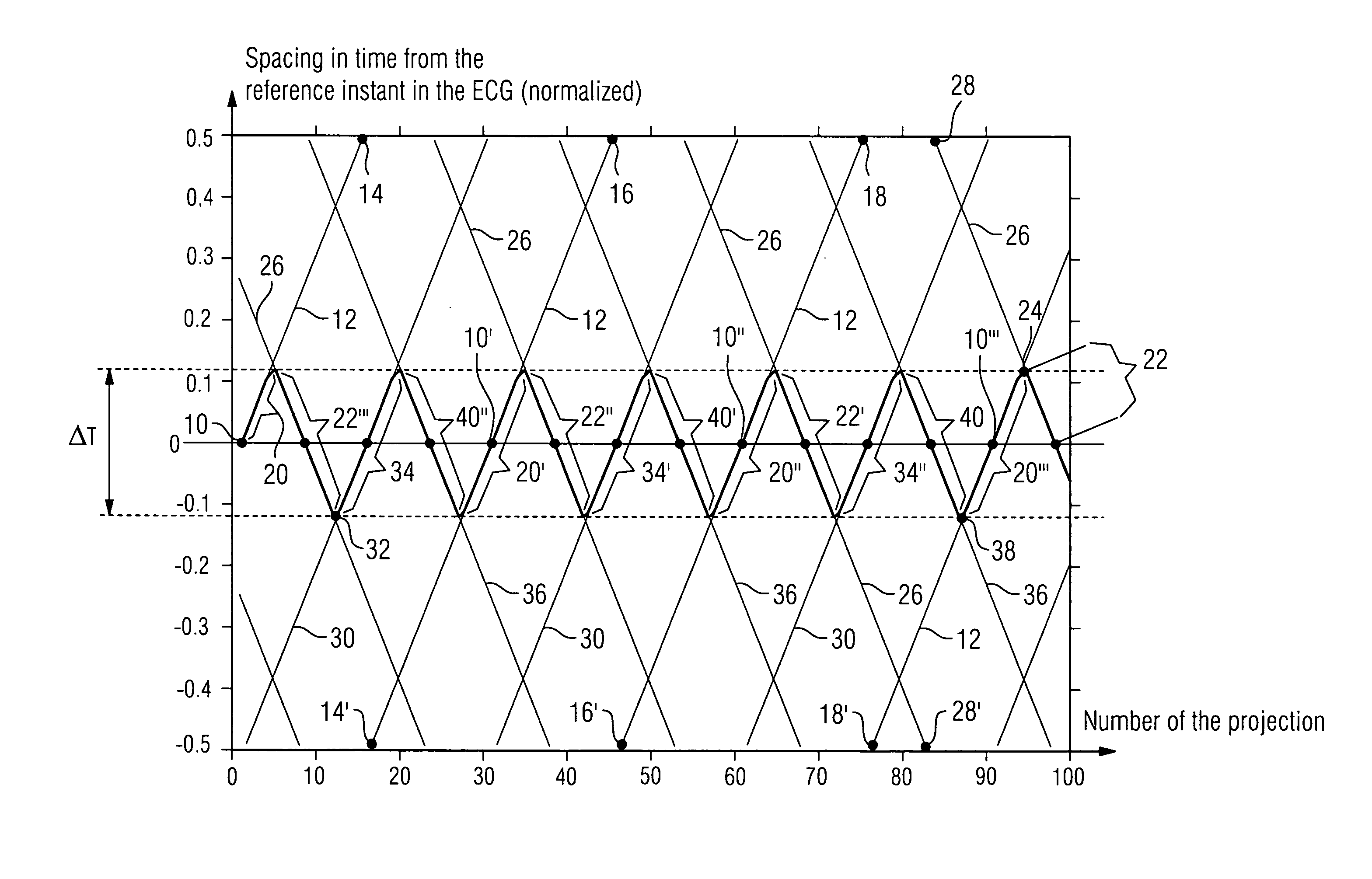
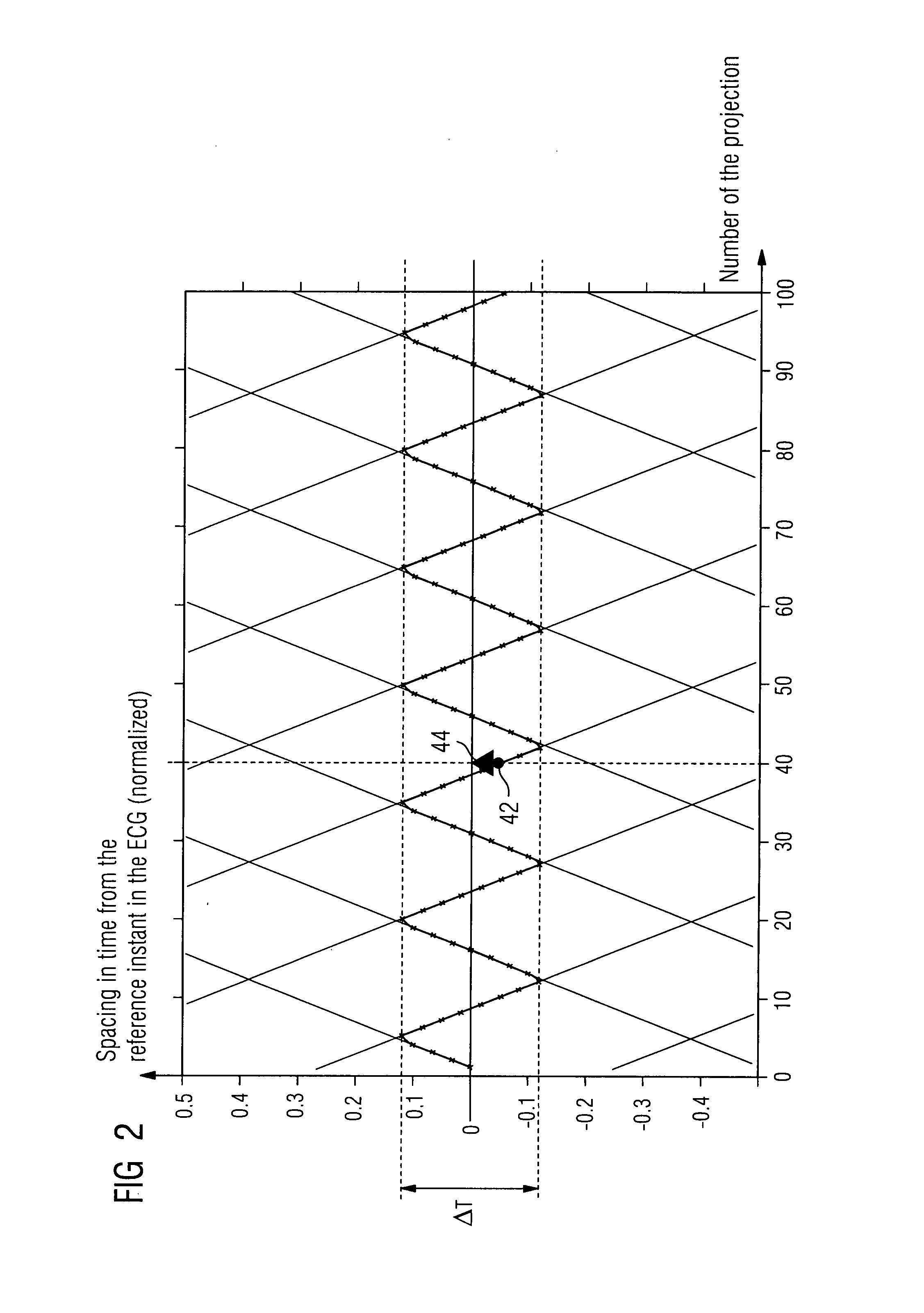
[0042]Assume that a patient is located in a manner known per se in a C-arm X-ray system. Attached at opposite points to the X-ray C-arm are an X-ray source and an X-ray detector. The X-ray C-arm can be moved to well-defined angular positions. An image from a specific angular position is referred to as a “projection”. The projections are in FIG. 1 counted successively along the x-axis. An electrocardiogram is taken of the patient. The regular heartbeat results, as is known, in regular structures in the electrocardiogram. A reference structure can be determined within each period and a cardiac phase determined in relation to said reference structure. The cardiac phase is the spacing in time from the respective reference instant which the reference structure has. Said spacing is in FIG. 1 plotted on the y-axis. The spacing in time between two such reference instants has here been normalized at “1”. If the spacing in time is in each case measured relative to the closest reference instan...


PUM


Abstract

X-ray images are recorded of a patient's heart and the heartbeat phase is registered as that is done. The heartbeat phases are coarsely divided into intervals and all X-ray images that have been assigned heartbeat phase from the interval are used for reconstructing a 3D image dataset. The movement fields of the other 3D image datasets are then calculated for one of said 3D image datasets. Movement fields are vector fields indicating the movements of similar structures from one local area to the other. A departure is then made from the coarse interval division, and for each heartbeat phase a movement field is interpolated individually or at least for fairly short intervals from the movement fields determined in advance, which field is used for generating a deformed 3D image dataset that has been imaged onto a reference heartbeat phase. The deformed 3D image datasets are then added together.

Description

CROSS REFERENCE TO RELATED APPLICATIONS[0001]This application claims priority of German application No. 10 2006 051 919.1 filed Oct. 31, 2006, which is incorporated by reference herein in its entirety.FIELD OF THE INVENTION[0002]The invention relates to a method for providing a 3D X-ray image dataset of a patient's heart.BACKGROUND OF THE INVENTION[0003]A 3D image dataset is a three-dimensional field of volumetric pixels (voxels) each of which has been assigned a gray or color value.[0004]To generate a 3D image dataset, a sequence of individual 2D images is usually recorded. The 3D image dataset is generated from the individual 2D images with the aid of what is termed a reconstruction process; with the aid of, for instance, filtered back-projection.[0005]A particular feature associated with cardiac imaging is that the heart is in constant motion. The time usually required to record a sequence of X-ray images is longer than the heartbeat period. The 2D X-ray images can not, therefore...

Claims


Application Information

Patent Timeline
IPC IPC(8): A61B6/00A61B5/352
CPCA61B5/7289A61B6/032A61B6/466G06T2207/30048A61B6/5288G06T7/20A61B6/503Y10S378/901
Inventor BOESE, JANHORNEGGER, JOACHIMLAURITSCH, GUNTERPRUMMER, MARCUS
Owner SIEMENS HEALTHCARE GMBH
Why Eureka
Try Eureka