Method to increase class i presentation of exogenous antigens by human dendritic cells

a human dendritic cell and exogenous technology, applied in the field of human dendritic cell class i presentation, can solve the problems of overcoming this natural immunity, inducing apcs, and often growing tumors, and achieve the effect of enhancing an existing immune response and enhancing mhc-class i processing

Inactive Publication Date: 2008-07-17
NORTHWEST BIOTHERAPEUTICS INC
View PDF1 Cites 12 Cited by
  • Summary
  • Abstract
  • Description
  • Claims
  • Application Information

AI Technical Summary

Benefits of technology

[0024]In another embodiment, the invention provides a method for producing a tumor cell proliferation growth inhibiting response, which comprises administering, to a cancer patient in need thereof, an effective amount of activated T cells, in which the T cells were activated in vitro. The in vitro activation includes exposure of human dendritic cells to a tissue associated, tissue specific, tumor associated, tumor specific antigen or antigenic fragments thereof in combination with BCG, either in combination with or without LPS, to enhance MHC-class I processing. In a further embodiment

Problems solved by technology

However, tumors frequently grow and metastasize, overcoming this natural immunity.
Various methods for immunotherapy directed to a number of particular cancers have been suggested to enhance this natural immune response, however, the primary difficulty has been inducing APCs to present soluble human tumor associated or tissue specific antigens via MHC-class I. Recent experiments have demonstrated in murine systems, that activation of CTLs in vitro can confer a potent protection from growth of syngeneic tumors in vivo (Fields et al., Proc. Natl. Acad. Sci.
However, experiments in the murine immune system are not completely predictive of human immune responses.
To date there are no therapeutic methods that successfully elicits an effective human CTL immunotherapeutic response against primary or metastatic cancer using APCs incubated with a soluble protein or proteinaceous antigen.
Although monocytes and B cells have been shown to be competent APC, their antigen presenting capacities in vitro appear to be limited to the re-activation of previously sensitized T cells.
Hence, they are not capable of directly activating functionally naive or unprimed T cel

Method used


Image

  • Method to increase class i presentation of exogenous antigens by human dendritic cells
  • Method to increase class i presentation of exogenous antigens by human dendritic cells
  • Method to increase class i presentation of exogenous antigens by human dendritic cells

Examples


example 1

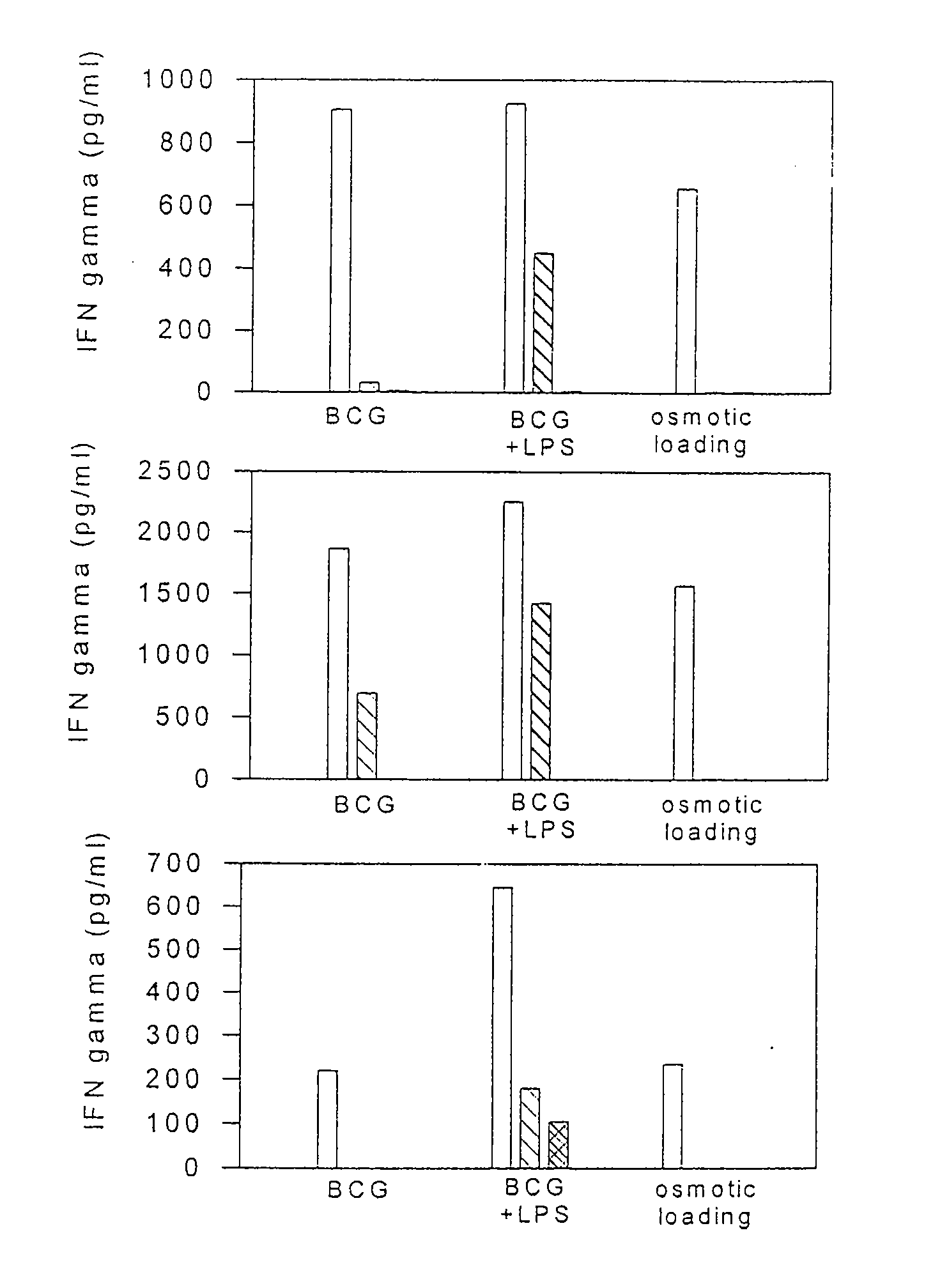
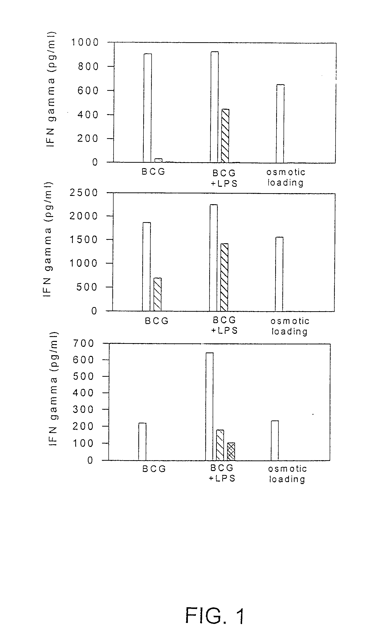
[0098]The following example describes the isolation and culturing of human dendritic cells. Isolated dendritic cells were contacted with tumor cell lysate, and partially purified tumor cell lysate in combination with BCG to demonstrate the stimulation of antigen-specific cytotoxic T cell response.

Culture of Patient Dendritic Cells

[0099]Cultures of human DCs were established as described previously herein and in U.S. Pat. No. 5,788,963 (incorporated herein by reference). Briefly, peripheral blood mononuclear cells (PBMC) were obtained from leukocyte-enriched “buffy coats” by standard centrifugation on Ficoll-Paque (Pharmacia, Uppsala, Sweden). Plastic-adherent PBMCs (about 1 hour at 37° C.) were cultured for 6 to 7 days in AIM-V either supplemented or not with 2% autologous serum, 50 U / ml penicillin, 50 μg / ml streptomycin, 2 mM L-glutamine, 10 mM HEPES, 0.1 mM non-essential amino acids, and 1 mM pyruvate (referred to as “culture medium”; all from Boehringer Ingelheim, Biowhittaker, V...

example 2

[0109]The present example examines whether dendritic cells retain their function after cryopreservation. This characteristic is particularly important because immunotherapy approaches involve multiple treatments and it is preferable that all the DCs for each patient be prepared and loaded with antigen during a single preparation, then aliquoted and cryopreserved for subsequent infusion. It was possible that the freezing and thawing of the DCs may limit their effectiveness as CD8+ T cell activators.

[0110]Dendritic cells were isolated from PBMC of a prostate cancer patient and cultured, as described above, for 7 days in the presence of 500 U / ml GM-CSF and 500 U / ml IL-4. On day 7, the isolated DC's were harvested and cryopreserved using 90% fetal calf serum and 10% dimethylsulfoxide. The cryopreserved DC's were subsequently stored frozen for a period of time, thawed in a 37° C. water bath and transferred to a 15 ml polypropylene tube and centrifuged at 1200 rpm for 5 min. The thawed DC...

example 3

[0113]In this example dendritic cells isolated from a cancer patient were isolated and treated with various concentrations of BCG. After several days of culture, the DCs were tested for 1) the capacity to uptake particles by pinocytosis, and 2) the surface expression of certain dendritic cell maturation markers, including HLA-DR, CD86, CD40, CD83, CD80 and HLA-class I.

[0114]Dendritic cells were isolated from patient 57 as described above. The isolated cells (1-5×10⁶) were cultured for about 6 days in eight T-75 flasks. BCG (1×10⁶ units / ml) was added to duplicate flasks to achieve a dilution of 1:250, 1:2,500, or 1:25,000. No BCG was added to the two remaining culture flasks. A first set of culture flasks comprising DCs with no BCG, or with 1:250; 1:2,500: or 1:25,000 BCG dilution was harvested after a 48 or 72 hour incubation. The duplicate set of culture flasks was harvested after a total of 72 hours in culture. Each DC culture was analyzed for: (1) the capacity to uptake FITC / Dext...


PUM


Abstract

Methods and compositions for use of human dendritic cells to activate T cells for immunotherapeutic responses against primary and metastatic cancer are disclosed. In one embodiment, human dendritic cells exposed to a tumor associated antigen, or an antigenic fragment thereof in combination with bacillus Calmette-Guerin (BCG), are administered to a cancer patient to activate a predominantly CD8+T cell response in vivo. In an alternate embodiment, human dendritic cells are exposed to a tumor associated antigen or a specific antigenic peptide in combination with BCG in vitro and incubated or cultured with primed or unprimed T cells to activate a predominantly CD8+T cell response in vitro. The activated T cells are then administered to a cancer patient. Antigen in combination with BCG is processed by dendritic cells through the MHC-CLASS I compartment which provides for a predominantly CD8+T cell response. The addition of LPS provides for a greater number of mature dendritic cells enhancing the T cell response to antigen. Methods and compositions for human dendritic cells with extended life span and cryopreserved dendritic cells are disclosed.

Description

CROSS REFERENCES TO RELATED APPLICATIONS[0001]This application claims priority to U.S. Provisional Patent Application No. 60 / 203,758, filed May 12, 2000, incorporated herein by reference in its entirety.BACKGROUND OF THE INVENTION[0002]It is well established that the immune system can function to kill tumor cells, including both primary and metastatic cancer cells. Indeed, evidence that the immune system recognizes the presence of neoplastic cancerous cells is supported by the existence of infiltrating lymphocytes in tumor tissues (Haskill et al., Contemp. Top. Immunobiol. 8:107-170 (1978); Vose and Moore, Semin. Hematol. 22:27-40 (1985)). Despite the presence of immune cells, tumors often prevail and not only survive but metastasize to distant sites with unrestricted growth.[0003]Recent advances in the understanding of T cell activation and recognition of target cells has permitted some progress in the development of T cell mediated cancer immunotherapy (Schwartz, Cell 71:1065-1068...

Claims


Application Information

Patent Timeline
IPC IPC(8): A61K35/12C12N5/06A61P43/00A61K35/50A61K39/00A61P35/00A61P35/04A61P37/04C12N5/0784
CPCA61K39/0011A61K2039/5158A61K2039/5154A61P13/08A61P35/00A61P35/04A61P37/04A61P43/00A61K39/001193A61K39/001194A61K39/001195
Inventor SALGALLER, MICHAELBOYNTON, ALTON
Owner NORTHWEST BIOTHERAPEUTICS INC
Why Eureka
Try Eureka