System and method for photoacoustic tomography of joints

a photoacoustic tomography and sample technology, applied in the field of system and method of photoacoustic tomography of samples, can solve the problems of low spatial resolution of traditional optical imaging modalities, irreversible structural injury, and demonstrating the time-integrated record of joint damag

Inactive Publication Date: 2008-07-24
RGT UNIV OF MICHIGAN
View PDF98 Cites 115 Cited by
  • Summary
  • Abstract
  • Description
  • Claims
  • Application Information

AI Technical Summary

Problems solved by technology

Traditional optical imaging modalities suffer from low spatial resolution in imaging subsurface biological tissues due to the overwhelming scattering of light in tissues.
Despite the hypervascularization, the rheumatic synovium appears to be a hypoxic environment that is thought to be caused by an imbalance between local metabolic rate and synovial vascular supply.
This modality, however, can only demonstrate the time-integrated record of joint damage that tends to develop late in the course of the diseases and which constitutes irreversible structural injury.
Furthermore, CR is fundamentally limited by its inherent inability to visualize articular soft tissues involved in the pathophysiology of arthritis.
The disadvantages of MRI include its high cost, lack of access compared to CR, lack of standardization, and poor reproducibility.
Contrast agents containing gadolinium, imperative in MRI imaging studies evaluating inflammatory arthritis, have been found to cause a very morbid condition called nephrogenic systemic fibrosis in patients with renal compromise, thus limiting its availability to this patient population.
Moreover, the long examination time with ensuing patient discomfort makes it difficult to use MRI repeatedly and, in some cases, impossible to use at all.
However, the mechanical contrast exhibited by US is not sensitive to the molecular conformation and functional changes in biological tissues (e.g., hemoglobin oxygenation).
Moreover, the performance of US is highly dependent on the skills of the operator and hence is difficult to repeat and standardize for clinical trials.
However, due to the overwhelming scattering of light in biological tissues, current optical technologies cannot delineate a joint as a whole organ with satisfactory imaging quality for clinical applications.
Confocal microscopy can achieve ˜1 micrometer spatial resolution, but its imaging depth is limited to ˜0.5 mm in biological tissues.
Both of these two techniques, as well as Laser Doppler imaging, are not able to provide optical information in subsurface synovial tissues in a joint when applied non-invasively.
However, the imaging resolution of DOT is poor and the reconstruction is ill posed (unstable) due to the diffusive nature of the imaging signals.
Up to now, optical imaging of joints based on DOT cannot achieve spatial resolution better than 5 mm, which is insufficient for evaluating the small joint structures of the hands and feet.

Method used


Image

  • System and method for photoacoustic tomography of joints
  • System and method for photoacoustic tomography of joints
  • System and method for photoacoustic tomography of joints

Examples


Embodiment Construction

[0033]As required, detailed embodiments of the present invention are disclosed herein; however, it is to be understood that the disclosed embodiments are merely exemplary of the invention that may be embodied in various and alternative forms. The figures are not necessarily to scale, some features may be exaggerated or minimized to show details of particular components. Therefore, specific structural and functional details disclosed herein are not to be interpreted as limiting, but merely as a representative basis for teaching one skilled in the art to variously employ the present invention.

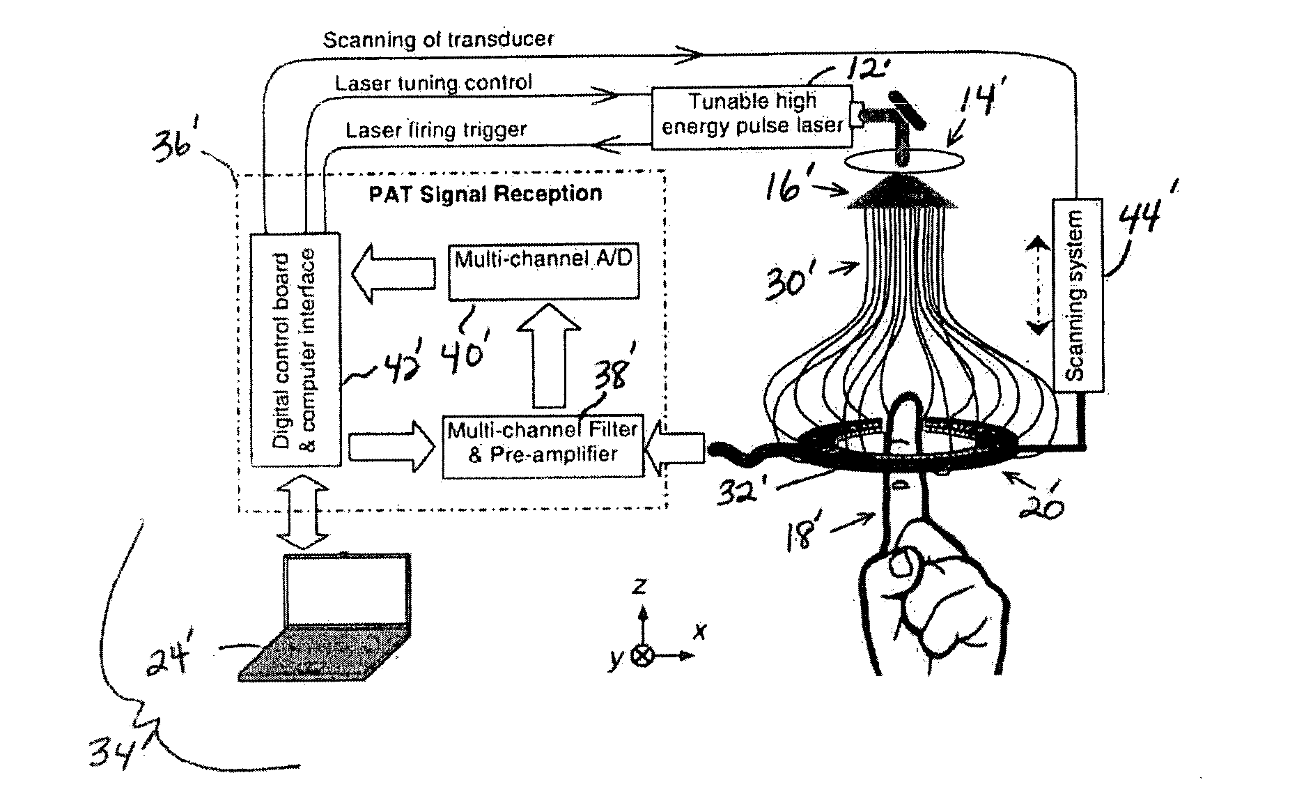
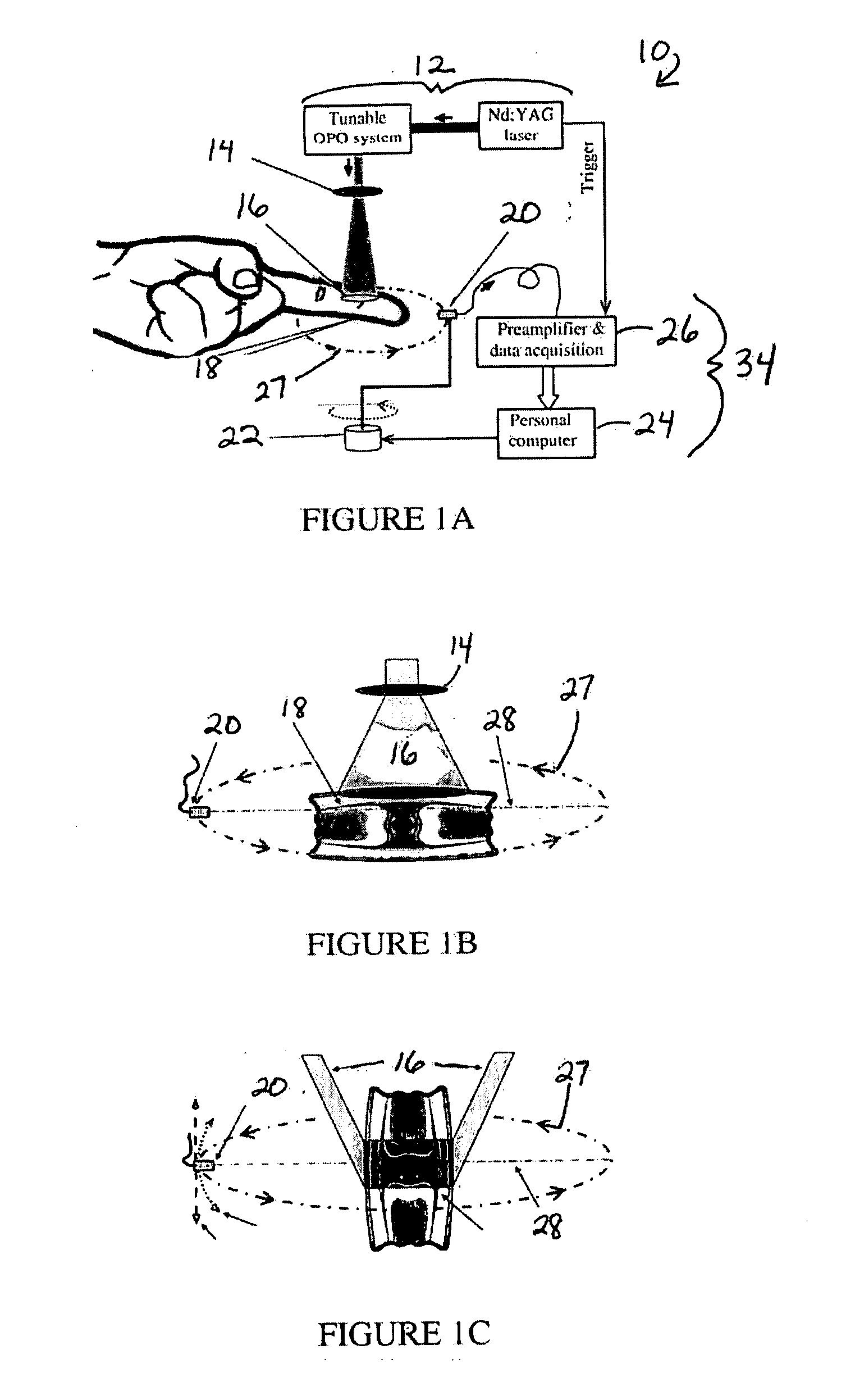
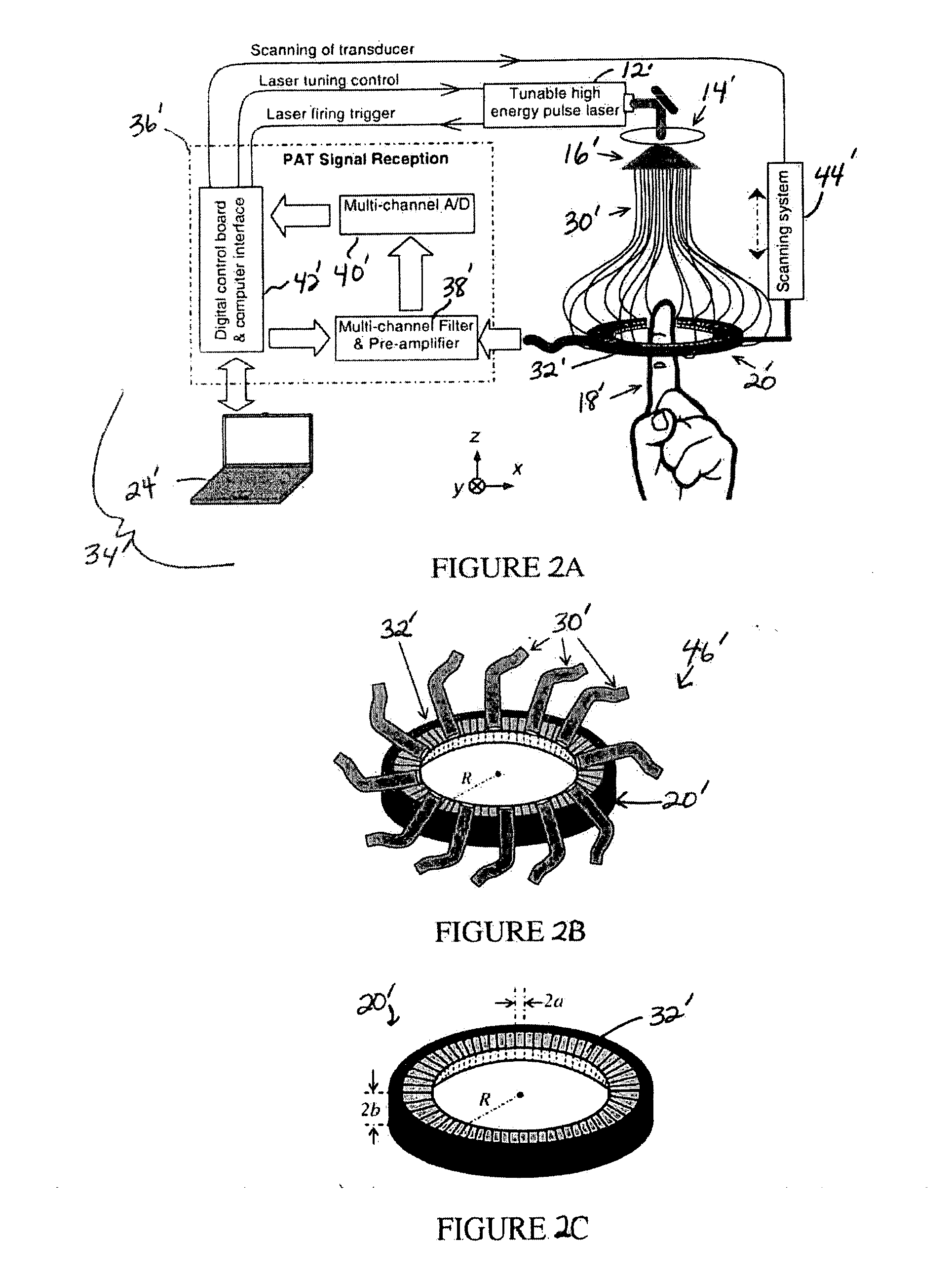
[0034]The present invention includes a system and method for PAT of joints. Optical signals employed in PAT to generate ultrasonic waves are sensitive to molecular conformations of biological tissues including both deoxy- and oxy-hemoglobin, as well as to soft tissue changes such as hypervascularization. Both abnormal oxygen state and, as a consequence of increased angiogenesis, hypervascularizat...


PUM

PropertyMeasurementUnit
imaging depthaaaaaaaaaa
tunable wavelengthaaaaaaaaaa

Abstract

A system and method for photoacoustic tomography of a sample, such as a mammalian joint, includes a light source configured to deliver light to the sample, an ultrasonic transducer disposed adjacent to the sample for receiving photoacoustic signals generated due to optical absorption of the light by the sample, a motor operably connected to at least one of the sample and the ultrasonic transducer for varying a position of the sample and the ultrasonic transducer with respect to one another along a scanning path, and a control system in communication with the light source, the ultrasonic transducer, and the motor for reconstructing photoacoustic images of the sample from the received photoacoustic signals.

Description

CROSS-REFERENCE TO RELATED APPLICATIONS[0001]This application claims the benefit of U.S. provisional application Ser. No. 60 / 881,123 filed Jan. 18, 2007, which is incorporated by reference herein.BACKGROUND OF THE INVENTION[0002]1. Field of the Invention[0003]This invention relates to a system and method for photoacoustic tomography of samples, such as mammalian joints.[0004]2. Background Art[0005]Photoacoustic tomography (PAT) may be employed for imaging tissue structures and functional changes, and describing the optical energy deposition in biological tissues with both high spatial resolution and high sensitivity. PAT employs pulsed electromagnetic signals to generate ultrasonic waves. In PAT, a short-pulsed electromagnetic source—such as a tunable pulsed laser source, pulsed radio frequency (RF) source, or pulsed lamp—is used to irradiate a biological sample. The photoacoustic (ultrasonic) waves excited by thermoelastic expansion are then measured around the sample by high sensi...

Claims


Application Information

Patent Timeline
Patent Type & Authority Applications(United States)
IPC IPC(8): G01N29/00
CPCA61B5/0073A61B5/4528A61B5/0095
Inventor WANG, XUEDINGCHAMBERLAND, DAVIDCARSON, PAULFOWLKES, BRIANBUDE, RONROESSLER, BLAKERUBIN, JONATHANKOTOV, NICHOLAS A.
Owner RGT UNIV OF MICHIGAN
Why Eureka
Try Eureka