Prevention of chlamydia infection using a protective antibody

a technology of chlamydia infection and protective antibody, which is applied in the field of new anti-chlamydia antibodies, can solve the problems of serious pneumonia, chlamydia infections are particularly serious health threats to newborns, and the risk of tubal obstruction and infertility in the pid, so as to inhibit the ability of chlamydia to bind and inhibit the binding of emp2

Inactive Publication Date: 2008-07-31
RGT UNIV OF CALIFORNIA
View PDF19 Cites 3 Cited by
  • Summary
  • Abstract
  • Description
  • Claims
  • Application Information

AI Technical Summary

Benefits of technology

[0025]In fourth aspect, the invention provides methods of identifying EMP2 Chlamydia inhibitors (e.g., EMP2 polypeptides, anti-EMP2 antibodies, and EMP2 siRNA) which would be of use in treating or preventing infection with Chlamydia. In some embodiments, a host cell which expresses EMP2 is contacted with the candidate agent and the effect of the agent on the ability of Chlamydia to enter or bind to the host cell is determine

Problems solved by technology

In women, the pathogen can cause pelvic inflammatory disease (PID) with a risk of tubal obstruction and infertility.
Chlamydial infections are a particularly serious health threat to newborns who contract occular infections at birth from infected birth canals of their mothers.
If untreated, almost 50% of these children develop inclusion conjunctivitis and 20% devel

Method used


Image

  • Prevention of chlamydia infection using a protective antibody
  • Prevention of chlamydia infection using a protective antibody
  • Prevention of chlamydia infection using a protective antibody

Examples


example 1

Methods for Studying Chlamydial Infectivity and EMP2 Chlamydial Inhibitors

[0138]A. Endometrial cell lines and Chlamydia strains. The human endometrial adenocarcinoma cell line HEC1A (HTB112, ATCC, Manassas, Va.) was cultured in McCoy 5a media (Invitrogen, Carlsbad, Calif.) supplemented with 10% fetal calf serum (Hyclone, Logan, Utah) at 37° C. in a humidified 5% CO2 and passaged every 7 days. EMP2-modulated HEC1A sublines were stable transfectants with expression plasmids for GFP, a human EMP2-GFP fusion protein, or a human EMP2-specific ribozyme (HEC1A-GFP, HEC1A-hEMP2, and HEC1A-hRV2). EMP2 protein expression levels relative to HEC1A-GFP were 1.0, 8.7, and 0.2, respectively.

[0139]An 8-strain mix of human C. trachomatis (serovars D, E, F, and K) and C. muridarum were purified, aliquoted, and stored at −80° C. until ready for use (see, Caldwell, H. D., et al., Infect Immun 31, 1161-76 (1981). All Chlamydia samples were made in Eagle MEM (Invitrogen) with 10% fetal calf serum (Atlant...

example 2

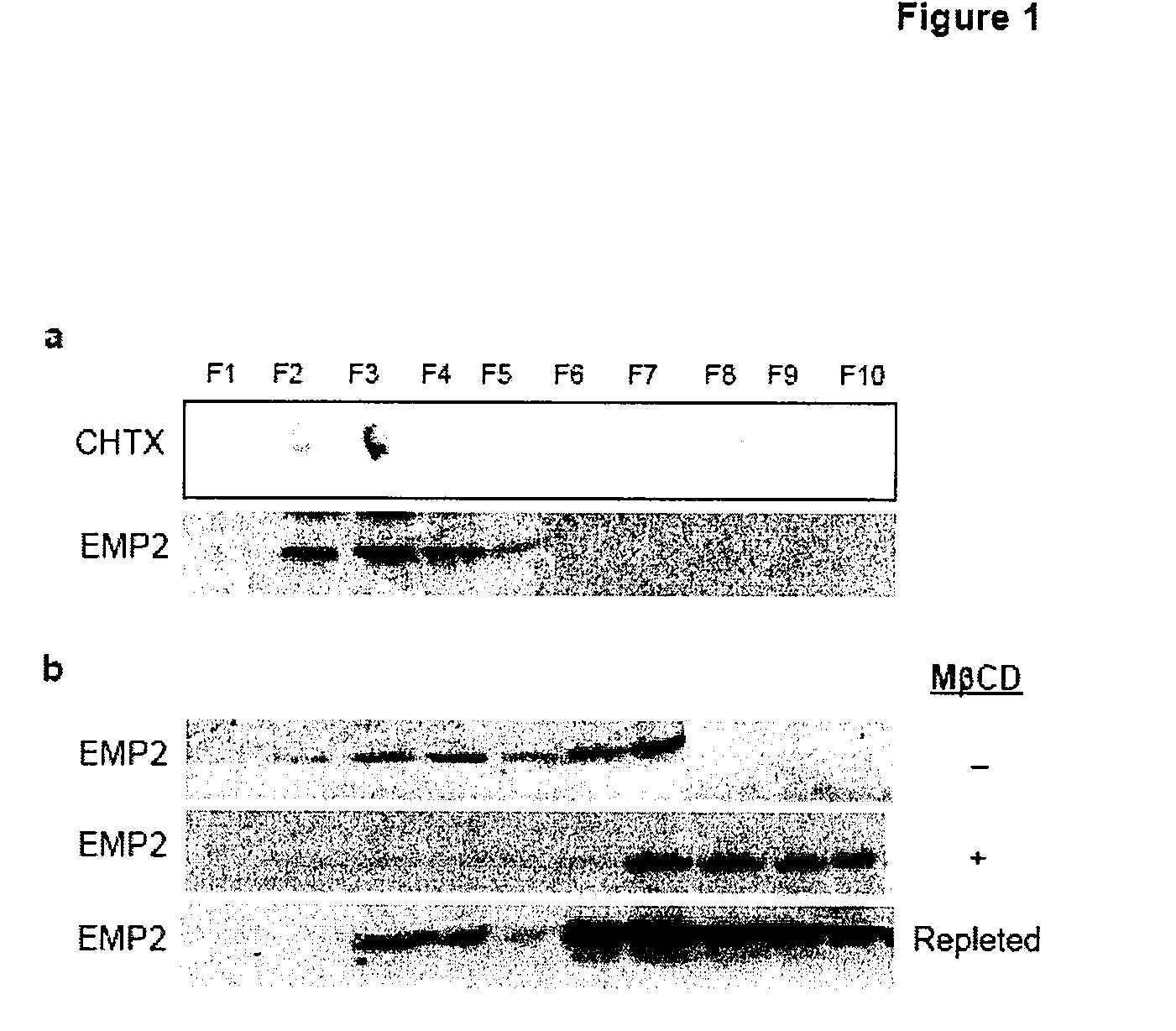
Localization of EMP2 to Lipid Rafts

[0147]To assess whether EMP2 is localized to lipid raft domains in endometrial cells (a Chlamydial host target), EMP2 was evaluated in the HEC1A human endometrial cancer cell line by lipid raft fractionation with Brij 58 and Triton X-100. In HEC1A cells, EMP2 localized to the light, detergent-resistant gradient fractions coinciding with GM1 ganglioside, a lipid raft component (FIG. 1a,b). To confirm the localization of EMP2 to lipid rafts, lysates were prepared in 1% Triton X-100 in the presence or absence of methyl-β-cyclodextrins (MβCD) that selectively deplete cholesterol from cellular membranes and causes loss of protein localization into lipid rafts. In 1% Triton X-100, EMP2 localized to both light, detergent-resistant fractions 3-4 as well as dense fractions 6-7 (FIG. 1c). When cells were incubated for 60 minutes with MβCD in serum free conditions, EMP2 expression completely shifted to soluble, dense fractions in the presence of 1% Triton X-1...

example 3

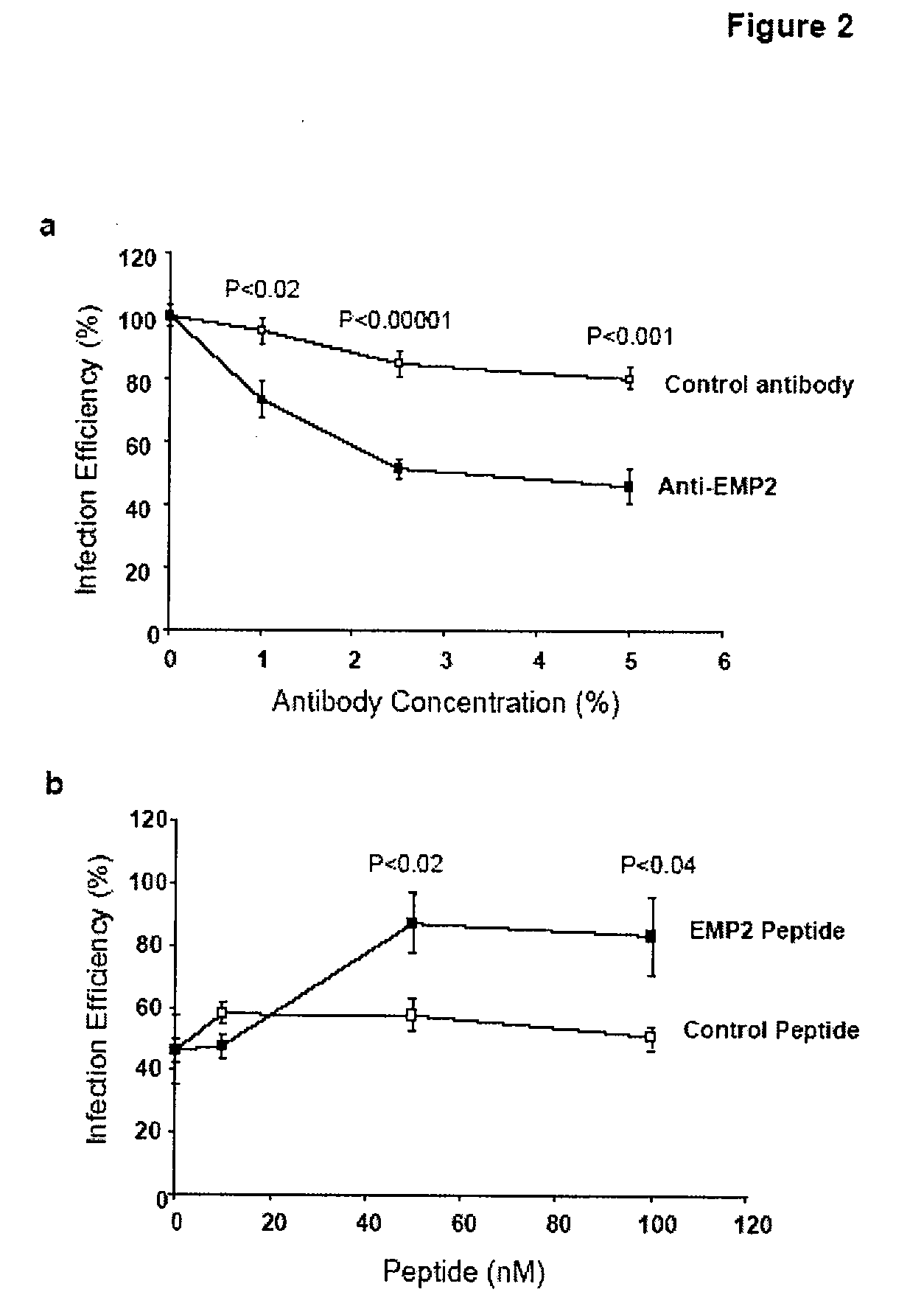
Effect of Anti-EMP2 Antibody on Chlamydia Infectivity

[0148]The lipid raft localization of EMP2 in endometrial cells and its control of lipid raft trafficking by integrins, caveolins, and glycosylphosphatidyl inositol-linked proteins (18, 20, 21), raised the possibility that EMP2 might directly or indirectly affect Chlamydial infectivity. To begin testing this hypothesis, anti-EMP2 antibody (specific for the 2nd extracellular loop of EMP2) was added to HEC1A cell cultures, then incubated with C. trachomatis, and infection was measured (Chlamydial inclusions, expressed as “infection efficiency”, % inclusions relative to HEC1A cells without antibody treatment) (FIG. 2a). Anti-EMP2 antibody produced a dose-dependent inhibition of infection efficiency (reaching less than 50% of HEC1A cells without antibody), at levels that were highly significant compared to control antibody. To determine if the observed inhibition was due to EMP2 specificity, anti-EMP2 was pre-incubated with the relevan...


PUM

PropertyMeasurementUnit
Lengthaaaaaaaaaa

Abstract

The present invention provides methods and compositions useful in the treatment or prevention of Chlamydia infections. The methods and compositions inhibit the entry of Chlamydia into a host cell expressing EMP2 by interfering with the interaction between the Chlamydia and EMP2. The compositions include EMP2 nucleic acids and polypeptides as well as anti-EMP2 antibodies.

Description

CROSS-REFERENCES TO RELATED APPLICATIONS[0001]This application is a continuation in part claiming priority benefit under 35 U.S.C. §365(c) of International Patent Application Serial No. PCT / US06 / 014238, filed Apr. 14, 2006, which designated the U.S., and which claims priority benefit of U.S. Patent Application Ser. No. 60 / 671,755, filed Apr. 15, 2005, the disclosure of each of which is incorporated by reference in its entirety.STATEMENT AS TO RIGHTS TO INVENTIONS MADE UNDER FEDERALLY SPONSORED RESEARCH AND DEVELOPMENT[0002]This work was supported by NIH HD48540-01, CA009120, GM007185, and CA016042 (UCLA Jonsson Comprehensive Cancer Center flow cytometry core); the Lalor Foundation, and the Giannini Family Foundation. The U.S. government has certain rights in this invention.REFERENCE TO A “SEQUENCE LISTING,” A TABLE, OR A COMPUTER PROGRAM LISTING APPENDIX SUBMITTED ON A COMPACT DISK[0003]Not applicableFIELD OF THE INVENTION[0004]This invention relates to novel pharmaceutical composit...

Claims


Application Information

Patent Timeline
IPC IPC(8): A61K39/395A61K38/02A61K31/7052A61K38/16A61K38/10G01N33/53C12N5/00C07K16/00
CPCC07K16/125C07K2317/622C07K2316/96C07K2317/76
Inventor BRAUN, JONATHANGORDON, LYNN K.SHIMAZAKI, KAORIWADEHRA-DHAWAN, MADHURIKELLY, KATHY A.
Owner RGT UNIV OF CALIFORNIA
Why Eureka
Try Eureka