Detection of Analytes in Samples Using Liposome-Amplified Luminescence and Magnetic Separation

a technology of luminescence and magnetic separation, applied in the field of assays, can solve the problems of inconvenient detection, low contamination rate, slow growth, etc., and achieve the effect of reducing the cost of conventional bacterial detection and reducing the cost of detection

Inactive Publication Date: 2008-07-31
CELSIS INT LTD (US) +1
View PDF7 Cites 21 Cited by
  • Summary
  • Abstract
  • Description
  • Claims
  • Application Information

AI Technical Summary

Problems solved by technology

Public health and quality control groups demand user-friendly detection methods with suitable levels of specificity and sensitivity, but few satisfactory methods exist.
If pathogenic or spoiling bacteria are present in commercially prepared products, then such contamination may occur in low numbers and may be slow-growing.
This problem can make conventional bacterial detection a lengthy process, often taking days to complete.
Conventional bacterial detection techniques typically rely upon visual detection of contaminating cells grown on agar plates which is very time consuming and labor intensive.
These increased cell numbers are usually achieved by laborious and time-consuming procedures involving selective enrichment and isolation steps.
Many of these methods are still restricted, however, by finite amounts of the components in the sample, and are therefore still reliant on some degree of cell growth to amplify the amounts of analyte(s) to detectable levels.
Despite its benefits, however, there are shortcomings to the technique that have hindered its adoption in other areas where specific detection is desired.
For example, because the technique is labor intensive and prone to contamination, it must be performed in a controlled environment and requires a certain level of technical skill on the part of the operator performing the assay.
Further, although costs have declined somewhat since its inception, PCR techniques are expensive to perform.
These shortcomings have limited the adoption of PCR techniques in industrial microbiology, where a large number of assays must be run every day, frequently in laboratories that are not highly trained, nor properly equipped to handle molecular biology methods.
In the case of inorganic, non-living analytes, such as pesticides, amplification through PCR or enrichment methods is not possible.
Most methods have been either time or labor-intensive or require additional, sophisticated equipment.
While rapid and easily performed, these reactions are sensitive only to the 10−12 mol / l level, and therefore, typically require a growth enrichment period where an absence / presence test is required.
This protocol can reduce, but usually does not eliminate, the dependency on a growth enrichment period.
The use of liposomes to provide signal amplification has been investigated with limited success.
The liposomes of the '355 patent, however, require filtration to isolate bound liposomes and the use of solid microtiter plates which in turn increases costs and is labor intensive.
The need for a strong extractant renders this product and protocol too difficult to use.
For example, acetone is a volatile material and is difficult to use with conventional instrumentation.
Further, strong extractants like acetone are detrimental to the luminescent signal generated by the reaction, and negatively impact assay sensitivity.
While convenient, these fluorescence protocols provide some signal amplification, but may not be sufficiently sensitive enough to determine if low levels of analytes are present in a sample.

Method used


Image

  • Detection of Analytes in Samples Using Liposome-Amplified Luminescence and Magnetic Separation
  • Detection of Analytes in Samples Using Liposome-Amplified Luminescence and Magnetic Separation
  • Detection of Analytes in Samples Using Liposome-Amplified Luminescence and Magnetic Separation

Examples


example 1

Encapsulation of ATP Into Liposomes

[0049]A total of six batches of liposomes were produced, in which ATP was encapsulated at four different concentrations. Liposomes containing 0 mM (one batch); 150 mM (three batches); 300 mM (one batch); and 400 mM (one batch) were prepared. It was found that, at ATP concentrations of 300 mM and 400 mM, significant aggregation of the liposomes occurred. Liposomes carrying 150 mM ATP remained unaggregated, and were suitable for use in the detection assays of the invention.

[0050]Generally, liposomes can be prepared by any method known to one skilled in the art. Several methods for encapsulating ATP and other luminescence-related amplificants are known. For example, Guo-Xing described and evaluated four methods for the encapsulation of ATP. (Guo-Xing et al, Adenosine Triphosphate Liposomes: Encapsulation and Distribution Studies, 7(5) Pharm. Res. 553-557 (1990)). Specifically, Guo-Xing described thin film-formed vesicles, reverse-phase evaporation ves...

example 2

Performance of ATP-Encapsulated Liposomes

[0057]Liposomes encapsulating 150 mM ATP were used for this example. In order to estimate the potential improvement in assay sensitivity possible by encapsulation of ATP, we compared the ability to detect liposomes encapsulating either 150 mM ATP, a bioluminescence-related amplificant, or 150 mM sulforhodamine B (SRB), fluorescence-related amplificant. Each set of liposomes was subjected to serial 10-fold dilutions and analyzed by using the assay shown in Table II to determine which population of liposomes could be detected at the highest possible dilution.

[0058]Both sets of liposomes were treated with either 60 mM n-octylglucopyranoside (OG, ‘Extractant 1’) or ‘Extractant 2’ in order to release the amplificant from the liposomes. It was found that the SRB-encapsulated liposomes could be detected at dilutions of 1 / 100,000 whether intact, or disrupted via Extractant 1 or Extractant 2 (Table III). The ATP-encapsulated liposomes, on the other ha...

example 3

Demonstration of Further Improvement in Performance Through the Use of Paramagnetic Beads

Immobilization of Oligonucleotides on Paramagnetic Bead Surface.

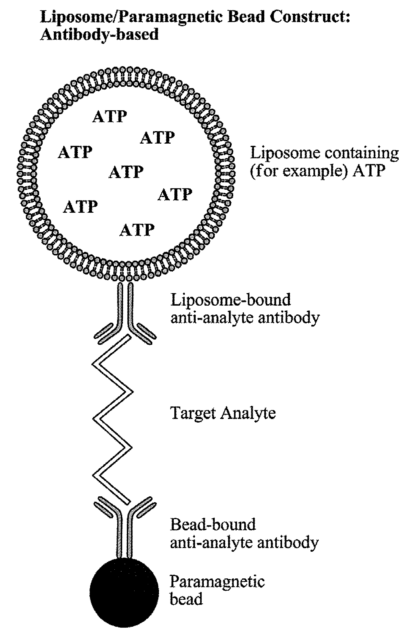
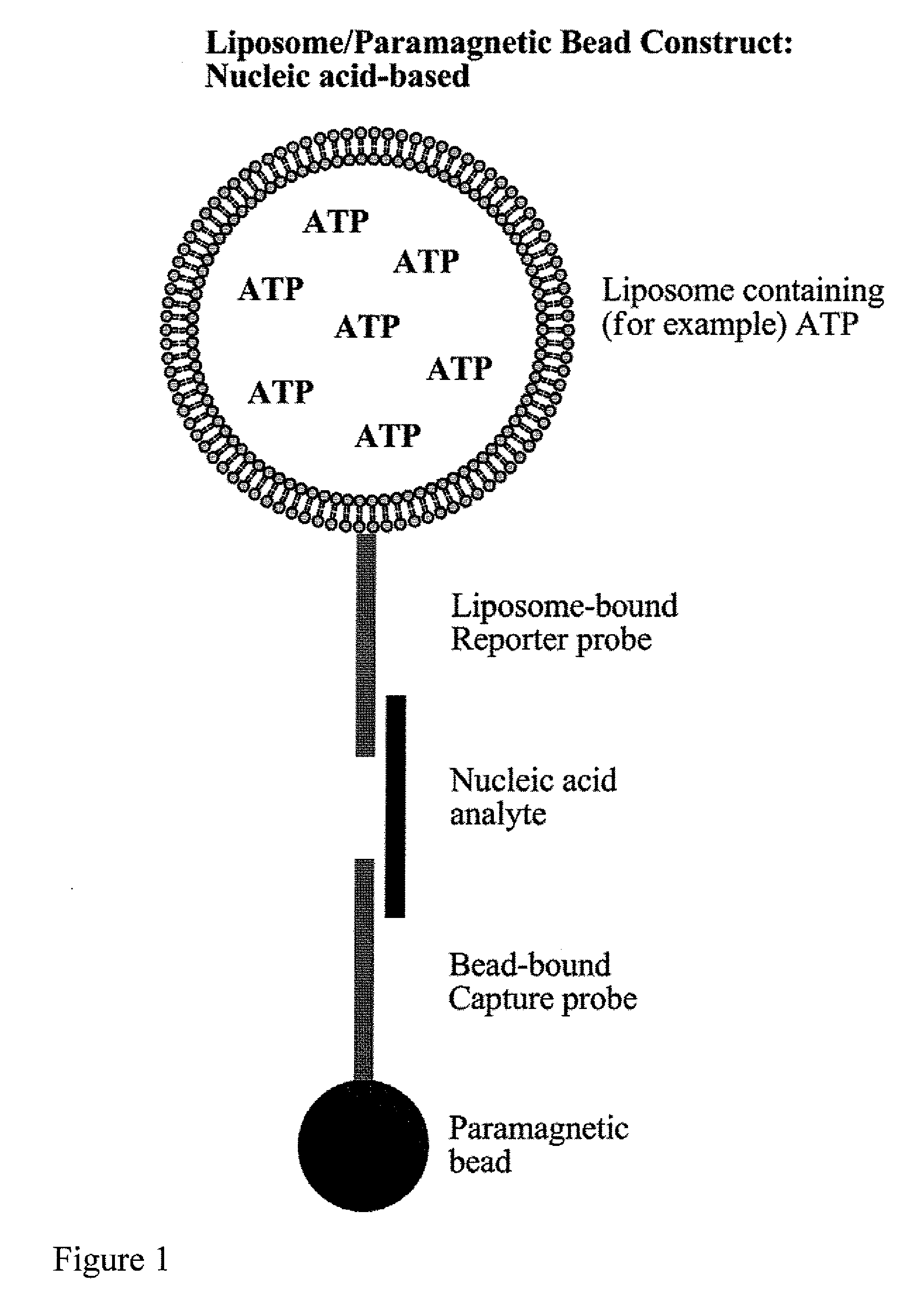
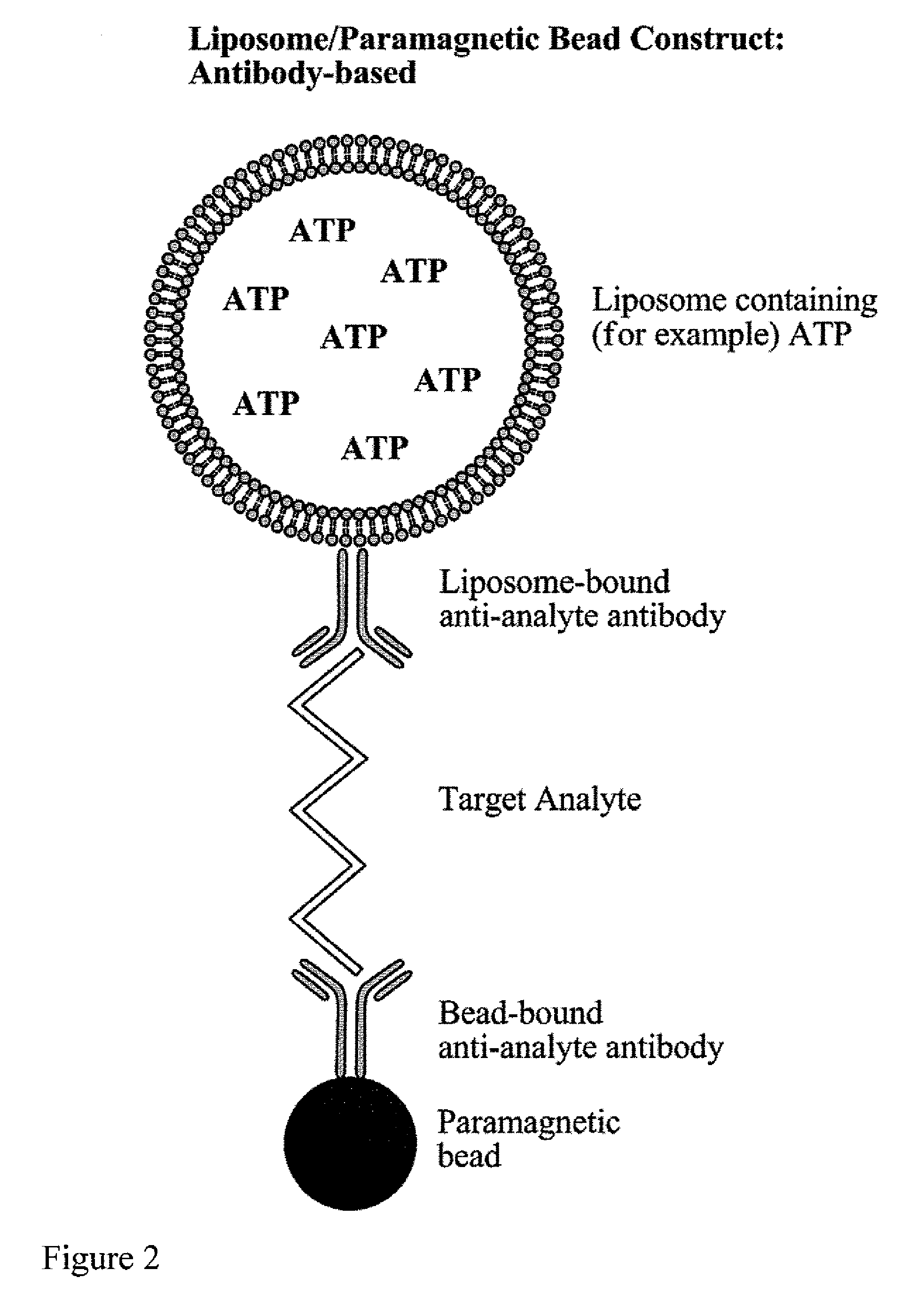
[0062]Biotinylated capture oligonucleotide probes were conjugated by methods known to those skilled in the art to streptavidin coated paramagnetic beads. After conjugation and washing, the labeled paramagnetic beads were stored at 4° C. for several months. It was noted that, while this particular example relates to the immobilization of oligonucleotides, antibodies or other compounds capable of binding to a sample can be also be used.

Determination of Detection Limits

[0063]The limits of detection of the assay system of the present invention were determined using a synthetic nucleotide sequence. A sandwich hybridization assay was performed in microplate wells using 15 μl of a 1:10 dilution of liposomes and 5 μl (1.25 μg) of paramagnetic beads. The bead-target-liposome complex was washed using two types of magnetic devices to retain th...


PUM

PropertyMeasurementUnit
diameteraaaaaaaaaa
diameteraaaaaaaaaa
diameteraaaaaaaaaa

Abstract

The invention relates to the encapsulation of luminescence-related molecules, including but not limited to, adenosine triphosphate (ATP), adenylate kinase (AK), alkaline phosphatase (ALP), luminol and luciferin / luciferase cocktails, within liposomes. These liposomes can be employed to enhance the luminescence detection of microorganisms and compounds in various products and samples. The liposomes containing the luminescence-related molecules can bear a probe which has a specific sequence or structure that, in turn can be used to hybridize to, or couple with, a portion of the target analyte. Within the same assay, paramagnetic beads can bear a probe having a specific sequence or structure that, can hybridize to, or couple with, a second portion of the target analyte to create a complex of analyte bound to paramagnetic beads and liposomes. This type of assay can be often referred to as a ‘sandwich’ assay. Once the probes hybridize to, or couple with, their targets, a complex can be formed of the paramagnetic beads, the analyte, or portion thereof, and the liposomes. This complex can then be washed to remove those components that are non-hybridized or non-coupled. Then, the paramagnetic bead-analyte-liposome complexes can be isolated from the sample using magnetic separation techniques and can be treated so as to release their encapsulated ATP, AK or other luminescence-related compounds. The resulting luminescence can then be determined in a chemical assay. This determination can be qualitative (i.e., an absence / presence assay) or quantitative (i.e., which can measure a specific amount of analyte present). Through the use of a cocktail of probe types, the assay can also qualitatively or quantitatively measure the presence of more than one analyte simultaneously. This type of assay can be of commercial importance in clinical and forensic applications, the personal care, pharmaceutical, food and beverage markets, as well as in environmental sample assays.

Description

FIELD OF THE INVENTION [0001]The invention relates to assays for detecting and determining the presence of specific analytes in a sample. Specifically, the invention relates to the use of sensitized liposomes having adenosine triphosphate (ATP), adenylate kinase (AK) or other luminescence-related compound encapsulated within for detecting and determining the presence of analytes such as bacteria, viruses, genetic material, haptens, immunogenic compounds, chemical compounds and other materials of interest. Liposomes are sensitized through the use of probes which may be oligonucleotides, antibodies or antigens with affinity for the target analyte(s). In addition, the process of specific detection is facilitated by the use of paramagnetic particles.BACKGROUND OF THE INVENTION [0002]Through recent innovations in the areas of both instrumentation and reagents, it is now feasible to perform new types of assays that were previously too difficult, too time consuming and / or too costly. Impro...

Claims


Application Information

Patent Timeline
Patent Type & Authority Applications(United States)
IPC IPC(8): C12Q1/70C12Q1/48G01N33/53G01N33/554C12Q1/68
CPCG01N33/5432
Inventor HEARN, ANDREWMADDEN, JUDITHSELLAPPAN, SUBRAMANIBAEUMNER, ANTJE J.ZAYTSEVA, NATALYA V.
Owner CELSIS INT LTD (US)
Why Eureka
Try Eureka