
Use of Myostatin (Gdf-8) Antagonists for Improving Wound Healing and Preventing Fibrotic Disease

a technology of myostatin and fibrotic disease, which is applied in the field of improving wound healing, can solve the problems of scarring and loss of original muscle function, loss of muscle tissue, and limited clinical practice of growth factors, and achieve the effect of improving the wound healing process

Inactive Publication Date: 2008-08-07
ORICO
View PDF1 Cites 19 Cited by
  • Summary
  • Abstract
  • Description
  • Claims
  • Application Information

AI Technical Summary

Benefits of technology

[0011]Surprisingly, the growth factor myostatin, a member of the TGF-beta family of growth factors, has been shown for the first time to be implicated in the wound healing process. Inhibition of myostatin activity has been found to significantly improve the wound healing process.
[0016](c) a decrease or inhibition of scar tissue formation,thereby resulting in improved functionality and cosmetic appearance of the treated tissue.
[0023]The present invention also provides for the use of one or more myostatin antagonists in the manufacture of a medicament for improving wound healing in a patient in need thereof.

Problems solved by technology

Whilst this process results in the gain of some muscle function at the wound site, muscle wounds invariably result in loss of muscle tissue, scarring and loss of original muscle function.
However, whilst these therapies are effective in stimulating and even accelerating the wound healing process, they still result in functional and / or cosmetic impairment at the wound site5.
However, to date growth factors have had a limited role in clinical practice.
The only currently available commercial product is PDGF which has been shown to reduce healing time, but which has not been successful in improving the cosmetic or functional aspect of wound healing2.

Method used


Image

  • Use of Myostatin (Gdf-8) Antagonists for Improving Wound Healing and Preventing Fibrotic Disease
  • Use of Myostatin (Gdf-8) Antagonists for Improving Wound Healing and Preventing Fibrotic Disease
  • Use of Myostatin (Gdf-8) Antagonists for Improving Wound Healing and Preventing Fibrotic Disease

Examples


example 1

Myostatin Antagonists Increase Inflammatory Response and Chemotaxis of Cells Involved in Muscle Wound Healing

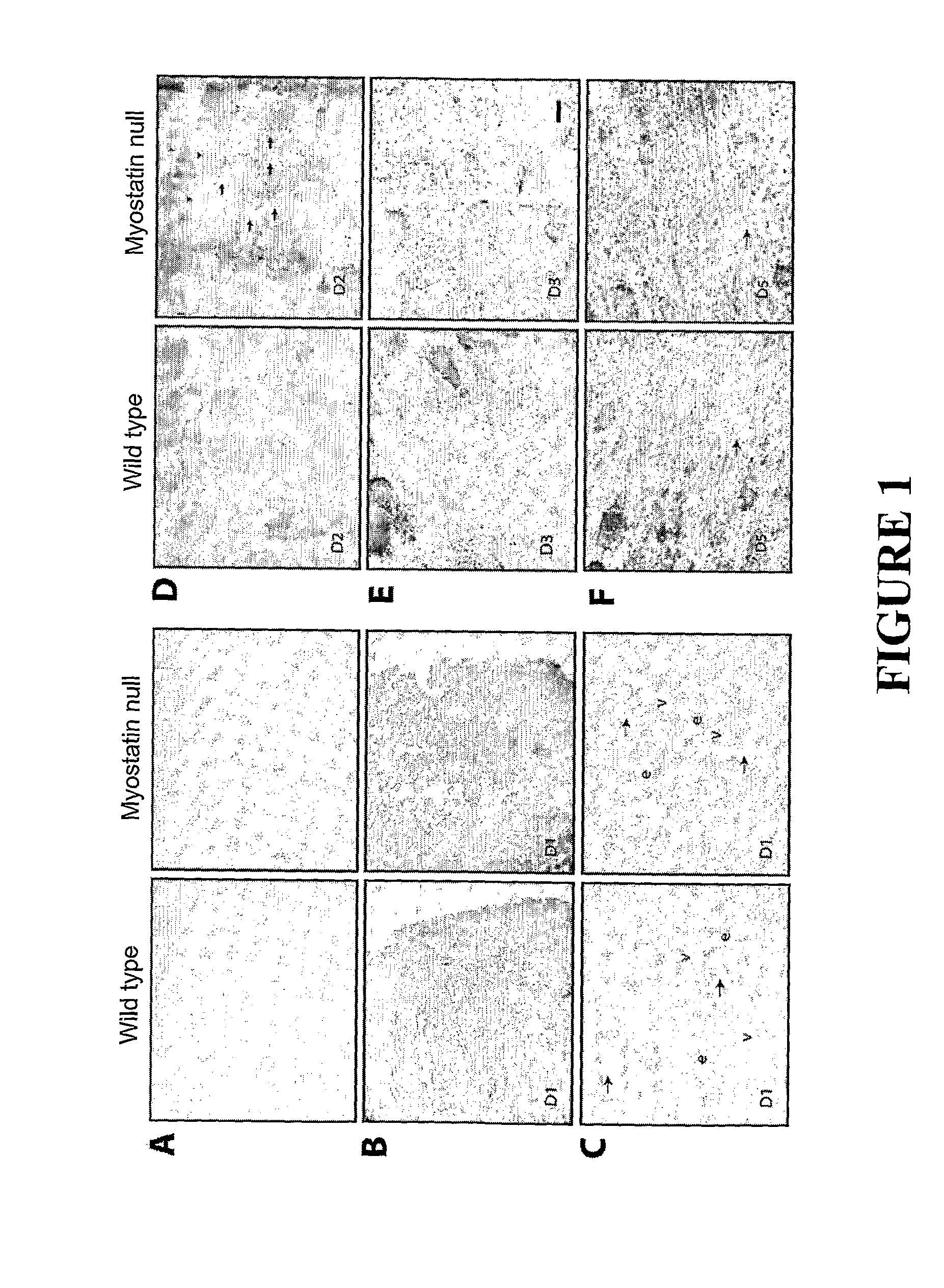
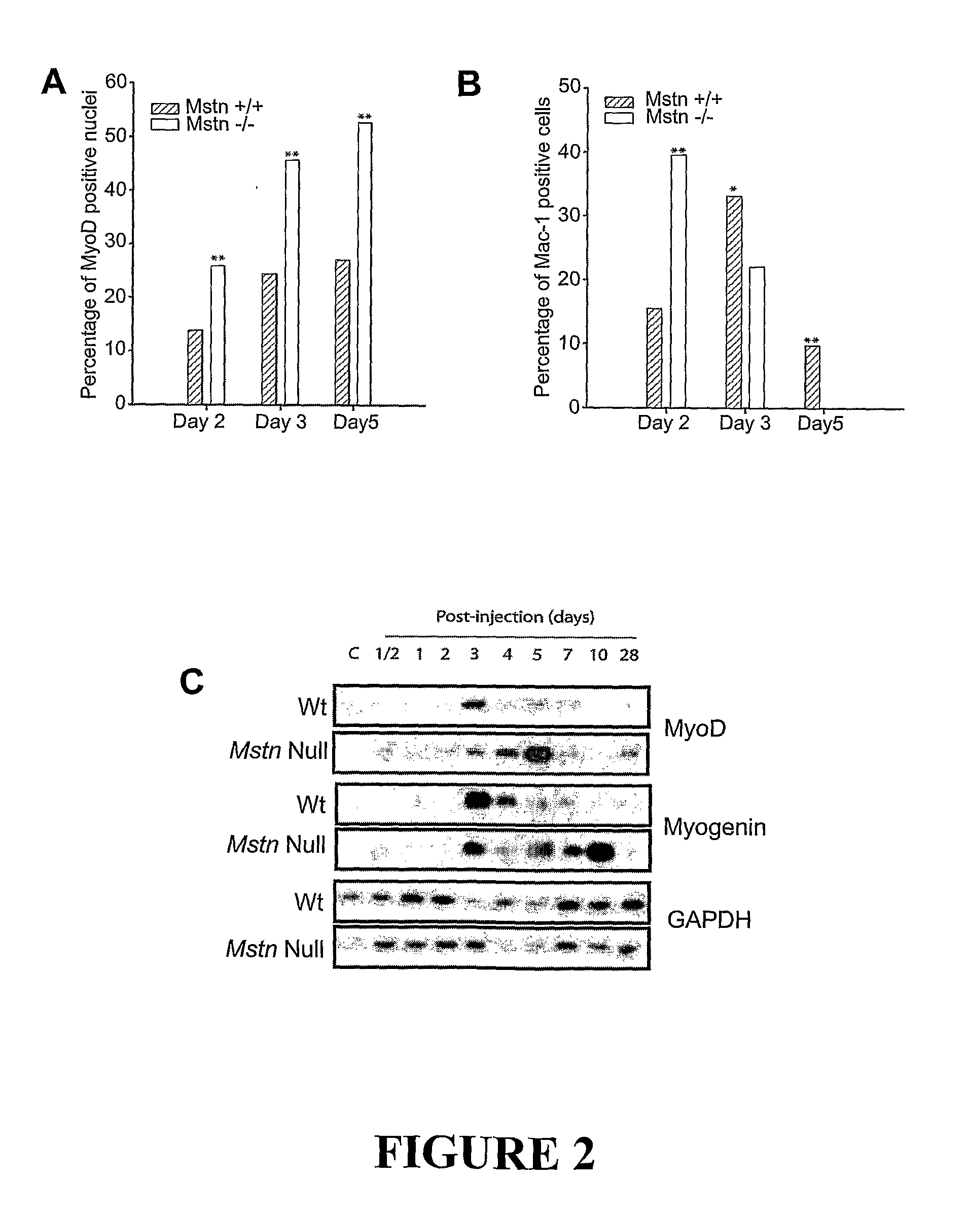
[0107]Wound healing is a highly ordered process; muscle tissue wounding results in immediate inflammatory response followed by chemotactic movement of myogenic precursor satellite cells. Here we have shown that myostatin actually inhibits the inflammatory response and the chemotactic movement of myogenic cells towards the wound site. Thus the beneficial effects of lack of myostatin or antagonists of myostatin on the speed and quality of wound healing are demonstrated.

Materials and Methods

Expression and Purification of 350

[0108]A cDNA corresponding to the 267-350 amino acids, of bovine myostatin (hereafter referred to as “350” or “350 protein”) was PCR amplified and cloned into pET16-B vector. Expression and purification of 350 protein was done according to the manufacturer's (Qiagen) protocol under native conditions.

Notexin Wounding Model

[0109]Six to eight week old male C57BL...

example 2

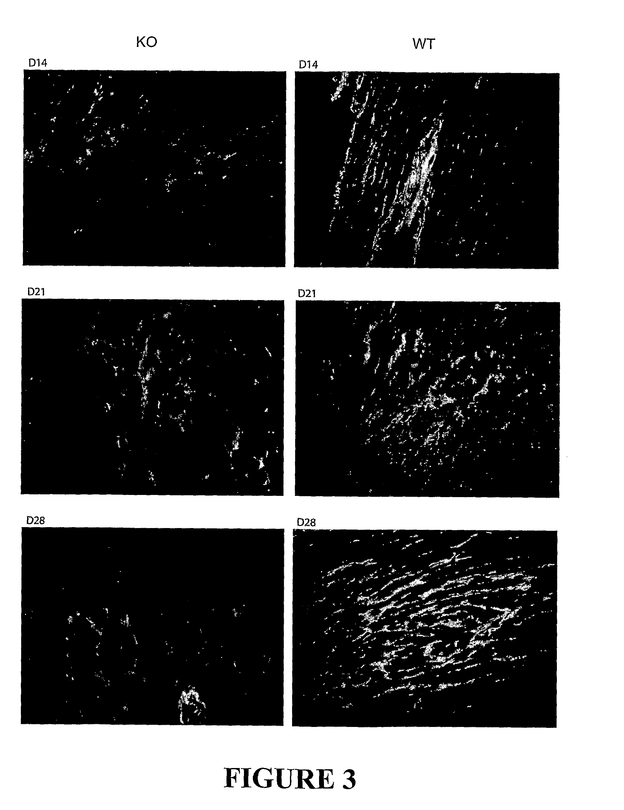
Antagonizing Myostatin Results in Reduced Fibrosis and Enhanced Muscle Healing

Methods

Cut Wound Model

[0125]A 3 mm transversal incision was made on the left tibialis anterior (TA) of each mouse (wild type and myostatin null). On days 0, 3, 5, and 7 after wounding the TAs of wild type were injected with either 350 protein at 2 μg / g body weight (total of 85 μg / mouse) or saline at the site of wounding (into the TA muscle). The uninjured right TA was used as control. The injured and control muscle were collected at day 2, 4, 7, 10 and 21 after wounding and their weights determined. The extent of collagen deposition in healing and healed cut wounds was also measured by Van Giesson staining.

SE Microscopy

[0126]The muscle samples were cleaned of fat and tendons and fixed in 10 ml of 0.1 M phosphate buffer (pH 7.4) containing 2.5% (v / v) glutaraldehyde for 48 hours with gentle rocking. The glutaraldehyde was washed off in PBS for 1 hour, before being transferred to 50 mls of 2 M NaOH, and incub...


PUM

PropertyMeasurementUnit
Fractionaaaaaaaaaa
Fractionaaaaaaaaaa
Fractionaaaaaaaaaa

Abstract

The present invention relates to a method of improving wound healing in a human or animal patient by inhibiting the activity of myostatin (GDF-8) using one or more myostatin antagonists. The present invention also relates to a method of treating fibrotic diseases or disorders comprising administering a myostatin antagonist.

Description

FIELD OF THE INVENTION[0001]The invention relates to methods and compositions for improving wound healing and in particular for preventing scar formation and thus loss of function that can occur in injured tissues during the natural wound healing process.BACKGROUND[0002]A wound is a disruption of tissue integrity that is typically associated with a loss of biological substance. Simple wounds include cuts and scrapes to the skin whilst deeper injuries to the muscle tissue, skeletal system or the inner organs are defined as complicated wounds1.[0003]Every wound undergoes a similar reparative process no matter what the wound type or the degree of tissue damage1, 2, 3. Three distinct phases of wound healing are recognised. Firstly the inflammatory or exudative phase for the detachment of deteriorated tissue and for wound cleansing; secondly a proliferative phase for the development of granulation tissue; and thirdly a differentiation or regeneration phase for maturation and scar formati...

Claims


Application Information

Patent Timeline
IPC IPC(8): A61K39/395A61K39/00A61K38/00A61K38/08A61K48/00A61K38/18A61P17/02
CPCA61K38/18A61K48/00A61K38/1808A61K38/1858A61K38/30A61K2300/00A61P17/00A61P17/02A61P21/00A61P29/00A61P41/00A61P43/00
Inventor KAMBADUR, RAVISHARMA, MRIDULAHENNEBRY, ALEXSENNA SALERNO DE MOURA, MONICA
Owner ORICO
Learn More