Method and Device For Representing the Microstructure of the Lungs

a microstructure and lung technology, applied in the field of lung microstructure imaging, can solve the problems of not providing global information, not allowing sufficient capture of details and their precise resolution, and available techniques that are not precise enough

Inactive Publication Date: 2008-08-28
JOHANNES GUTENBERG UNIV
View PDF4 Cites 8 Cited by
  • Summary
  • Abstract
  • Description
  • Claims
  • Application Information

AI Technical Summary

Benefits of technology

[0015]The present invention provides a non-invasive method or device, with which the images of the lung can be captured and reproduced, also in regional areas, in detailed resolution. Accordingly, the current invention provides means with which even the smallest constrictions or distensions of the lung and / or the airways can be imaged at an early stage and can thereby be detected, thereby providing an opportunity to prevent, or to therapeutically intervene, as early as possible, lung diseases like COPD (chronic obstructive pulmonary disease) and lung emphysema. In addition, the high costs and expenditure associated with the methods and devices that are known from the current state of the art are avoided.

Problems solved by technology

In many instances the available techniques are not precise enough or do not permit sufficient capture of details and their precise resolution.
However, spirometry testing only provides global information and does not deliver detailed images with detailed resolution.
The processing of the gas into a highly polarized contrast gas is very expensive and requires special handling during transportation and application since the artificial polarization condition is destroyed by oxygen and magnetic field exposure.
In addition, the costs and the technical expenditure of the methods by way of hyperpolarized noble gases are extremely high.
It is certain that the diffusion of clusters of this type cannot be proven in any form whatsoever in any feasible imaging situation.

Method used


Image

  • Method and Device For Representing the Microstructure of the Lungs
  • Method and Device For Representing the Microstructure of the Lungs

Examples


Embodiment Construction

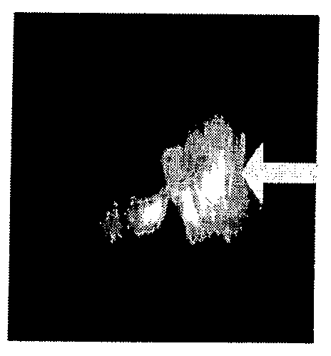
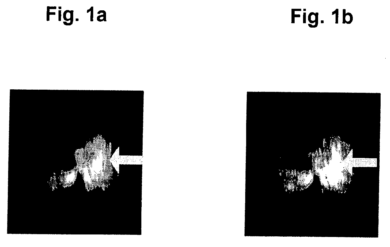
[0062]Referring now to the drawings, in the diffusion-weighted image in FIG. 1a, a signal reduction can be noted when compared with a non-diffusion-weighted image with a magnetic resonance tomograph as illustrated in FIG. 1b. This is a result of the diffusion of the C4F8, which is being brought into the airways of the lung in this example. This enables the apparent diffusion coefficients to be determined with which a realistic partial image of a lung, including the microstructures can be produced in high resolution.

[0063]The advantages of the inventive science are multifaceted: A method and a device are provided in accordance with the current invention which, compared to the current state avoid the high cost of noble gases, and the technological expenditure necessary to polarize these gases.

[0064]With the inventive non-invasive method and non-invasive device the smallest airway constrictions and distensions are regionally captured and images provided at a high resolution by way of u...


PUM


Abstract

The invention relates to an apparatus and a method for imaging the microstructure of an animal or human lung by way of introducing a fluoric contrast gas into the lung which is to be imaged; definition of the apparent diffusion coefficient of the contrast gas by way of diffusion weighted 19fluorine magnetic resonance tomography and based on the determined apparent diffusion coefficients; and imaging of the lung's microstructure.The current invention also describes a device for the implementation of the inventive method. The science of the current invention allows for the first time the production of a high resolution image of the microstructures of the lung through non-invasive measures, by way of fluorinated gases.

Description

BACKGROUND OF THE INVENTION[0001]1. Field of the Invention[0002]The invention relates to a method and device for imaging the microstructure of the lung.[0003]2. Description of the Related Art[0004]Different methods are available for the imaging of lung and / or respiratory tract diseases where attempts are made to obtain an original image of the lung and the respiratory tracts. In many instances the available techniques are not precise enough or do not permit sufficient capture of details and their precise resolution. It is therefore an ongoing objective in medical research to find ways to gain better insight into normal or defective developments / conditions of the human body and to aid a physician in his diagnosis by providing data which is as precise as possible.[0005]Various methods are known for the imaging of the microstructure of the lung: pathological changes of the lung are symptomatic for obstructive pulmonary disease COPD (chronic obstructive pulmonary disease) and pulmonary ...

Claims


Application Information

Patent Timeline
Patent Type & Authority Applications(United States)
IPC IPC(8): A61B5/055A61K49/08
CPCA61K49/06G01R33/56341G01R33/5601
Inventor SCHREIBER, WOLFGANGWOLF, URSULASCHOLZ, ALEXANDER-WIGBERTHEUSSEL, CLAUS-PETER
Owner JOHANNES GUTENBERG UNIV
Why Eureka
Try Eureka