Method and apparatus for performing maxillary sinus elevation

a maxillary sinus and elevation technology, applied in the field of osseous regeneration of the maxillary sinus, can solve the problems of limited dental implants placement in the anterior lower jaw, inferior dental bridge alternatives in comparison to dental implants, and considerable challenges in the replacement of the maxillary posterior teeth

Inactive Publication Date: 2008-09-04
HOCHMAN MARK N
View PDF13 Cites 40 Cited by
  • Summary
  • Abstract
  • Description
  • Claims
  • Application Information

AI Technical Summary

Benefits of technology

[0027]A method and device is disclosed that provides a new approach to the placement of dental implants when inadequate bone mass exists in patients' upper and lower jaws. The method and device enables dental implants to be placed more effectively within bone tissues of the oral cavity.
[0028]Moreover, a method and device is described to improve the quality and quantity of bone during the placement of dental implants. This innovation is particularly well suited to allow an increase of bone mass, including bone quality and quantity in the posterior maxilla when the elevation of the maxillary sinus floor is required. It is also well suited when poor bone quality exists in either jaw of the oral cavity.
[0029]Briefly, a system for increasing bone mass in the posterior maxilla in accordance with this invention includes one or more sleeves having various lengths and diameters. One or more of these sleeves are used in each procedure. Each sleeve is preferably cylindrical and includes a tip used to cut or otherwise form a hole into the maxillary sinus but under the subantral membrane. The sleeve is hollow to allow a fluid to be inserted under the membrane to cause it to be gently lifted from the sinus floor and form a cavity by such fluid. The cavity is then filled with a suitable material such as a material that promotes bone growth. In one embodiment, the sleeve also has cross-channels that allow the same material to be disbursed in the alveolar ridge itself to change the quality of the bone type from a less dense to a more dense bone density type.

Problems solved by technology

Initially the placement of dental implants were limited to the anterior lower jaw, as this region provided sufficient bone quantity, quality and strength to support and hold a dental implant having an effective length.
The previous alternative of dental bridges is now considered an inferior alternative in comparison to dental implants.
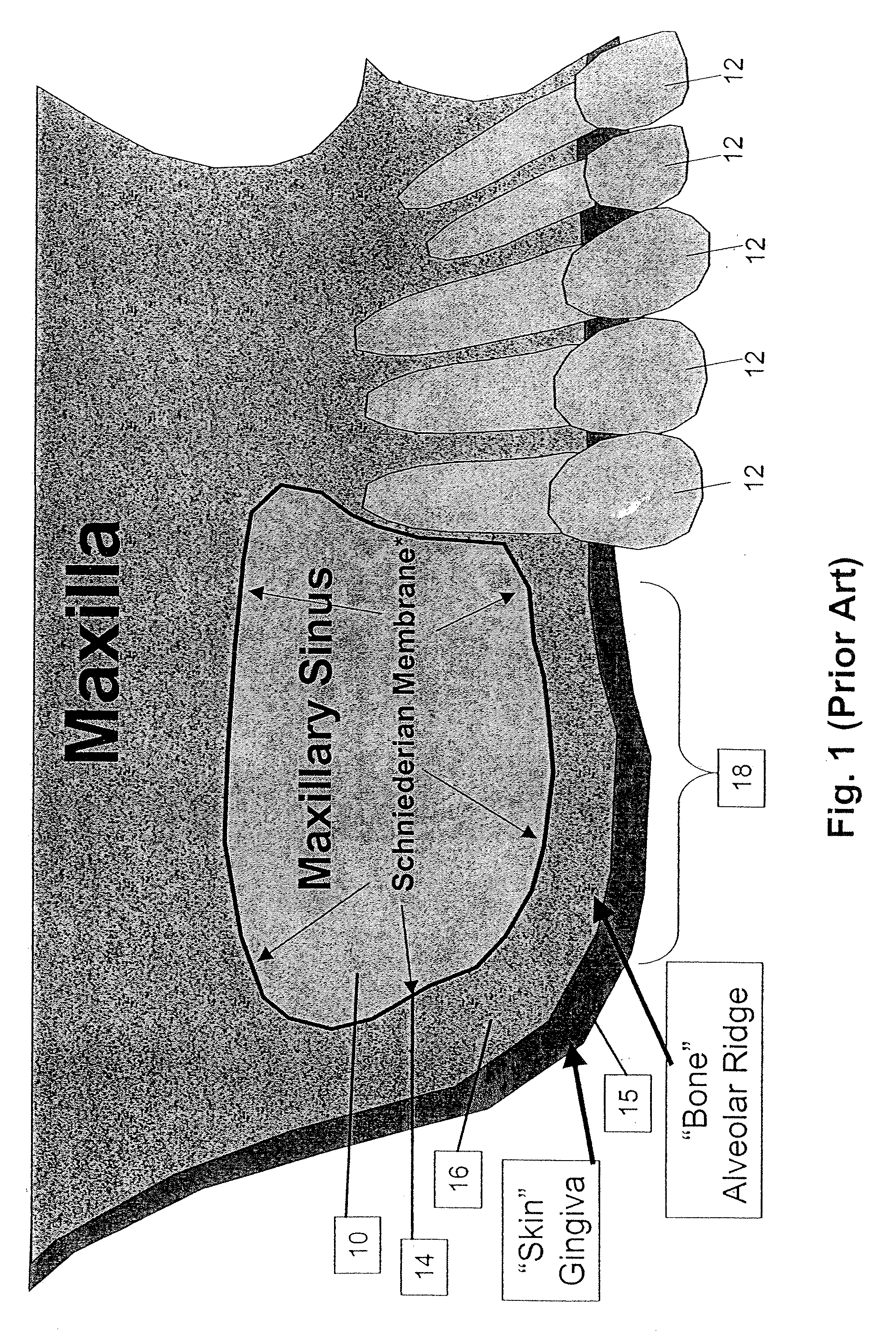
To that end, the replacement of the maxillary posterior teeth have presented a considerable challenge because, after the loss of maxillary posterior teeth the quality and quantity of the remaining supporting bone may be insufficient to support implants properly or reliably.
Once a tooth is removed from the complex, the surrounding alveolar bone is frequently resorbed because of the lack of physical stimulation and support of the teeth.
This leads to a loss of bone mass and a corresponding reduction in the effective height and thickness of the bone of the maxillary complex.
In addition, with the loss of teeth from the upper jaw (and the maxillary complex) the compensatory enlargement of the maxillary sinus occurs that also reduces the vertical height of the remaining bone of the maxillary complex.
These effects further compromise the option for dental implants to be used.
If the membrane is not properly dissected from the bone, bone augmentation may not occur, or may not be sufficient.
Unintentional perforation of the subantral membrane may also lead to undesirable short and long-term consequences.
A lack of integrity of the membrane can also lead to the migration of regenerative bone materials leading to long-term chronic infections.
This technique is fraught with many risks and complications because of the limitations of healing potential in the maxillary sinus.
While, this technique is safer, an overzealous use of an osteotome during the placement of the regenerative material can result in the perforation of the subantral membrane with disadvantages discussed above.
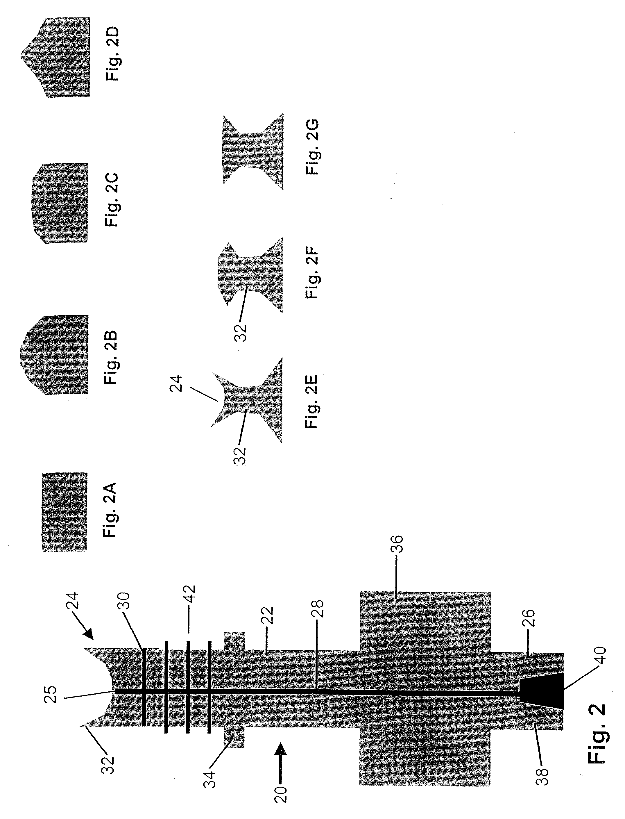
The new technique also uses an inferior approach to the membrane via the alveolar ridge.
This technique has many deficiencies owing to the inability to properly adapt a standard medical syringe to an opening created in the bone.
Unfortunately, tearing or ripping of the subantral membrane may still occur and it is difficult, if not impossible, to detect it during the inflation of the balloon.
This technique has multiple deficiencies owing to the inability to control the pressure and forces applied to the subantral membrane owing to the lack of precision of this technique.
The deficiencies and limitations of current techniques for sinus elevation relate to primarily: (1) the inability of the operator to control the infracture (“green-stick fracture) of the bony floor and lateral window of the maxillary sinus, (2) the inability to carefully separate the membrane from its physical adherence off the floor of the maxillary sinus and (3) a lack of feedback indication or confirmation for the surgeon of a perforation or tearing of the membrane prior to the placement of regenerative materials.
1. Traumatic invasive surgical procedures that include extensive mucogingival flap elevation methods to gain access to the surgical site.
2. The use of instrumentation that comes into direct physical contact with the subantral membrane with the risk of perforating or tearing the membrane during separation of membrane from the floor of the sinus.
3. The need to use a infracture, i.e. “green-stick” fracture of the floor of the sinus or lateral aspect of the sinus that could once again perforate or tear the delicate subantral membrane leading to failure.
4. Using previous methods there is a lack of subjective indication or feedback to the operator that the membrane has been torn or perforated during is separation and elevation from the floor of the sinus.
5. Inability to precisely control the infracture of the bone that is required to gain access to the floor of the sinus using previous methods.
6. Inability to precisely control the force used to raise the subantral membrane.
7. Inability to precisely control the delivery of the regenerative material and to determine weather a tear or perforation of the membrane has occurred during the placement of such regenerative material.
8. Difficulty in accessing the integrity of the membrane prior to the placement of the regenerative material that will be placed on the floor of the sinus.
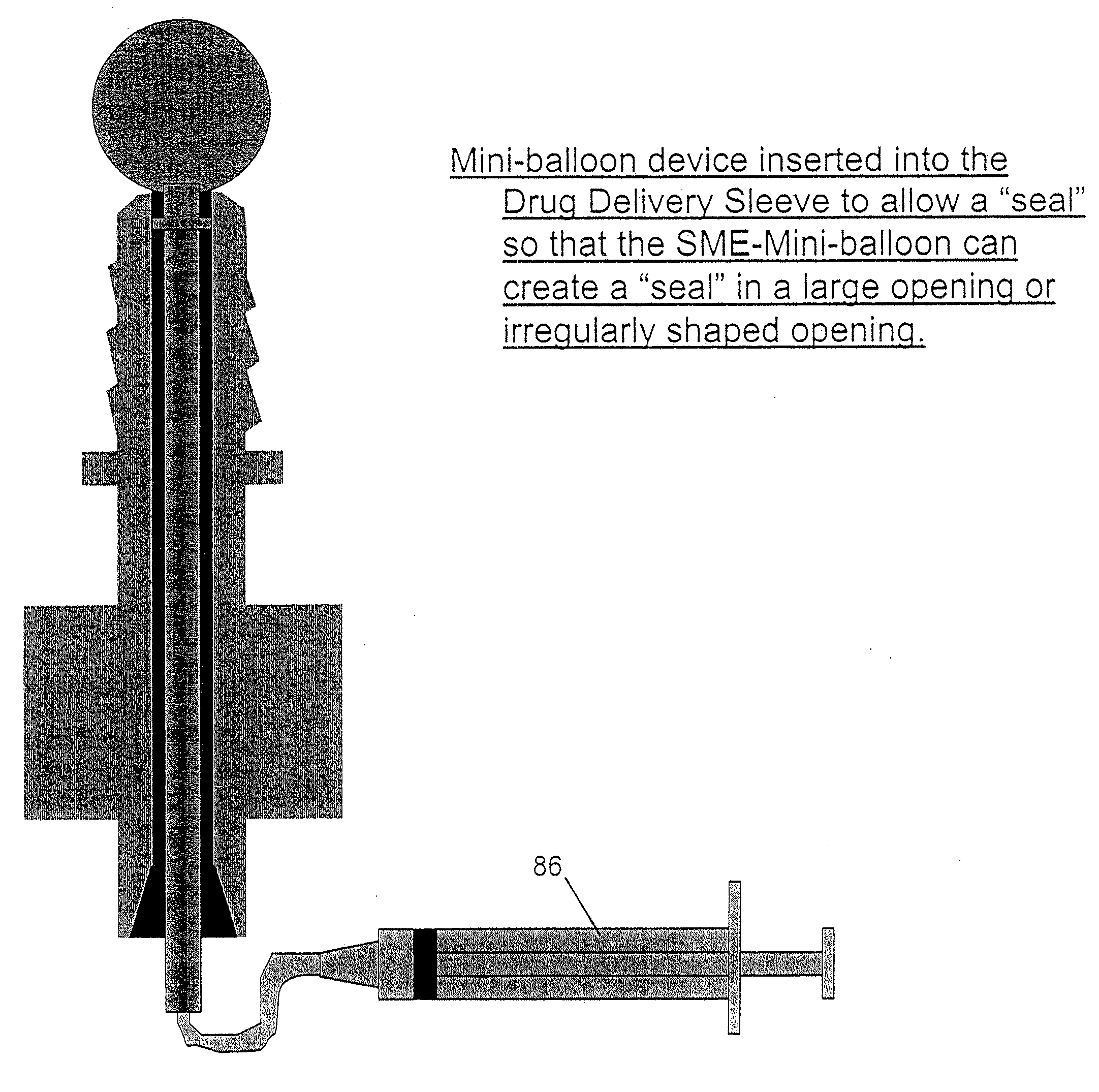
9. Inability to create a seal between the delivery device, such as a syringe and the prepared bony site during the elevation of the membrane and delivery of regenerative materials beneath the subantral membrane.
Failure to contain regenerative materials in the maxillary sinus often lead to the need for additional surgeries to retrieve such materials and may require extensive medical follow-up to corrective this iatrogenic outcome.

Method used


Image

  • Method and apparatus for performing maxillary sinus elevation
  • Method and apparatus for performing maxillary sinus elevation
  • Method and apparatus for performing maxillary sinus elevation

Examples


Embodiment Construction

[0041]FIG. 1 shows a typical cross section of the maxillary sinus 10. Several teeth 12 are seen in the Figure in a normal configuration. The maxillary sinus 10 is separated from the mouth by the alveolar ridge 16 formed of bone. The bone is covered on top by the subantral membrane 14 and on the bottom by the gingiva 15. As shown in the Figure, because this person has lost several molars, the floor of the maxillary sinus has been depressed or lowered in the area 18. As a result, the alveolar ridge 16 in this region has lost considerable bone mass and quality. Frequently, the bone in this area is porous and is not strong enough to support an implant of sufficient length and an associated final dental prosthesis. Typically, in a health person the ridge 16 in this area may be about 8 to 14 mm however, in persons who have lost their molars, the thickness of the ridge 16 may be no more than about 4 mm.

[0042]FIG. 2 shows a first embodiment of a sleeve 20 constructed in accordance with this...


PUM


Abstract

A method and apparatus is disclosed for providing implants in the upper jaws of a person. A sleeve is inserted through the alveolar ridge to the maxillary sinus. The sleeve is used to raise the subantral membrane and form a cavity. A filler, such as a bone growth stimulant is injected through the sleeve into the cavity. In the process, the sleeve also can cut and / or condense the bone around itself so that the bone can hold an implant. Optionally, the bone growth stimulant is also introduced into the bone surrounding the sleeve. During the injection, the pressure within the sleeve or the cavity is monitored to detect and prevent the rupture of the subantral membrane.

Description

RELATED APPLICATIONS[0001]This application claims priority to U.S. provisional application Ser. No. 60 / 619,542 filed Oct. 15, 2004 and incorporated herein by reference.BACKGROUND OF THE INVENTION[0002]A. Field of Invention[0003]This invention relates generally to improvements to the delivery of drugs, particularly for osseous regeneration for the maxillary sinus. More specifically this invention provides a method and device to the elevation of the floor of the maxillary sinus to increase the amount of bone available based on using hydro-dissection to raise the subantral membrane floor of the sinus (a.k.a. the Schneiderian membrane) allowing placement of osseous regenerative materials.[0004]B. Description of the Prior Art[0005]Dental implants have been used in dentistry for about 20 years. They offer a tremendous benefit to patients by allowing the replacement of missing teeth. Prof. Per-ingvaar Branemark introduced the use of titanium dental implants that showed that predictable, st...

Claims


Application Information

Patent Timeline
Patent Type & Authority Applications(United States)
IPC IPC(8): A61C8/00
CPCA61C8/0092A61C8/0033
Inventor HOCHMAN, MARK N.
Owner HOCHMAN MARK N
Why Eureka
Try Eureka