Method of detection and compensation for respiratory motion in radiography cardiac images synchronized with an electrocardiogram signal

a radiography and cardiac image technology, applied in the field of medical imaging, can solve the problems of high degree of motion, inconvenient operation, and inconvenient monitoring, and achieve the effects of reducing tracking time, improving the success rate of catheter ablation procedures, and improving accuracy and simplicity

Active Publication Date: 2008-10-02
GENERAL ELECTRIC CO
View PDF15 Cites 40 Cited by
  • Summary
  • Abstract
  • Description
  • Claims
  • Application Information

AI Technical Summary

Benefits of technology

[0020]Respiratory motion is fairly simple. As an initial approximation, the respiratory motion may be considered to be a rigid motion without deformation of the heart. It may even be considered to be an essentially translational motion. It is therefore theoretically possible to calculate this motion and compensate for it in order to obtain efficient registration with 3D cardiac images acquired at a given phase of the cardiac cycle. As a second approximation, the respiratory motion may be considered to be a more complex motion with a deformation of the heart. This is implemented by a finer modeling of respiratory motion.
[0022]The invention is thus particularly suited to an ablation operation procedure in which the ablation catheter is used to “burn” the tissue of the cardiac wall in order to modify electrical conduction on its surface. Indeed, through an initial registration and then an additional registration, the 3D cardiac images are put into in a state of concordance with the projection images of the fluoroscopy system. As a consequence, pulmonary veins in other regions involved in the launching and tracking of an auricular fibrillation may be identified with greater precision and simplicity in the radiography images, thus improving the success rate of a catheter ablation procedure.
[0023]The embodiments of the invention presented here also constitute a system and method by which 3D images of anatomical structures, for example the coronary sinus and the left ventricle, may be put into concordance with projection images of the radiography system, thus enabling the navigation and positioning of the stimulation electrodes at the place most suited in the case of biventricular stimulation procedures.
[0024]The fact of having perfect concordance between the two images enables, for example, a cardiologist to carry out the real-time tracking of the progress of a vascular tool during an operation or again the tracking of catheters whenever they may be.

Problems solved by technology

However, this type of solution has drawbacks, for iodized compounds are often sources of allergies in the patient and are also toxic for the kidneys.
However, this type of approach has drawbacks.
Indeed, the main problem encountered in the specific part of the anatomy, namely the heart, is that it undergoes a high degree of motion due to the patient's heart cycle and respiration.
However, the respiratory motion is always present in X-ray images, and therefore does not enable precise registration with the pre-operation images.

Method used


Image

  • Method of detection and compensation for respiratory motion in radiography cardiac images synchronized with an electrocardiogram signal
  • Method of detection and compensation for respiratory motion in radiography cardiac images synchronized with an electrocardiogram signal

Examples


Embodiment Construction

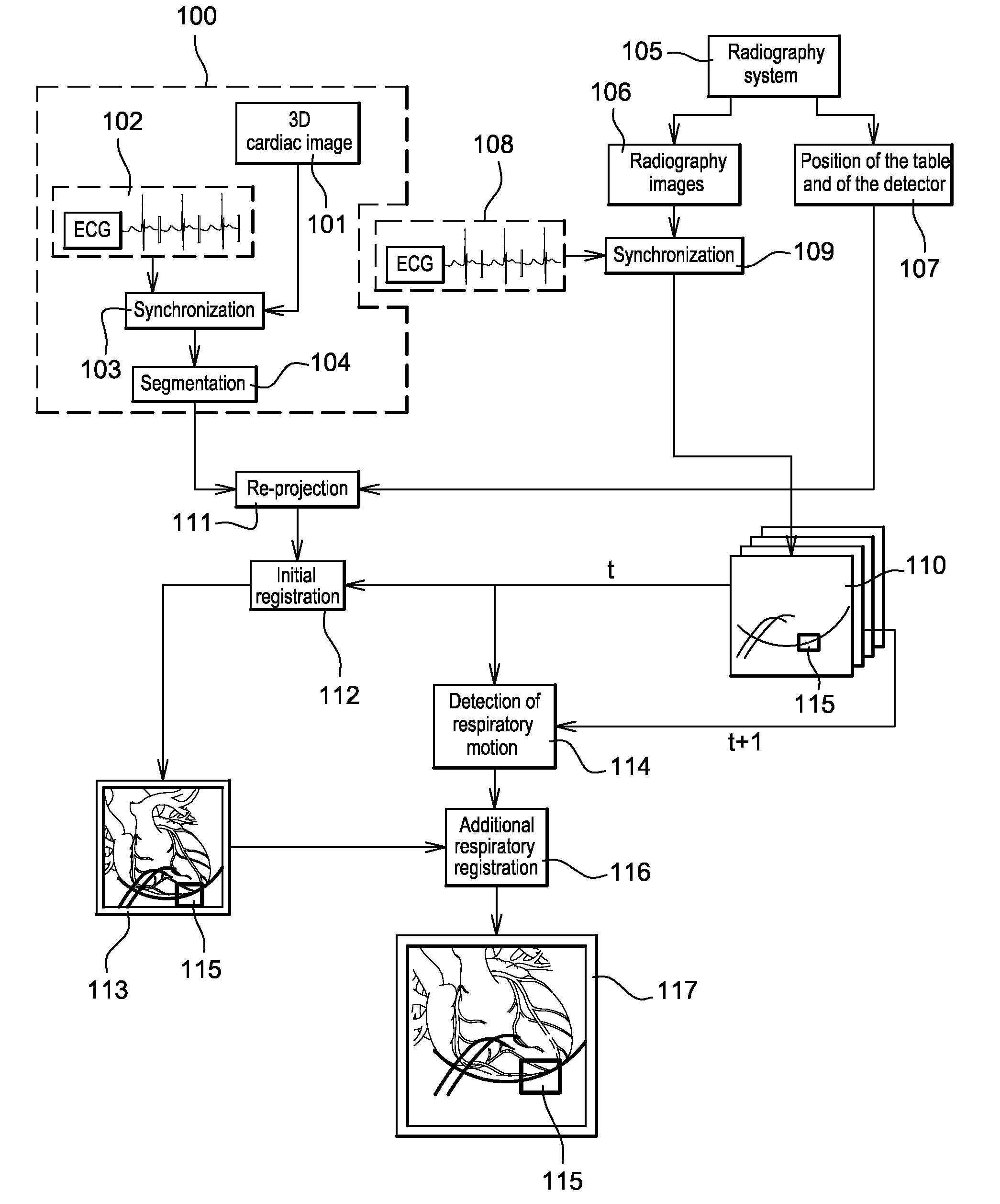
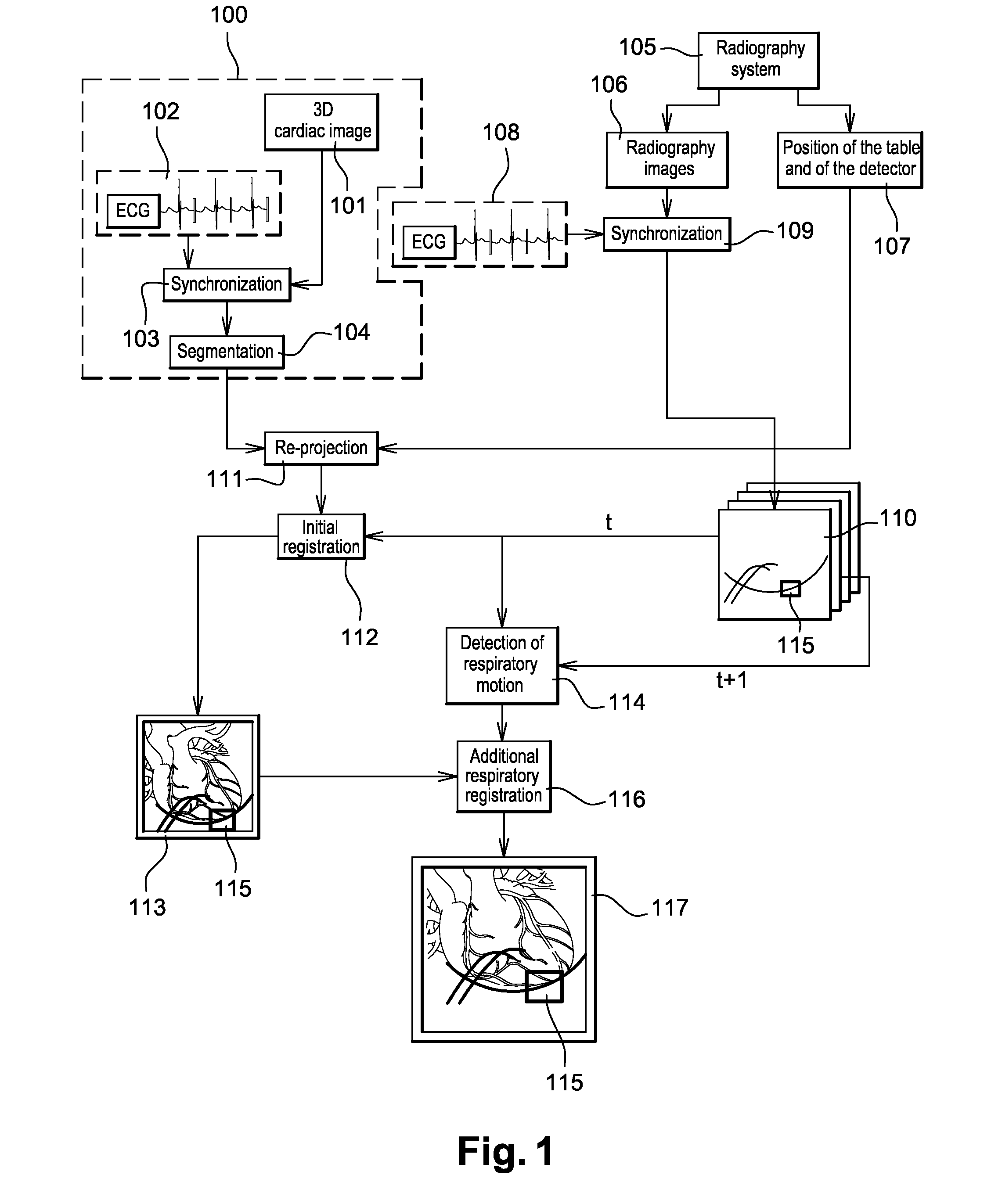
[0041]FIG. 1 illustrates means implementing the method for detecting respiratory motion in a cardiac image. The example of FIG. 1 can be used in a medical intervention procedure such as for example a procedure for the ablation of auricular fibrillation or a biventricular procedure.

[0042]In the example of FIG. 1, the X-ray apparatus (not shown) comprises both a 3D imaging system and a radiography imaging system. The radiography imaging system may be distinct or it may be included in the 3D imaging system.

[0043]The respiratory motion detection algorithm of the invention is implemented by a processor (not shown) of the X-ray apparatus.

[0044]The 3D imaging system may be, inter alia, a computerized tomography machine, a radiography system taking 3D images by rotation, magnetic resonance systems, positron-emission tomography (PET), ultrasonic systems, nuclear medicine systems and 3D radiography systems.

[0045]Before or during a medical intervention, the phase 100 is performed. This phase 1...


PUM


Abstract

A method of detection and compensation for respiratory motion improves the registration between a 3D pre-operation image and X-ray images acquired during a cardiac intervention. By synchronizing the X-ray images with the electrocardiogram, the disclosed method thus eliminates the motion related to the heart cycle from these images, thus isolating the contribution of the respiratory motion. From this point, an algorithm is proposed capable of attributing the motion remaining in the radiography images to respiration. The algorithm also enables this motion to be detected and compensated for in order to obtain a registration between 3D cardiac images and radiography images.

Description

CROSS-REFERENCE TO RELATED APPLICATIONS[0001]This application claims the right of priority, under 35 USC 119(a)-(d), to the earlier filing date of co-pending French patent application serial number 0754070, filed 27 Mar. 2007, which is herein incorporated by reference in its entirety. The basis for this claim of right of priority is France's membership in the World Trade Organization.BACKGROUND[0002]1. Field of the Invention[0003]Embodiments of the present invention find particularly advantageous but not exclusive application in the field of medical imaging, and more particularly in the field of X-ray imaging in cardiology.[0004]2. Prior Art[0005]X-ray imaging is now widely used for the diagnosis and processing of cardiac pathologies. The treatment may include coronaroplasty, valve replacement, electrophysiology etc.[0006]In a certain number of interventional procedures, the practitioner must pass catheters and / or guides into vessels or cavities of the heart. These interventions ena...

Claims


Application Information

Patent Timeline
Patent Type & Authority Applications(United States)
IPC IPC(8): G06K9/00
CPCA61B5/7289A61B6/032A61B6/12A61B6/503A61B6/5217A61B6/5235A61B6/541A61B8/5238G01R33/48G01R33/4812G01R33/56509G01R33/5673G16H50/30
Inventor SOUBELET, ELISABETHLIENARD, JEANGAVIT-HOUDANT, LAURENCEVAILLANT, REGIS
Owner GENERAL ELECTRIC CO
Why Eureka
Try Eureka