
Preventing Arrhythmias Associated with Cell Transplantation

a cell transplantation and arrhythmia technology, applied in the field of cell transplantation, can solve the problems of slowing of conduction velocity, congestive heart failure, and high risk of complications, and achieve the effect of improving electrical conductivity

Inactive Publication Date: 2008-10-23
THE JOHN HOPKINS UNIV SCHOOL OF MEDICINE
View PDF15 Cites 2 Cited by
  • Summary
  • Abstract
  • Description
  • Claims
  • Application Information

AI Technical Summary

Benefits of technology

The patent text describes an assay system for simulating cardiac arrhythmias using a co-culture of cardiac myocytes and skeletal muscle myoblasts. The system measures the electrical coupling of cells using a special technique. The patent also describes methods for treating myoblasts to improve their ability to communicate with other cells in the body. These techniques can be used to develop better cell-based therapies for treating heart disease.

Problems solved by technology

Congestive heart failure is a major public health problem in the United States.1 Cellular myoplasty represents a novel therapy for congestive heart failure, but is fraught with potential pitfalls.
Hence, we hypothesized that a mixture of myoblasts and myocytes would result in slowing of conduction velocity and greatly increase tissue heterogeneities.

Method used


Image

  • Preventing Arrhythmias Associated with Cell Transplantation
  • Preventing Arrhythmias Associated with Cell Transplantation
  • Preventing Arrhythmias Associated with Cell Transplantation

Examples


example 1

Materials and Methods

Lentivirus

[0032]The lenti-vectors pLV-CAG-GFP and pLV-CAG-Cx43-GFP were generated from second generation lentiviral vector, pLV-CAG SIN-18 (Trono lab) under the control of the promoter CAG. Recombinant lentiviruses were generated by co-transfecting HEK293T cells with the plasmids pLV-CAG-GFP or pLV-CAG-Cx43-GFP, pMD.G and pCMVAR8.91 using Lipofectamine 2000 (Invitrogen). Lentiviral particles were harvested at 24 and 48 hrs post-transfection and titered by FACS analysis. For transduction, lentiviruses were added to the myoblasts (MOI=10), with 8 μg / ml polybrene to facilitate transduction. Lentiviral transduction was confirmed by examining GEP expression under fluorescence microscopy (Nikon) and by immunostaining and western blot for Cx43.

Immunostaining

[0033]Cells were fixed with 4% paraformaldehyde for 5 min at room temperature and then permeabilised with 0.075% saponin. Cx43 was detected using a monoclonal mouse anti-Cx43 antibody (Chemicon) and an Alexa Fluor-c...

example 2

Lack of Electrical Coupling Between Adjacent Cultures

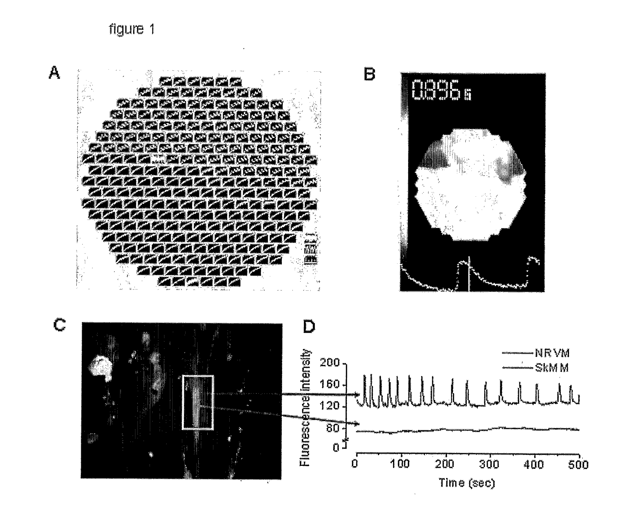
[0044]One likely contributor to arrhythmias following myoblast transplantation is the predicted absence of electrical coupling between NRVMs and myotubes. Indeed, mathematical simulations have shown that, with decreased gap junction coupling, conduction is very slow but, paradoxically, very robust (due to an increase in the safety factor for propagation), increasing the tendency for reentry.17 We confirmed the lack of electrical coupling at a syncytial level by optical mapping of co-cultures plated with SkMs on one half and NRVM on the other half of the coverslip. Stimulation on the NRVM half resulted in a propagated wave-front that blocked at the NRVM / SkM interface (FIG. 1a, b). The absence of electrical coupling was confirmed at a single-cell level by measuring lack of propagation of calcium transients between neighboring myocytes and myotubes using Rhod-2 AM (5 μM) as the calcium indicator. (FIG. 1c, d).

example 3

Lack of Electrical Coupling in Mixed Co-Cultures

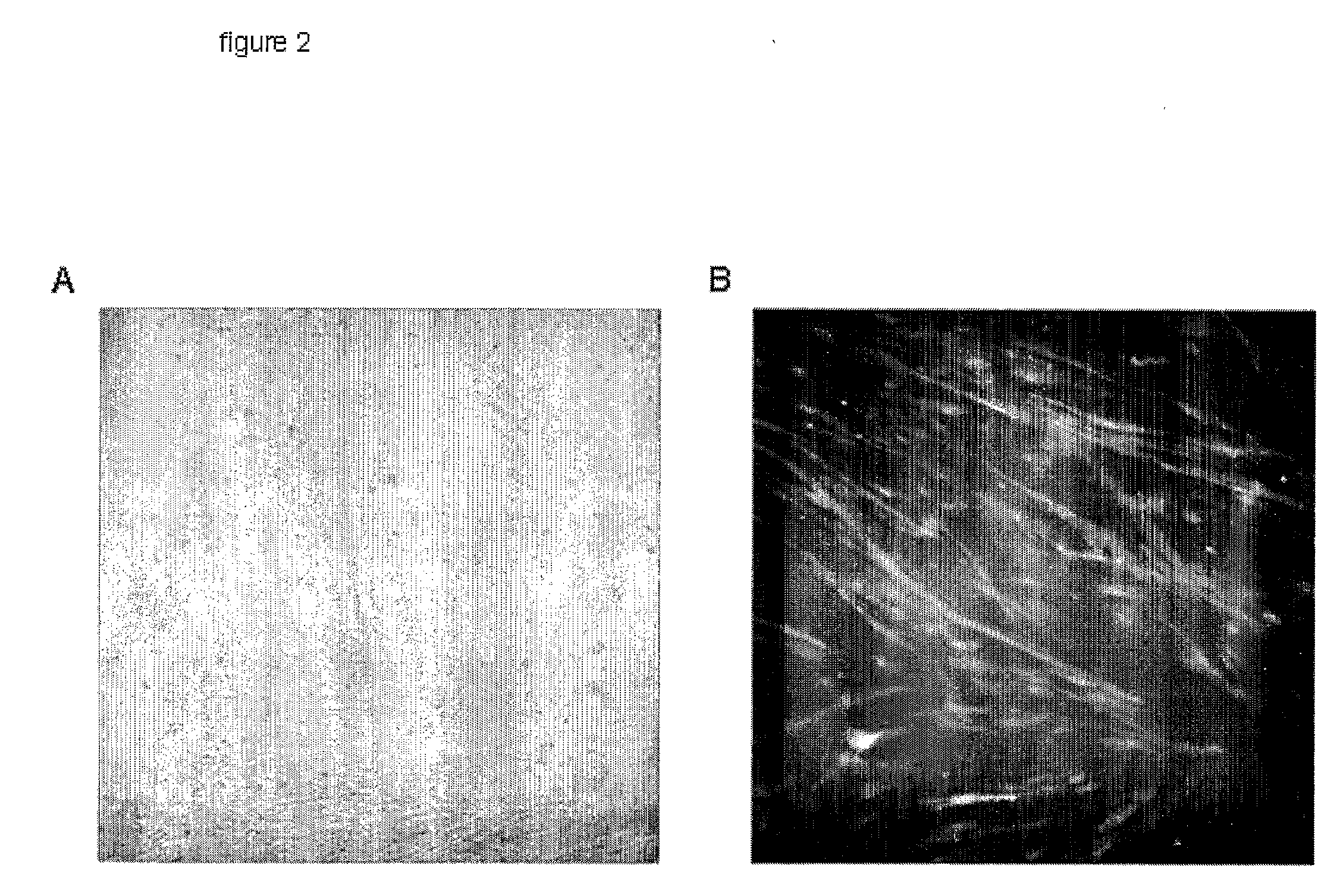
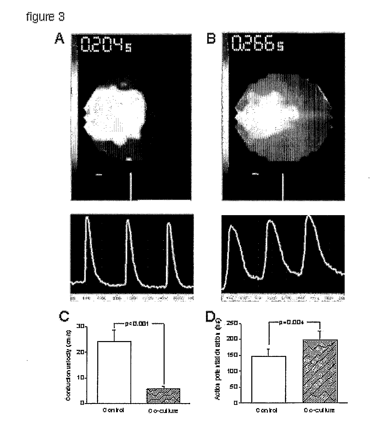
[0045]We next proceeded to characterize mixed co-cultures, a situation that mimics the engraftment of SkM in hearts in vivo.6 Light (FIG. 2a) and fluorescence microscopy (FIG. 2b) revealed that myotubes tend to grow in linear irregular patterns. The electrically-uncoupled myotubes interspersed among NRVMs would be expected to behave as localized barriers to propagation, resulting in slowing of overall conduction and predisposing to irregularities in the wave-front, source-load mismatch, wave-break and reentry.18-20 Indeed, optical mapping of mixed SkM / NRVM co-cultures revealed greatly decreased conduction velocity in all SkM: NRVM co-cultures, compared to control (NRVM-only) cultures. FIG. 3a, b shows conduction velocity in co-cultures compared to control. Additionally, action potential duration (APD80) in co-cultures was prolonged. This unanticipated delay of cardiac repolarization represents a novel pro-arrhythmic effect21 of SkM co-...


PUM

PropertyMeasurementUnit
electricalaaaaaaaaaa
voltage-sensitiveaaaaaaaaaa
fluorescentaaaaaaaaaa

Abstract

Skeletal myoblasts are an attractive cell type for transplantation since they are autologous and resistant to ischemia. However, clinical trials of myoblasts transplantation in heart failure have been plagued by ventricular tachy-arrhythmias and sudden cardiac death. The pathogenesis of these arrhythmias is poorly understood, but may be related to the fact that skeletal muscle cells, unlike heart cells, are electrically isolated by the absence of gap junctions. An in vitro model of myoblasts transplantation into cardiomyocyte monolayers can be used to investigate the mechanisms of transplant-associated arrhythmias. Co-cultures of human skeletal myoblasts and rat cardiomyocytes result in reentrant arrhythmias (spiral waves) that reproduce the features of ventricular tachycardia seen in patients receiving myoblasts transplants. These arrhythmias can be terminated by nitrendipine, an L-type calcium channel Mocker, but not by the Na channel blocker lidocaine. Genetic modification of myoblasts to stably express the gap junction protein connexin 43 decreases arrhythmogenicity in co-cultures. It similarly can be used to increase the safety of myoblasts transplantation in patients.

Description

[0001]This application claims the benefit of provisional applications Ser. Nos. 60 / 555,125 filed, Mar. 22, 2004, the disclosure of which is expressly incorporated herein.TECHNICAL FIELD OF THE INVENTION[0002]This invention is related to the area of cell transplantation. In particular, it relates to transplantation into organs that are contractile or electrically responsive.BACKGROUND OF THE INVENTION[0003]Congestive heart failure is a major public health problem in the United States.1 Cellular myoplasty represents a novel therapy for congestive heart failure, but is fraught with potential pitfalls. Skeletal myoblasts (SkM) are attractive donor cells for myoplasty: they have a contractile phenotype, can be harvested for autologous transplantation, and are resistant to ischemia.2 In ongoing phase 2 clinical trials, SkMs are harvested from individual patients via muscle biopsy, grown in culture for 2-4 weeks, and then transplanted by injection into the heart.3;4 Despite reports of impr...

Claims


Application Information

Patent Timeline
Patent Type & Authority Applications(United States)
IPC IPC(8): A61K48/00G01N33/483C12N5/10G01N27/26
CPCA61K48/005C12N5/0657C12N5/0658C12N15/86C12N2501/11C12N2740/16043A61P9/06
Inventor MARBAN, EDUARDOABRAHAM, MARIA ROSELLE
Owner THE JOHN HOPKINS UNIV SCHOOL OF MEDICINE
Learn More