

Wound healing compositions containing keratin biomaterials
a biomaterial and wound technology, applied in the field of keratin biomaterials, can solve the problems of ineffective dissolving of proteins using keratin, scientists confused by the behavior of some purified proteins, etc., and achieve the effect of promoting wound closure and inhibiting wound conversion
- Summary
- Abstract
- Description
- Claims
- Application Information
AI Technical Summary
Problems solved by technology
Method used
Image

Examples
example 1
Keratin Derivatives / Fractions
[0121]Keratose fractions were obtained using a method based on that of Alexander and coworkers. However the method was substantially modified to minimize hydrolysis of peptide bonds. Briefly, 50 grams of clean, dry hair that was collected from a local barber shop was reacted with 1000 mL of an aqueous solution of 2 w / v % peracetic acid (PAA) at room temperature for 12 hr. The oxidized hair was recovered using a 500 micron sieve, rinsed with copious amounts of DI water, and the excess water removed. Keratoses were extracted from the oxidized hair fibers with 1000 mL of 100 mM Trizma® base. After 3 hours, the hair was separated by sieve and the liquid neutralized by dropwise addition of hydrochloric acid (HCl). Additional keratoses were extracted from the remaining hair with two subsequent extractions using 1000 mL of 0.1M Trizma® base and 1000 mL of DI water, respectively. Each time the hair was separated by sieve and the liquid neutralized with HCl. All ...
example 2
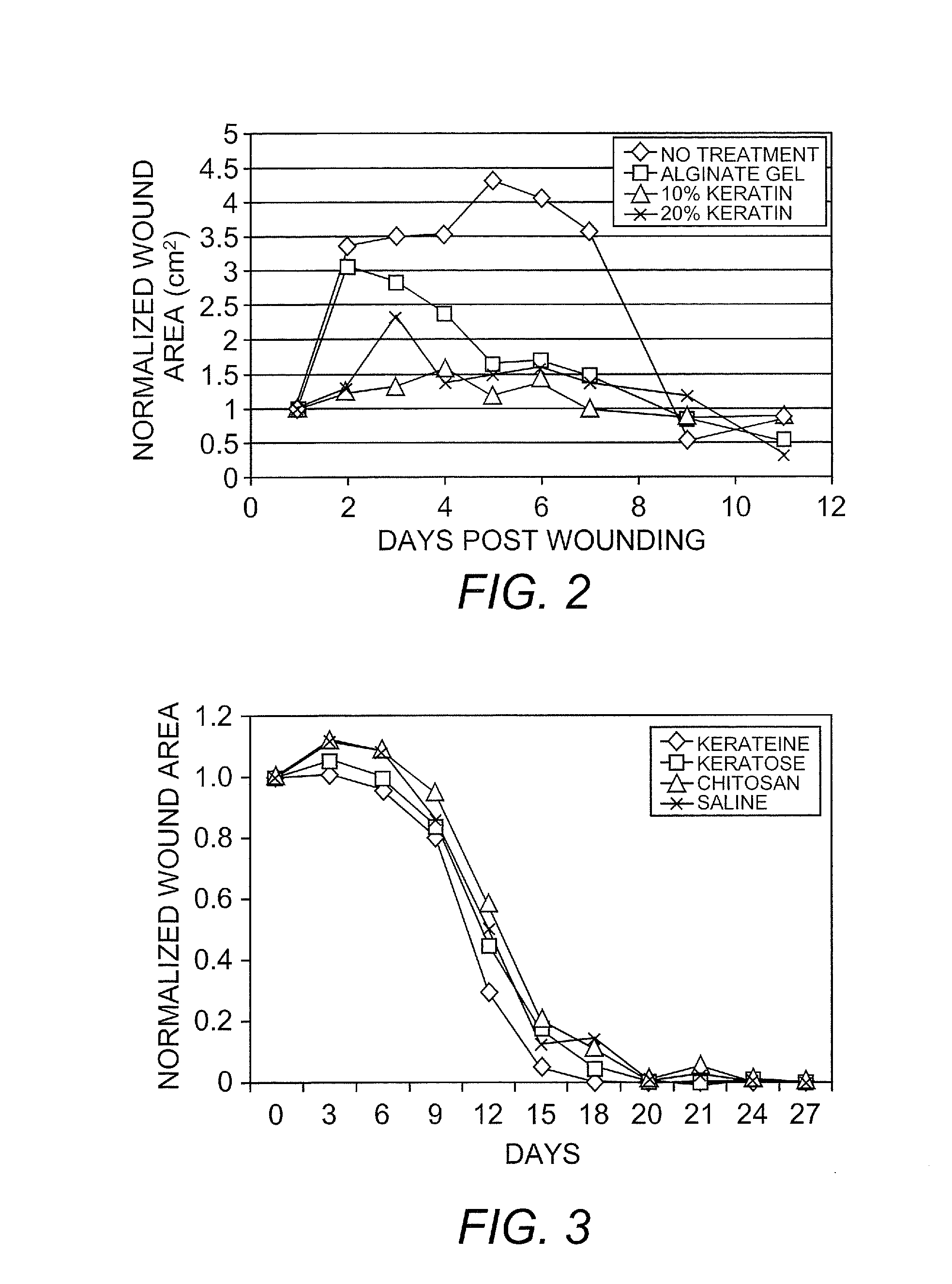
Cell Proliferation and Wound Healing in Animal Models
[0126]Several in vitro and in vivo studies were conducted to demonstrate the biological activity of keratin biomaterials. They involved the use of keratin proteins derived from human hair using oxidation and reduction reactions to break down the tertiary structure of the cortex and extract soluble proteins according to the following methods.
[0127]Keratin biomaterials derived from human hair mediated the growth behavior of skin component cells. In cell culture experiments, certain types of keratins were mitogenic toward fibroblasts and keratinocytes. Keratin-based hydrogels were shown to be capable of passivating chemical and thermal burns in a mouse and pig model, respectively.
[0128]Keratose: Clean, dry hair was cut into small fibers and oxidized with peracetic acid. Free proteins were extracted using a denaturing solution, neutralized, purified by dialysis, concentrated, and isolated by lyophilization. A hydrogel was formed by re...
example 3
Control of Bleeding in an Animal Model
[0133]The hemostatic potential of keratin gel was evaluated in a modestly challenging animal model. The keratin gel comprised unfractionated kerateine (alpha+gamma). Liver injuries are notoriously problematic as both the size of the liver and of the wound increase. This rabbit model can produce both profuse and lethal hemorrhage. Controlled liver transaction was used as a means to establish a consistent set of conditions that would result in exsanguination in the absence of treatment (negative control), yet provide for the recovery of test animals when a conventional hemostat was applied (positive control). It should be noted that the hemostats used as positive controls in this study are indicated for topical wounds and require concomitant pressure; they were applied without compression in this study. This was done to avoid the confounding contribution compression would add as it was not used with the keratin gel.
[0134]A total of 16 New Zealand ...
PUM
| Property | Measurement | Unit |
|---|---|---|
| molecular weights | aaaaa | aaaaa |
| pH | aaaaa | aaaaa |
| temperature | aaaaa | aaaaa |
Abstract
Description
Claims
Application Information