Dopominergic neurons differentiated from embryonic cells for treating neurodegenerative diseases

a technology of embryonic cells and dopominergic neurons, applied in the direction of artificial cell constructs, peptides, immunoglobulins, etc., can solve the problems of heavy dependence on unstable and problematic sources of fetal tissue in the processing process, and achieve the effect of reducing or eliminating the symptoms of neurodegenerative diseases

Active Publication Date: 2008-11-06
THE MCLEAN HOSPITAL CORP
View PDF10 Cites 2 Cited by
  • Summary
  • Abstract
  • Description
  • Claims
  • Application Information

AI Technical Summary

Benefits of technology

[0007]The invention generally provides a method for generating dopaminergic neurons in vitro by inhibiting one or more pathway components of a TGF-β signaling pathway in pluripotent cells that overexpress one or more cell fate-inducing polypeptides. The invention also provides methods and compositions for treating a neurodegenerative disease in a patient by transplanting these dopaminergic neurons into the brain of the patient, such that the transplanted dopaminergic neurons are sufficient to reduce or eliminate the symptoms of the neurodegenerative disease.

Problems solved by technology

Recently, significant progress has been achieved with transplants in Parkinson's disease (PD), but the process is heavily dependent on an unstable and problematic source of fetal tissue.

Method used


Image

  • Dopominergic neurons differentiated from embryonic cells for treating neurodegenerative diseases

Examples


example 1

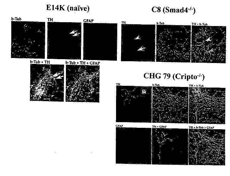
In Vitro Differentiation Neuronal Differentiation of Smad4− / − and Cripto− / − Embryonic Stem Cells

[0098]Smad4 and Cripto are key TGF-β signaling pathway components that control multiple aspects of embryogenesis, including mesodermal and epidermal cell development. Both Smad4− / − and Cripto− / − mice die prior to embryonic day 7.5 with severe developmental defects in gastrulation and axial organization, respectively.

[0099]ES Cell Culture and In Vitro Differentiation.

[0100]Wild type (WT) E14K and Smad4− / − (C8-13A1) ES cells were kindly provided by Drs. C. Sirard and Tak W. Mak, University of Toronto, Toronto, Canada). Wild type TC 1 and the Cripto− / − (clones CHG51 and CHG79) were kindly provided by Dr. M. M. Shen, UMDNJ-Robert Wood Johnson Medical School, New Jersey, USA. The ES cells were propagated and in vitro differentiated as described previously (Lee et al. Nat. Biotechnol. 18: 675-679, 2000; Chung et al. Eur. J. Neurosci. 16: 1829-1838, 2002).

[0101]Immunocytochemistry

[0102]Cells wer...

example 2

Transplantation of Cultured Smad4− / − and Cripto− / − Embryonic Stem Cells into the Mammalian Brain

[0118]Preparation of ES Cells for Transplantation

[0119]ES cells were cultured for four days in the absence of LIF on 100 mm Fisher brand bacteriological grade petri dishes to form embryoid bodies (EB). EBs were then transferred to 15 ml sterile culture tubes, spun at 1000 rpm for 5 min, and rinsed once in Ca2+ and Mg2+-free Dulbecco's Phosphate-Buffered Saline (D-PBS, Gibco / BRL). After rinsing, D-PBS was removed and 1.5 ml of trypsin solution was added. The cells were incubated for 5 minutes at 37° C. and triturated with fire polished Pasteur pipettes with decreasing aperture size to fully dissociate the cells. Finally, ES cells were spun at 1000 for 5 min and the trypsin solution replaced with 200 μl culture media. The viability and concentration of the ES cells was determined using a hemocytometer after staining with acridine orange and ethidium bromide.

[0120]Transplantation of Cultured...


PUM

PropertyMeasurementUnit
anatomical structuresaaaaaaaaaa
nucleic acidaaaaaaaaaa
lengthaaaaaaaaaa

Abstract

Disclosed herein are methods for generating dopaminergic neurons in vitro by inhibiting a pathway component of a TGF-β signaling pathway and overexpressing one or more cell fate-inducing polypeptides in pluripotent cells, causing differentiation of the pluripotent cells into dopaminergic neurons. Also disclosed are methods for treating a neurodegenerative disease in a patient by generating dopaminergic neurons in vitro, and transplanting them into the brain of the patient, such that the dopaminergic neurons are sufficient to reduce or eliminate the symptoms of the neurodegenerative disease.

Description

[0001]This application claims benefit of the filing date of the copending U.S. Provisional Application No. 60 / 432,128 (filed Dec. 9, 2002), hereby incorporated by reference.BACKGROUND OF THE INVENTION[0002]Neurodegenerative disorders such as Parkinson's, Alzheimer's, and Huntington's disease are becoming ever more prominent in our society. A direct approach towards therapeutic treatment of these diseases is through replacement therapy where undamaged tissue is transplanted into the nervous system. Recently, significant progress has been achieved with transplants in Parkinson's disease (PD), but the process is heavily dependent on an unstable and problematic source of fetal tissue. Embryonic stem (ES) cells may be a tissue / cell source for developing the therapeutic potential of neural transplantation. Self-renewing and multipotent stem cells provide a source of transplantable material to replace post-mitotic neurons that do not spontaneously regenerate after injury.[0003]PD is a prog...

Claims


Application Information

Patent Timeline
Patent Type & Authority Applications(United States)
IPC IPC(8): C12N5/06C12N5/08C12N15/87C12N5/02C12N5/0793
CPCC12N5/0619C12N2501/15C12N2501/40C12N2506/02
Inventor ISACSON, OLEBJORKLUND, LARS
Owner THE MCLEAN HOSPITAL CORP
Why Eureka
Try Eureka