Cell-Image Analysis Method, Cell-Image Analysis Program, Cell-Image Analysis Apparatus, Screening Method, and Screening Apparatus

Inactive Publication Date: 2008-11-13
OLYMPUS CORP
View PDF3 Cites 81 Cited by
  • Summary
  • Abstract
  • Description
  • Claims
  • Application Information

AI Technical Summary

Benefits of technology

[0089]According to the present invention, it is possible to perform detailed analysis of cell populations. By investigating the correlations of feature changes, it is possible to perform, for example, quantitative measurement and characterization of cellular changes caused by a specific compound and to efficiently compare the effects of a plurality of compounds.
[0090]By analyzing the positions of cells on an image, it is possible to predict the influence of inter-cell interactions on t

Problems solved by technology

In particular, in experiments on cancer cells, it is frequently the case that the properties that characterize them as cancer cells are lost; therefore, it is necessary from the outset to check whether these cel

Method used


Image

  • Cell-Image Analysis Method, Cell-Image Analysis Program, Cell-Image Analysis Apparatus, Screening Method, and Screening Apparatus
  • Cell-Image Analysis Method, Cell-Image Analysis Program, Cell-Image Analysis Apparatus, Screening Method, and Screening Apparatus
  • Cell-Image Analysis Method, Cell-Image Analysis Program, Cell-Image Analysis Apparatus, Screening Method, and Screening Apparatus

Examples


first embodiment

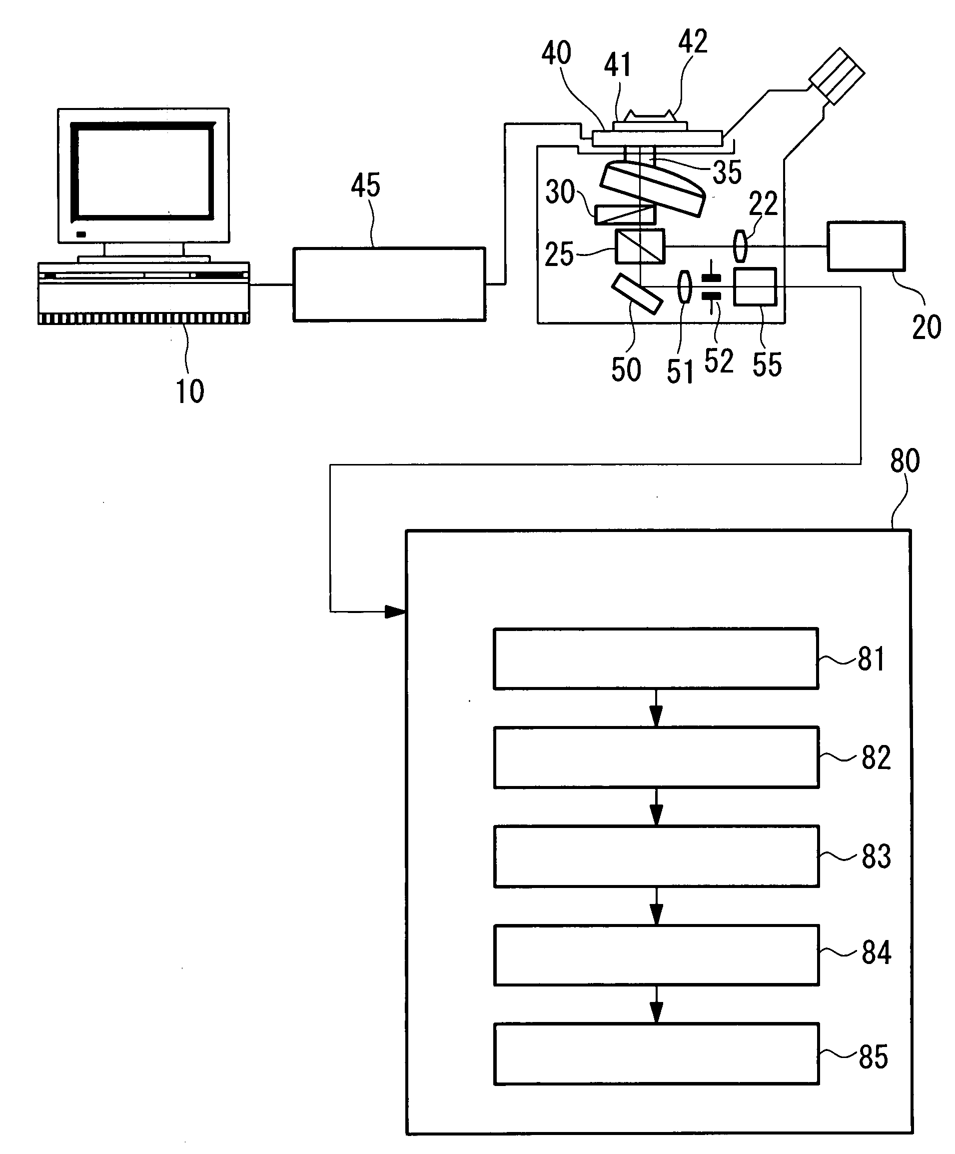
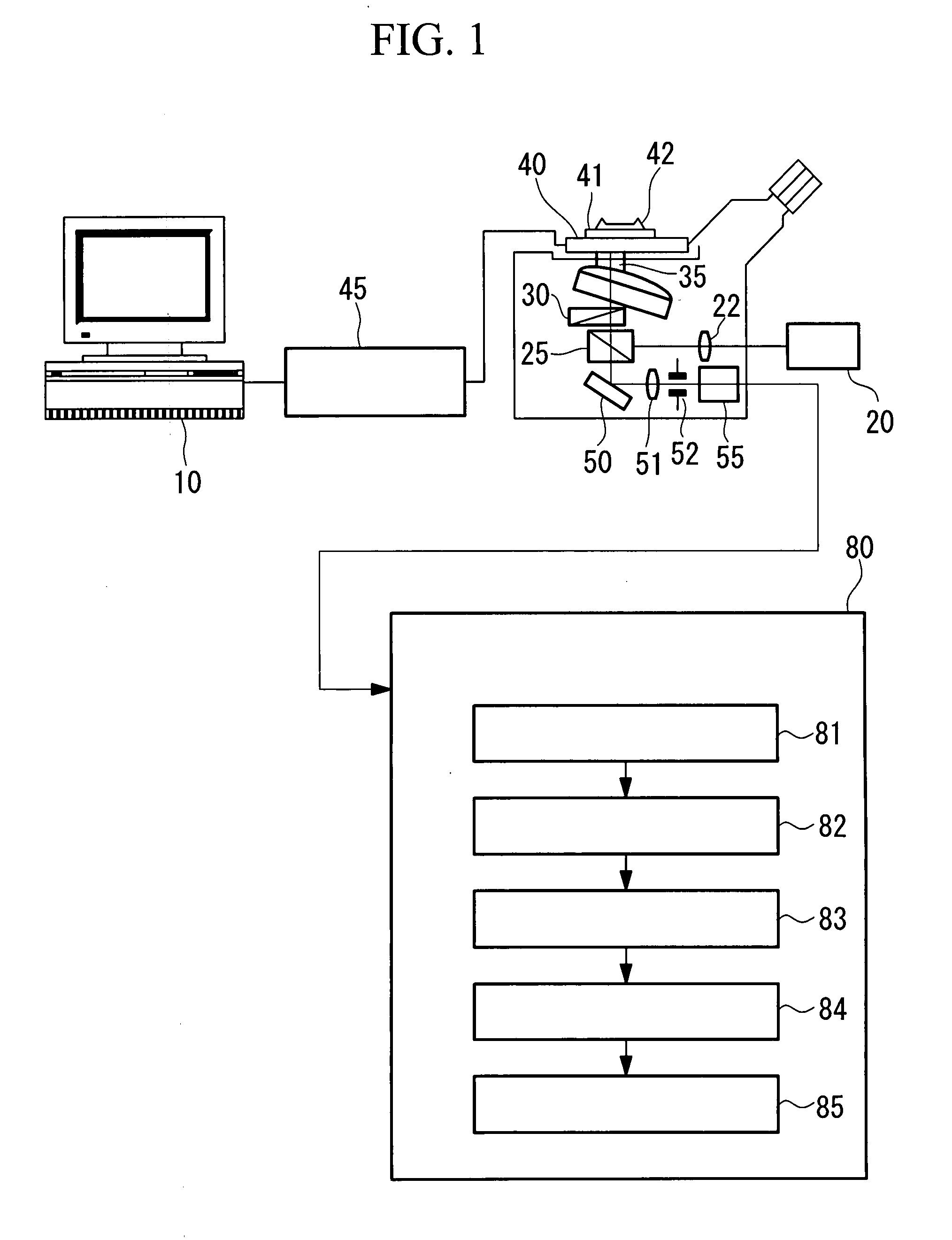
[0119]A first embodiment of the present invention will be described with reference to the drawings. The basic configuration for realizing the present invention will be described using FIG. 1. FIG. 1 is a block diagram showing, in outline, the configuration of a cell-image analysis apparatus according to a first embodiment of the present invention, in which a scanning confocal optical microscope is illustrated as an example of the basic configuration of the cell-image analysis apparatus according to this embodiment.

[0120]An argon laser with a wavelength of, for example, 488 nm is used as a light source 20. The beam diameter of the laser light emitted from the light source 20 is expanded by a collimator lens 22 to be converted to a collimated beam, and is guided to a dichroic mirror 25. The laser light guided to the dichroic mirror 25 is reflected at the dichroic mirror 25, is incident on an XY scanner 30, and is scanned in the X and Y directions at the scanner 30. The laser light pas...

second embodiment

[0194]A second embodiment of the present invention will be described below with reference to FIGS. 7 to 11, and 15 to 17.

[0195]FIG. 7 shows the configuration of a cell-image analysis apparatus according to the second embodiment of the present invention.

[0196]As shown in FIG. 7, the cell-image analysis apparatus according to this embodiment includes a stage 101 for mounting a sample plate 1; an excitation light source 102; a power supply unit 103 connected to the excitation light source 102; a shutter unit 104 for turning the light from the excitation light source 102 on and off; a lens 105 for converting the light to collimated light; a filter unit 106 for selecting the wavelength of excitation light to be made incident on the sample; a mirror set 107 for reflecting the excitation light to make it incident on the sample and for transmitting fluorescence; an objective lens 108 for focusing the excitation light onto a sample; an image-forming lens 109 for focusing the fluorescence col...

third embodiment

[0218]Next, a third embodiment of the present invention will be described below with reference to the drawings.

[0219]In the description of these embodiments, parts having the same configuration as those in the second embodiment described above are assigned the same reference numerals, and a description thereof is omitted.

[0220]The sample plate 1 used in this embodiment is the same as that used in the second embodiment; it has 15 wells 1a, with a diameter of 20 mm and a depth of 15 mm, arranged in three rows by five columns. Cells stained with fluorescent dye, serving as the objects to be examined, are inoculated in each of these wells 1a. The fluorescent dye used in this embodiment is a dye that can emit fluorescence while the cells are kept alive, for example, GFP, and dyes the cell nucleus.

[0221]Screening of an anti-cancer drug is described as an example.

[0222]Flowcharts of the cell-image analysis method according to this embodiment are shown in FIGS. 16 and 18.

[0223]The examiner ...


PUM


Abstract

In compound screening using cell images, compounds having an effective action on cells are searched for by automated processing, to more perform existing methods for compound searching more effectively. There is provided an apparatus including an image-acquisition unit for capturing a plurality of cell images under different image-acquisition conditions; and an analysis unit for extracting a cell position and at least one feature on the basis of the plurality of cell images and for performing statistical analysis with the extracted features serving as parameters.

Description

TECHNICAL FIELD[0001]The present invention relates to a cell-image analysis method and a cell-image analysis apparatus for performing extraction of specific data by image processing images of plant and animal cells.[0002]Furthermore, the present invention relates to a cell-image analysis method, a cell-image analysis program, and a cell-image analysis apparatus used in pathological diagnosis of cancer etc., drug screening, and so forth in which living cells are used.[0003]Furthermore, the present invention relates to a screening method and a screening apparatus using image processing, and in particular, to a screening method and a screening apparatus which compare changes in the morphology of cells with reference to cell images captured by a microscope.BACKGROUND ART[0004]In the fields of biotechnology and pharmaceutical development, work is carried out to check the effect or influence of substances, such as various pharmaceuticals and drugs, on plant and animal cells or microorgani...

Claims


Application Information

Patent Timeline
IPC IPC(8): G06K9/00
CPCG01N15/1475G01N21/6458G01N33/5008G01N33/5026G06K9/0014G06T7/0012G06T2207/30024G06V20/695
Inventor MATSUO, YUICHIROTAKAGI, KOSUKESHIMADA, YOSHIHIROKAWAI, TSUYOSHI
Owner OLYMPUS CORP
Why Eureka
Try Eureka