

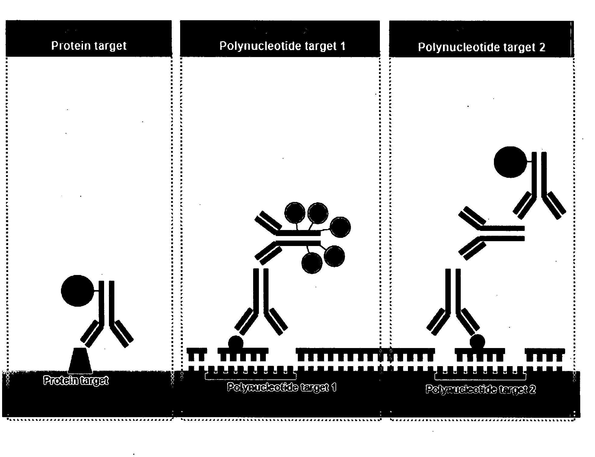
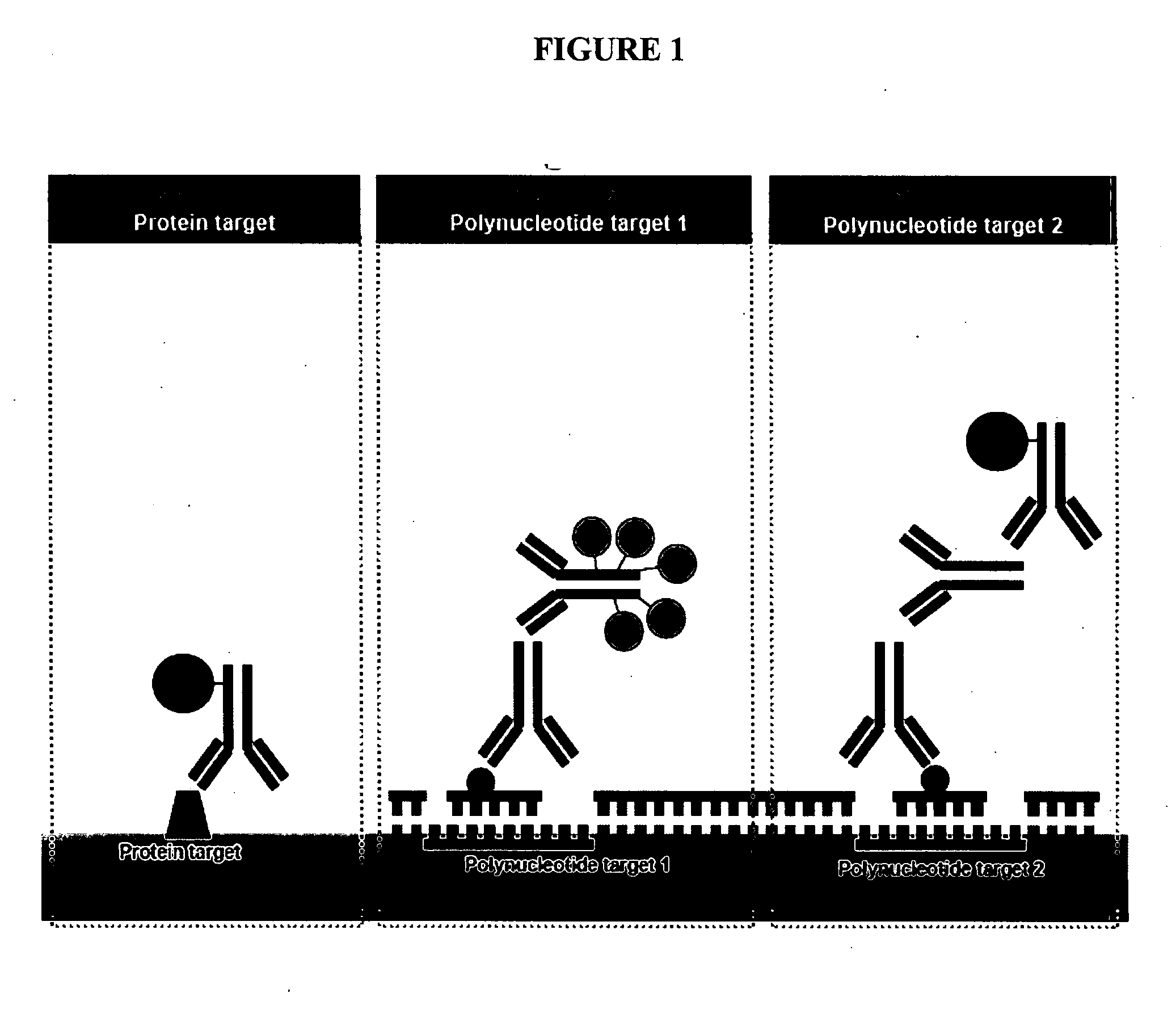
Multicolor chromogenic detection of biomarkers
a biomarker and color technology, applied in the field of multicolor chromogenic detection of biomarkers, can solve the problems of fish not always ideal for clinical diagnostic practice, cumbersome and limited resolution, etc., and achieve the effect of more sensitive and/or rapid detection of targets
- Summary
- Abstract
- Description
- Claims
- Application Information
AI Technical Summary
Benefits of technology
Problems solved by technology
Method used
Image

Examples
example 1
Detection of Single Copy of HER2 Gene in Tissue Using SISH Detection
[0168]In this example, the method of the present invention was used to sensitively and selectively detect a single copy of a gene in situ, such as HER2 gene in normal as well as in breast cancer tissue, using Silver in situ hybridization (“SISH”).
[0169]Formalin-fixed, paraffin-embedded (FFPE) human breast carcinoma containing normal breast epithelium was used as a positive control. The slides containing the FFPE tissue were prepared and stained automatically by using an automated staining system BenchMark™ XT (Ventana Medical Systems, Inc., Tucson, Ariz.) operated in accordance with standard procedures.
[0170]The pre-programmed protocol “XT SISH iVIEW™ SILVER” was used to prepare and stain all tissues via automated in situ hybridization. The deparaffinization option was selected for 20 minutes at 75° C. under the EZ Prep™ solution (Ventana P / N 950-102). The solution is applied through the rinse nozzles which leaves a...
example 2
Detection of Single Copy of HER2 Gene in Tissue with SISH
[0178]In yet another experiment, the method of the present invention was used to sensitively and selectively detect a single copy of a gene in situ, such as normal HER2 genes as well as amplified HER2 genes, using a repeat-depleted HER2 probe with SISH detection.
[0179]Formalin-fixed, paraffin-embedded (FFPE) human breast carcinoma cell line xenograft tumors were used for assay optimization. The slides containing the FFPE tumor sections were prepared and stained using an automated staining system BenchMark™ XT (Ventana Medical Systems, Inc., Tucson, Ariz.) operated in accordance with standard procedures.
[0180]The pre-programmed protocol “XT SISH iVIEW™ SILVER” was used to prepare and stain all tissues via automated in situ hybridization. The deparaffinization option was selected for 20 minutes at 75° C. under the EZ Prep™ solution (Ventana P / N 950-102). The solution was applied through the rinse nozzles which leaves approximate...
example 3
Co-Detection of HER2 and Chromosome 17 Centromere in Tissue with Sequential Hybridization Steps
[0186]In this example, the method of the present invention was used to sensitively and selectively detect copy numbers of.the HER2 gene in combination with a Chromosome 17 centromere (CEP 17) label in situ, in formalin-fixed, paraffin-embedded xenograft tumor tissue sections using a double stranded DNA probe (HER2) and a single stranded oligoprobe (CEP 17) with different stringency characteristics. Inclusion of CEP 17 detection allows for the relative copy number of the HER2 gene to be determined. For example, normal samples will have a HER2 / CEP 17 ratio of less than 2, whereas samples in which the HER2 gene is reduplicated will have a HER2 / CEP 17 ratio of greater than 2.0.
[0187]The assay was completely automated on a Ventana BenchMark™ XT. The slides with the fixed cells were baked / heated at 65° C. for 20 minutes and deparaffinized using EZ Prep (Ventana P / N 950-102). The slides were furt...
PUM
| Property | Measurement | Unit |
|---|---|---|
| distance | aaaaa | aaaaa |
| distance | aaaaa | aaaaa |
| distance | aaaaa | aaaaa |
Abstract
Description
Claims
Application Information