Method for detecting a target using enzyme directed deposition of elemental metal

a technology of elemental metal and detection method, which is applied in the field of detection method using enzyme directed deposition of elemental metal, can solve the problems of low sensitivity of other enzyme methods, less popular type of detection, and the inability to detect targets, etc., and achieves easy calibration of reaction rates, more sensitive detection, and inherent amplification steps

Inactive Publication Date: 2011-02-15
NANOPROBES
View PDF20 Cites 12 Cited by
  • Summary
  • Abstract
  • Description
  • Claims
  • Application Information

AI Technical Summary

Benefits of technology

[0034]Gold nanoparticles have extinction coefficients about 1,000 times higher than highly colored compounds or fluorophores. Therefore, their use substantially increases sensitivity.
[0035]No expensive fluorescent optics, light sources or filters are required. Site-specific and selective deposition of elemental metal in the targeted site or molecules not facilitates chromogenic detection of the signals, allows cell morphology and in situ hybridization (ISH) signal to be viewed at the same time, and provides accurate results using standard equipment, such as bright field-microscopes. There is no fading of the sample upon observation or storage or exposure to room lights. Autofluorescence from cells and other molecules does not create any interference. Standard stains (such as nuclear fast red, hematoxylin and eosin) can be used and seen simultaneously with the enzyme deposited metals, making visualization of landmarks of a tissue simple.
[0036]In one aspect, the enzymatic metal deposition of the invention allows deposition of silver metal in the presence of an enzyme and activating agents with high sensitivity combined with high resolution and minimal background for chromogenic in situ hybridization (CISH) detection, and visualization in the conventional bright field microscope without the need for oil immersion. Such an assay is herein termed as “Silver In Situ Hybridization” (SISH). In particular, the enzymatic metal deposition of the invention allows detection of a single copy of a target gene in a chromosome by a conventional bright field microscope with requiring oil immersion. The invention also enables detection of gene copies with a resolution that allows for individual enumeration of signals, such as discrete metal deposit dots for individual gene copies. In a variation of the embodiment, the invention allows for detection of at least 2, 3, 4, 5, 6, 7 or 8 copies of a target gene per nucleus, such as HER2 gene in human chromosome 17, as discrete metal deposit dots.
[0058]The invention disclosed herein utilizes an enzyme reaction for deposition or product formation. The deposition rates are determined by enzyme kinetics, which result in linear or easily calibrated rates of reaction. This makes the invention much more suitable for quantitative detection than conventional autometallographic methods of depositing metal on nucleating particles. Additionally the invention does not suffer from irregular rates of deposition from particle to particle as is known to occur with conventional autometallographic methods.
[0059]The present invention has an inherent amplification step since an enzyme catalyst is used. Thus the invention can provide more sensitive detection than is possible with non-enzymatic methods, such as direct immunolabeling with a fluorophore or chromophore.
[0060]Compared with, for example, deposition of an organic molecule; enzymatic metal deposition makes possible many additional detection methods including by x-rays, electron microscopy, electrochemical, optical, magnetic, and many others. Many of the additional test methods useful with enzymatic metal deposits are exceedingly sensitive, and permit assays more sensitive and / or rapid than is possible with conventional test methods.

Problems solved by technology

Enzymes control most body processes, and even cancer involves improper levels of certain enzymes regulating cell growth and death.
A disadvantage of these calorimetric substrates is that they are generally of lower sensitivity than other enzyme methods.
Although sensitive, this type of detection is becoming less popular due to the risks of handling and disposing of radioactive material, and since other methods now rival or exceed its sensitivity.
Radioactive labeling for histochemical uses and autoradiography, typically require months to expose films, due to low specific activity, which is another disadvantage.
A number of drawbacks, however, come with use of fluorescent substrates.
Fluorescence fades upon observation, sample storage or even exposure to room lights, thus making permanent or quantitative data difficult to achieve.
Autofluorescence (fluorescence coming from certain compounds found naturally in many living organisms) from cells and other molecules can interfere with the test result.
Standard tissue stains (such as nuclear fast red, hematoxylin, and eosin) cannot be seen simultaneously with the very different optics and illumination required for fluorescence detection, thus making visualization of landmarks of a tissue difficult.
Unfortunately, fluorescence is viewed using different illumination and sharp bandpass filters, so that only the fluorescent label is visible, and the general view of the stained tissue is not simultaneously available.
Detection requires expensive equipment or necessitates film development.
Sensitive detection often requires lengthy light integration times of hours or even a day.
Standard stains (such as nuclear fast red, hematoxylin, and eosin) cannot be seen simultaneously, thus making visualization of landmarks of a tissue difficult, for example.
Additionally, all of the conventional detection schemes have some practical limitations for sensitivity.
Another limitation on detection method sensitivity is the amount of signal produced.
For example, if an enzyme is used that has a low turnover, and produces only relatively few products, these few products will be harder to detect, and sensitivity will be worse than if a more efficient enzyme producing more products was used.
Of course there are practical limitations to enzyme amplification, such as the enzyme losing activity, the long times necessary to accumulate product, side reactions or other sources of background limit detection.
Further, at some point the product produced may interfere with enzyme activity, either by shifting the reaction equilibrium, depositing products so as to impede flow to the active site of the enzyme or otherwise inhibiting the enzyme.
Although this type of assay has been used quite successfully, it has several drawbacks, including: expense of reagents, insufficient amplification, background problems, localization at the ultrastructural level (using electron microscopy) can be diffuse, and the limitations using fluorophores or chromophores mentioned previously.
Similarly, the enzyme / device interface in other types of known biosensors is often prone to similar non-specific signals.
Detection is usually in the 10−4 to 10−2 M region, and therefore the above method generally lacks the accuracy and precision required for many analytes.
However, these metal ions do not serve as substrates, are not linked to substrates and do not deposit as metal.
Unfortunately, this use has not been widely employed since the background goes up substantially, and little improvement in signal-to-noise ratio is generally found.

Method used


Image

  • Method for detecting a target using enzyme directed deposition of elemental metal
  • Method for detecting a target using enzyme directed deposition of elemental metal
  • Method for detecting a target using enzyme directed deposition of elemental metal

Examples


example 1

Enzymatic Deposition of Silver Metal

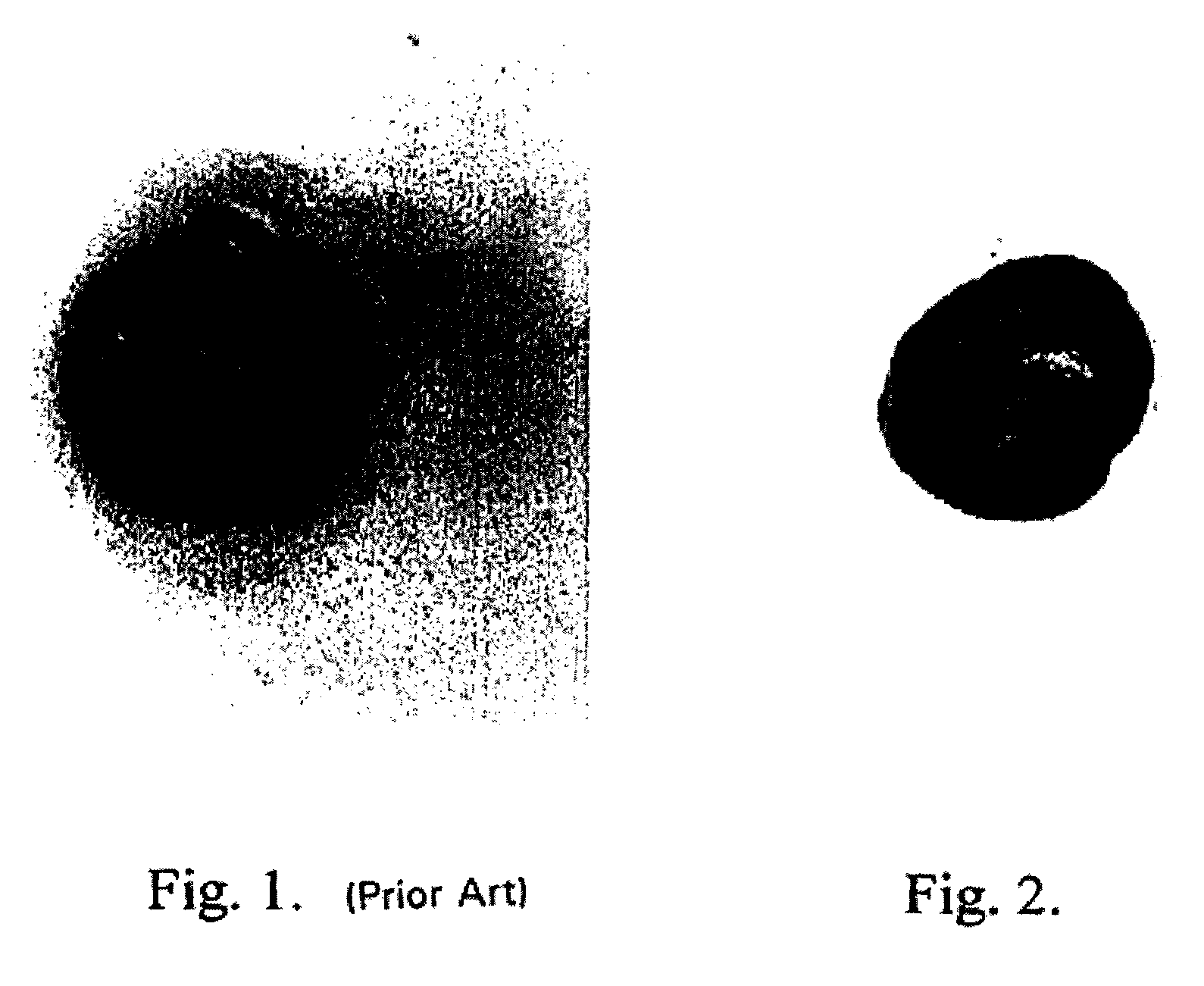
[0184]One microgram of horseradish peroxidase was applied to a nitrocellulose membrane and allowed to dry. The membrane was then optionally blocked using 4% bovine serum albumin and washed. A solution containing 2.5 mg / ml hydroquinone and 1 mg / ml silver acetate in a 0.1 M citrate buffer, pH 3.8 was applied. Next, hydrogen peroxide was added and mixed to a final concentration of 0.03 to 0.06%. Silver deposition selectively occurred at the peroxidase spot as evidenced by a black product. No deposit occurred if the hydrogen peroxide was omitted.

example 2

Enhanced Enzymatic Deposition of Silver Metal; Pretreatment with Silver Ions

[0185]One microgram of horseradish peroxidase was applied to a nitrocellulose membrane and allowed to dry. The membrane was then optionally blocked using 4% bovine serum albumin and washed. A solution of 2 mg / ml silver acetate in water was applied for three to five minutes, then washed with water. A solution containing 2.5 mg / ml hydroquinone and 1 mg / ml silver acetate in a 0.1 M citrate buffer, pH 3.8 was applied. Next, hydrogen peroxide was added and mixed to a final concentration of 0.03 to 0.06%. Silver deposition immediately and selectively occurred at the peroxidase spot as evidenced by an intense black product. No silver deposit occurred if the hydrogen peroxide was omitted.

example 3

Enhanced Enzymatic Deposition of Silver Metal; Pretreatment with Gold Ions

[0186]One microgram of horseradish peroxidase was applied to a nitrocellulose membrane and allowed to dry. The membrane was then optionally blocked using 4% bovine serum albumin and washed. A solution of 0.1 mg / ml potassium tetrabromoaurate in water was applied for five minutes, then briefly washed with water. A solution containing 2.5 mg / ml hydroquinone and 1 mg / ml silver acetate in a 0.1 M citrate buffer, pH 3.8 was applied. Next, hydrogen peroxide was added and mixed to a final concentration of 0.03 to 0.06%. Silver deposition immediately and selectively occurred at the peroxidase spot as evidenced by an intense brown-black product.


PUM

PropertyMeasurementUnit
distanceaaaaaaaaaa
diameteraaaaaaaaaa
pHaaaaaaaaaa

Abstract

Disclosed embodiments concern site-specific, enzymatic-directed deposition of elemental metal for in-situ analysis. Enzyme substrates are contacted with metal ions and subsequently direct the deposition of elemental metals. Sensitive and selective detection of target molecules, such as biomarkers in various biological samples, can be obtained using various methods, such as in-situ chromogenic immunohistochemical (IHC) detection with bright field light microscopy.

Description

CROSS-REFERENCE TO RELATED APPLICATIONS[0001]This application is a continuation-in-part application of U.S. patent application Ser. No. 11 / 714,682, filed Mar. 5, 2007 now U.S. Pat. No. 7,691,598, which is a continuation-in-part of U.S. patent application Ser. No. 11 / 627,735, filed Jan. 26, 2007 now U.S. Pat. No. 7,592,153, which is a divisional application of U.S. patent application Ser. No. 10 / 658,609, filed Sep. 8, 2003, now U.S. Pat. No. 7,183,072, issued Feb. 27, 2007, which is a continuation-in-part of U.S. patent application Ser. No. 09 / 822,131, filed Mar. 30, 2001, now U.S. Pat. No. 6,670,113, issued Dec. 30, 2003, the contents of which are incorporated by reference in their entirety.BACKGROUND OF THE INVENTIONEnzymesTheir Function and Uses[0002]Enzymes are proteins, usually derived from living organisms, that are also catalysts for various metabolic or chemical reactions. Enzymes are therefore essential to all life. Recently, enzymes have been isolated, studied, altered, com...

Claims


Application Information

Patent Timeline
Patent Type & Authority Patents(United States)
IPC IPC(8): C12Q1/26
CPCC12N9/0065C12P3/00C12Q1/001C12Q1/28G01N33/543G01N33/5438G01N33/581
Inventor HAINFELD, JAMES F.LIU, WENQIUPOWELL, RICHARD D.JOSHI, VISHWAS N.
Owner NANOPROBES
Why Eureka
Try Eureka